Proteins
By the end of this section, you will be able to:
- Describe the functions proteins perform in the cell and in tissues
- Discuss the relationship between amino acids and proteins
- Explain the four levels of protein organization
- Describe the ways in which protein shape and function are linked
Proteins are one of the most abundant organic molecules in living systems and have the most diverse range of functions of all macromolecules. Proteins may be structural, regulatory, contractile, or protective. They may serve in transport, storage, or membranes; or they may be toxins or enzymes. Each cell in a living system may contain thousands of proteins, each with a unique function. Their structures, like their functions, vary greatly. They are all, however, amino acid polymers arranged in a linear sequence.
Types and Functions of Proteins
Enzymes, which living cells produce, are catalysts in biochemical reactions (like digestion) and are usually complex or conjugated proteins. Each enzyme is specific for the substrate (a reactant that binds to an enzyme) upon which it acts. The enzyme may help in breakdown, rearrangement, or synthesis reactions. We call enzymes that break down their substrates catabolic enzymes. Those that build more complex molecules from their substrates are anabolic enzymes, and enzymes that affect the rate of reaction are catalytic enzymes. Note that all enzymes increase the reaction rate and, therefore, are organic catalysts. An example of an enzyme is salivary amylase, which hydrolyzes its substrate amylose, a component of starch.
Hormones are chemical-signaling molecules, usually small proteins or steroids, secreted by endocrine cells that act to control or regulate specific physiological processes, including growth, development, metabolism, and reproduction. For example, insulin is a protein hormone that helps regulate the blood glucose level. The table below lists the primary types and functions of proteins.
Protein Types and Functions
| Type | Examples | Functions |
|---|---|---|
| Digestive Enzymes | Amylase, lipase, pepsin, trypsin | Help in food by catabolizing nutrients into monomeric units |
| Transport | Hemoglobin, albumin | Carry substances in the blood or lymph throughout the body |
| Structural | Actin, tubulin, keratin | Construct different structures, like the cytoskeleton |
| Hormones | Insulin, thyroxine | Coordinate different body systems’ activity |
| Defense | Immunoglobulins | Protect the body from foreign pathogens |
| Contractile | Actin, myosin | Effect muscle contraction |
| Storage | Legume storage proteins, egg white (albumin) | Provide nourishment in early embryo development and the seedling |
Proteins have different shapes and molecular weights. Some proteins are globular in shape; whereas, others are fibrous in nature. For example, hemoglobin is a globular protein, but collagen, located in our skin, is a fibrous protein. Protein shape is critical to its function, and many different types of chemical bonds maintain this shape. Changes in temperature, pH, and exposure to chemicals may lead to permanent changes in the protein’s shape, leading to loss of function, or denaturation. Different arrangements of the same 20 types of amino acids comprise all proteins. Two rare new amino acids were discovered recently (selenocysteine and pyrrolysine), and additional new discoveries may be added to the list.
Amino Acids
Amino acids are the monomers that comprise proteins. Each amino acid has the same fundamental structure, which consists of a central carbon atom, or the alpha (α) carbon, bonded to an amino group (NH₂), a carboxyl group (COOH), and to a hydrogen atom. Every amino acid also has another atom or group of atoms bonded to the central atom known as the R group (below).
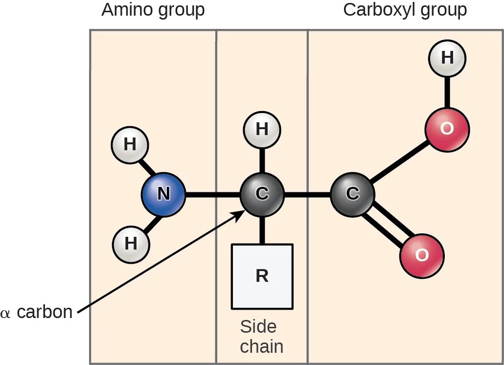
Scientists use the name “amino acid” because these acids contain both amino group and carboxyl-acid-group in their basic structure. As we mentioned, there are 20 common amino acids present in proteins. Nine of these are essential amino acids in humans because the human body cannot produce them and we obtain them from our diet. For each amino acid, the R group (or side chain) is different (below).
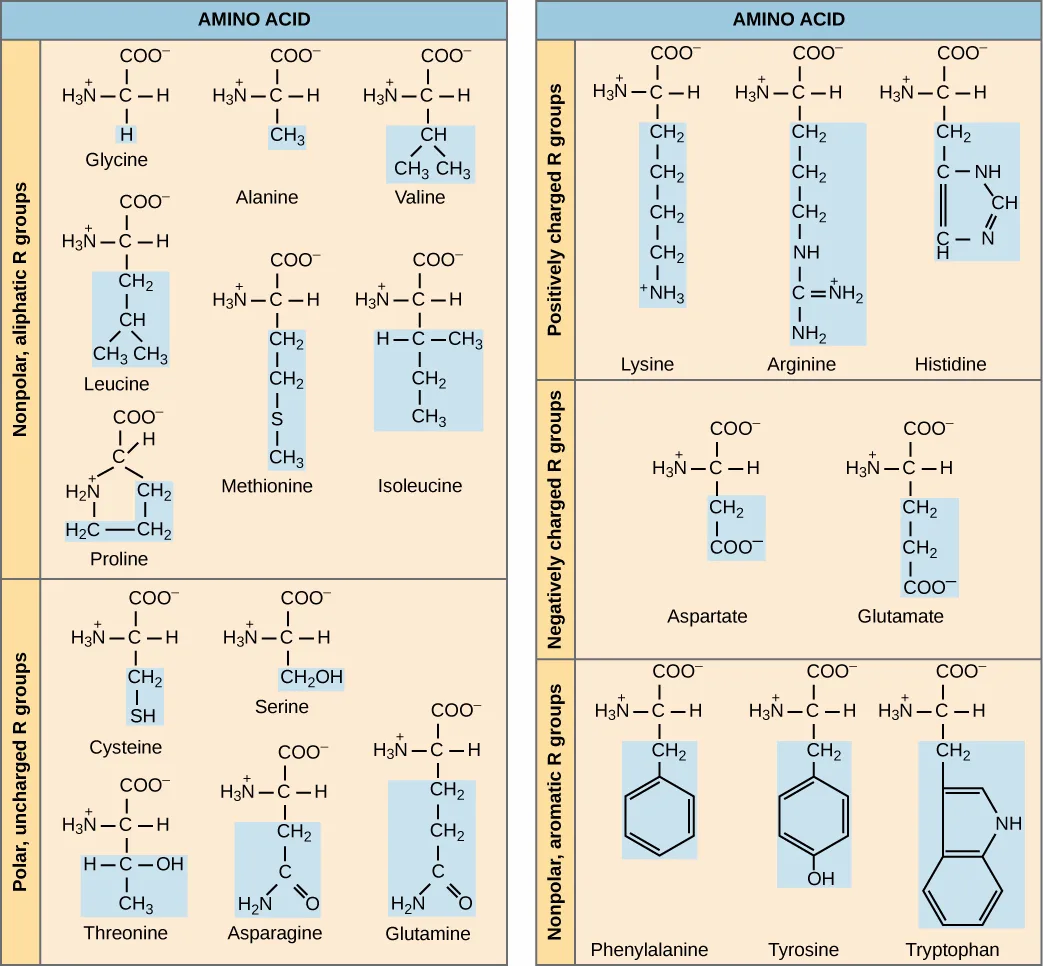
Extended description
Two columns of boxed amino acid structures grouped by side-chain chemistry. Left column, top to bottom: nonpolar, aliphatic R groups — glycine, alanine, valine, leucine, methionine, isoleucine, and proline; then polar, uncharged R groups — cysteine, serine, threonine, asparagine, and glutamine. Right column, top to bottom: positively charged R groups — lysine, arginine, and histidine; negatively charged R groups — aspartate and glutamate; and nonpolar, aromatic R groups — phenylalanine, tyrosine, and tryptophan. Each amino acid is drawn as its full backbone structure (a central carbon bonded to an amino group and a carboxyl group) with its distinct side chain attached — for example, glycine’s side chain is a single hydrogen, while alanine’s is a methyl group.
Which categories of amino acid would you expect to find on the surface of a soluble protein, and which would you expect to find in the interior? What distribution of amino acids would you expect to find in a protein embedded in a lipid bilayer?
Show model answer
Did your answer mention:
The chemical nature of the side chain determines the amino acid’s nature (that is, whether it is acidic, basic, polar, or nonpolar). For example, the amino acid glycine has a hydrogen atom as the R group. Amino acids such as valine, methionine, and alanine are nonpolar or hydrophobic in nature, while amino acids such as serine, threonine, and cysteine are polar and have hydrophilic side chains. The side chains of lysine and arginine are positively charged, and therefore these amino acids are also basic amino acids. Proline has an R group that is linked to the amino group, forming a ring-like structure. Proline is an exception to the amino acid’s standard structure since its amino group is not separate from the side chain (above).
A single upper case letter or a three-letter abbreviation represents amino acids. For example, the letter V or the three-letter symbol val represent valine. Just as some fatty acids are essential to a diet, some amino acids also are necessary. These essential amino acids in humans include isoleucine, leucine, and lysine. (Source note: the source lists cysteine here; this book’s own table of essential amino acids in the Nutrition and Energy Production section places cysteine among the amino acids the body makes and lysine among those that must be consumed.) Essential amino acids refer to those necessary to build proteins in the body, but not those that the body produces. Which amino acids are essential varies from organism to organism.
The sequence and the number of amino acids ultimately determine the protein’s shape, size, and function. A covalent bond, or peptide bond, attaches to each amino acid, which a dehydration reaction forms. One amino acid’s carboxyl group and the incoming amino acid’s amino group combine, releasing a water molecule. The resulting bond is the peptide bond (below).
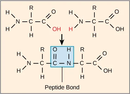
The products that such linkages form are peptides. As more amino acids join to this growing chain, the resulting chain is a polypeptide. Each polypeptide has a free amino group at one end. This end is called the N terminal, or the amino terminal, and the other end has a free carboxyl group, also called the C or carboxyl terminal. While the terms polypeptide and protein are sometimes used interchangeably, a polypeptide is technically a polymer of amino acids, whereas the term protein is used for a polypeptide or polypeptides that have combined together, often have bound non-peptide prosthetic groups, have a distinct shape, and have a unique function. After protein synthesis (translation), most proteins are modified. These are known as post-translational modifications. They may undergo cleavage, phosphorylation, or may require adding other chemical groups. Only after these modifications is the protein completely functional.
Evolution Connection. The Evolutionary Significance of Cytochrome c. Cytochrome c is an important component of the electron transport chain, a part of cellular respiration, and it is normally located in the cellular organelle, the mitochondrion. This protein has a heme prosthetic group, and the heme’s central ion alternately reduces and oxidizes during electron transfer. Because this essential protein’s role in producing cellular energy is crucial, it has changed very little over millions of years. Protein sequencing has shown that there is a considerable amount of cytochrome c amino acid sequence homology among different species. In other words, we can assess evolutionary kinship by measuring the similarities or differences among various species’ DNA or protein sequences.
Scientists have determined that human cytochrome c contains 104 amino acids. For each cytochrome c molecule from different organisms that scientists have sequenced to date, 37 of these amino acids appear in the same position in all cytochrome c samples. This indicates that there may have been a common ancestor. On comparing the human and chimpanzee protein sequences, scientists did not find a sequence difference. When researchers compared human and rhesus monkey sequences, the single difference was in one amino acid. In another comparison, human to yeast sequencing shows differences at 44 positions. (Source note: the source says “a difference in the 44th position”; human and yeast cytochrome c differ at 44 of the 104 positions (Kimball, Biology, “Taxonomy”), which is what the paragraph’s contrast with the single rhesus-monkey difference requires.)
Protein Structure
As we discussed earlier, a protein’s shape is critical to its function. For example, an enzyme can bind to a specific substrate at an active site. If this active site is altered because of local changes or changes in overall protein structure, the enzyme may be unable to bind to the substrate. To understand how the protein gets its final shape or conformation, we need to understand the four levels of protein structure: primary, secondary, tertiary, and quaternary.
Primary Structure
Amino acids’ unique sequence in a polypeptide chain is its primary structure. For example, the pancreatic hormone insulin has two polypeptide chains, A and B, and they are linked together by disulfide bonds. The N terminal amino acid of the A chain is glycine; whereas, the C terminal amino acid is asparagine (below). The amino acid sequences in the A and B chains are unique to insulin.
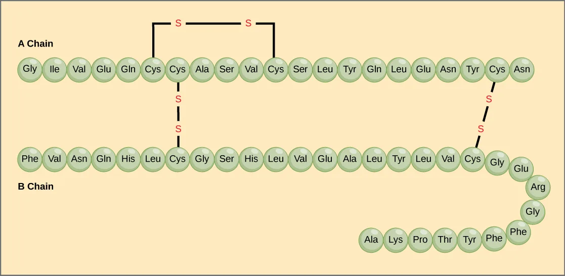
Extended description
Two horizontal rows of amino-acid circles, each labeled with its three-letter code. The A chain (top, 21 residues) begins with glycine and ends with asparagine. The B chain (bottom, 30 residues) begins with phenylalanine and curls into a short second row ending in alanine. Three disulfide (S-S) bonds, drawn as bars linking cysteine residues, hold the structure together: one within the A chain connecting its two internal cysteines, and two between the A and B chains.
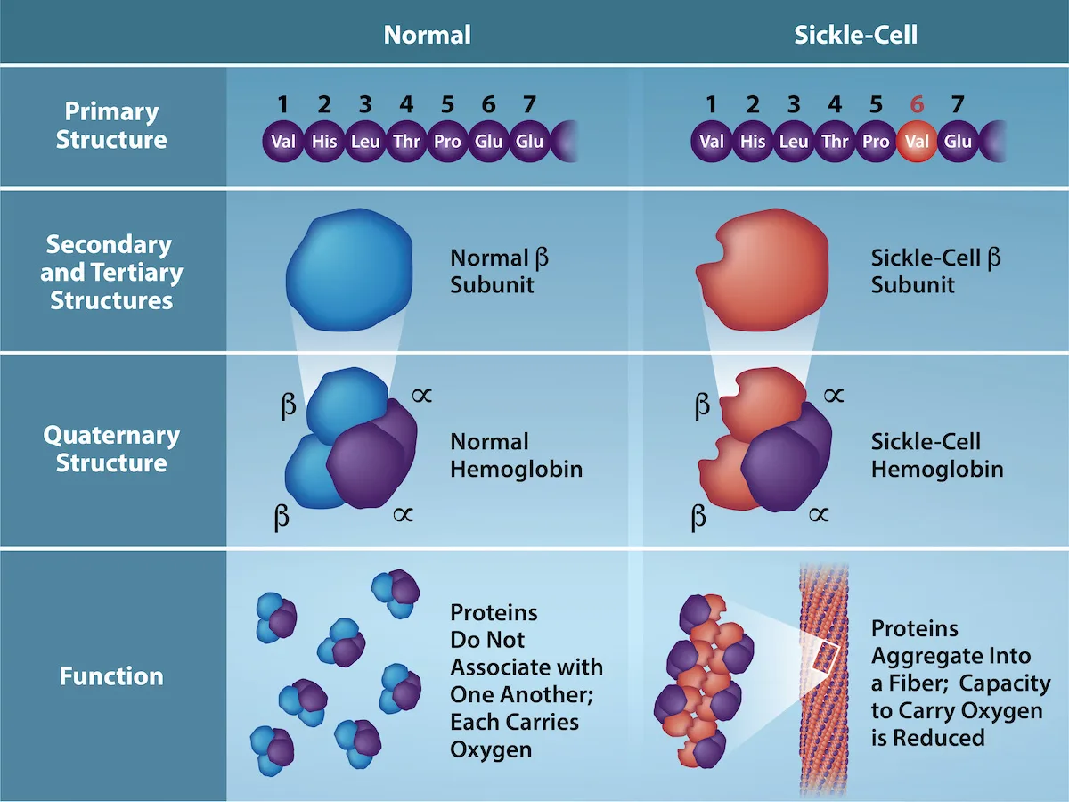
The gene encoding the protein ultimately determines the unique sequence for every protein. A change in nucleotide sequence of the gene’s coding region may lead to adding a different amino acid to the growing polypeptide chain, causing a change in protein structure and function. In sickle cell anemia, the hemoglobin β chain (a small portion of which we show below) has a single amino acid substitution, causing a change in protein structure and function. Specifically, valine in the β chain substitutes the amino acid glutamic. What is most remarkable to consider is that a hemoglobin molecule is comprised of two alpha and two beta chains that each consist of about 150 amino acids. The molecule, therefore, has about 600 amino acids. The structural difference between a normal hemoglobin molecule and a sickle cell molecule—which dramatically decreases life expectancy—is a single amino acid of the 600. What is even more remarkable is that three nucleotides each encode those 600 amino acids, and a single base change (point mutation), 1 in 1,800 bases causes the mutation.
Extended description
A four-row comparison of normal and sickle-cell hemoglobin, arranged in two columns. Row 1, Primary Structure: both columns show the same seven-residue stretch; position 6 is glutamate (Glu) in the normal column and valine (Val), highlighted, in the sickle-cell column. Row 2, Secondary and Tertiary Structures: the normal β subunit is a rounded blue shape; the sickle-cell β subunit is a similarly shaped but reddish, misshapen form. Row 3, Quaternary Structure: normal hemoglobin shows two blue β subunits and two purple α subunits assembled into a compact molecule; sickle-cell hemoglobin shows the same arrangement but with the β subunits in red. Row 4, Function: normal hemoglobin molecules are shown as separate, unassociated units, each free to carry oxygen; sickle-cell hemoglobin molecules are shown aggregating into a long fiber, with reduced oxygen-carrying capacity noted.
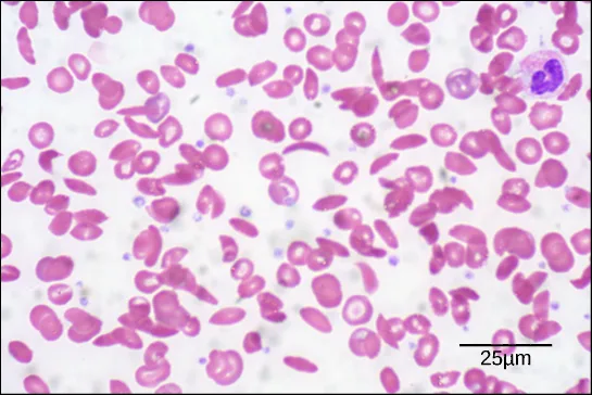
Because of this change of one amino acid in the chain, hemoglobin molecules form long fibers that distort the biconcave, or disc-shaped, red blood cells and causes them to assume a crescent or “sickle” shape, which clogs blood vessels (below). This can lead to myriad serious health problems such as breathlessness, dizziness, headaches, and abdominal pain for those affected by this disease. William Warrick Cardozo showed that sickle-cell anemia is an inherited disorder, meaning that the difference in the specific gene’s encoding region is passed down from parents to children. As you will learn in the genetics unit, the inheritance of such traits is determined by a combination of genes from both parents, and these very small differences can have significant impacts on organisms.
Secondary Structure
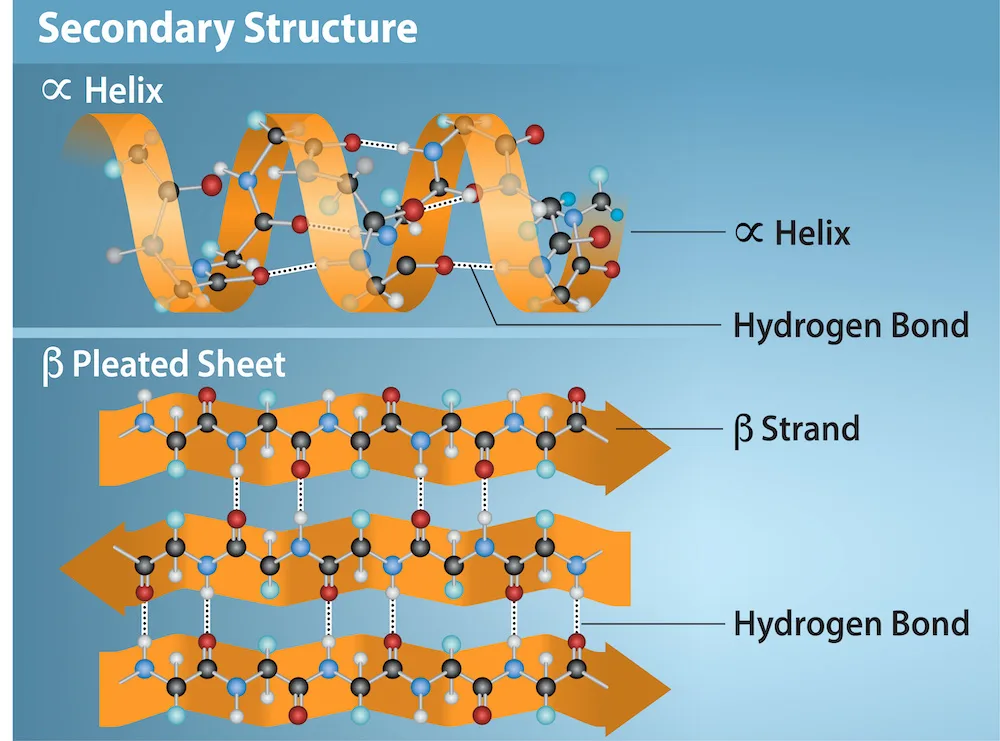
The local folding of the polypeptide in some regions gives rise to the secondary structure of the protein. The most common are the α-helix and β-pleated sheet structures (below). Both structures are held in shape by hydrogen bonds. The hydrogen bonds form between the oxygen atom in the carbonyl group in one amino acid and another amino acid that is four amino acids farther along the chain.
Extended description
Two labeled panels under the heading Secondary Structure. Top panel, α Helix: a ribbon-and-ball-and-stick model of a coiled polypeptide backbone forming a spring-like helix, with dotted lines labeled Hydrogen Bond connecting turns of the coil. Bottom panel, β Pleated Sheet: three stacked ribbon strands, one labeled β Strand, running side by side in an accordion-fold pattern, with dotted lines labeled Hydrogen Bond connecting atoms on adjacent strands.
Every helical turn in an alpha helix has 3.6 amino acid residues. The polypeptide’s R groups (the variant groups) protrude out from the α-helix chain. In the β-pleated sheet, hydrogen bonding between atoms on the polypeptide chain’s backbone form the “pleats”. The R groups are attached to the carbons and extend above and below the pleat’s folds. The pleated segments align parallel or antiparallel to each other, and hydrogen bonds form between the partially positive hydrogen atom in the amino group and the partially negative oxygen atom in the peptide backbone’s carbonyl group. The α-helix and β-pleated sheet structures are in most globular and fibrous proteins and they play an important structural role.
Tertiary Structure
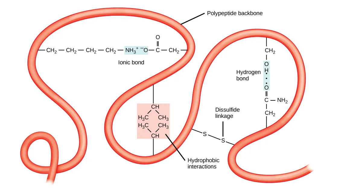
The polypeptide’s unique three-dimensional structure is its tertiary structure (below). This structure is in part due to chemical interactions at work on the polypeptide chain. Primarily, the interactions among R groups create the protein’s complex three-dimensional tertiary structure. The nature of the R groups in the amino acids involved can counteract forming the hydrogen bonds we described for standard secondary structures. For example, R groups with like charges repel each other and those with unlike charges are attracted to each other (ionic bonds). When protein folding takes place, the nonpolar amino acids’ hydrophobic R groups lie in the protein’s interior; whereas, the hydrophilic R groups lie on the outside. Scientists also call the former interaction types hydrophobic interactions. Interaction between cysteine side chains forms disulfide linkages in the presence of oxygen, the only covalent bond that forms during protein folding.
Extended description
A looping red ribbon represents the polypeptide backbone folding back on itself, with four labeled chemical interactions shown along it: an ionic bond, where a positively charged amino group on one part of the chain pairs with a negatively charged oxygen-bearing group on another; hydrophobic interactions, where two branched, all-carbon side chains cluster together away from water; a hydrogen bond, drawn as a dotted line between a hydroxyl group on one side chain and a carbonyl-amide group on another; and a disulfide linkage, where two sulfur atoms from separate cysteine side chains bond covalently.
All of these interactions, weak and strong, determine the protein’s final three-dimensional shape. When a protein loses its three-dimensional shape, it may no longer be functional.
Quaternary Structure
In nature, some proteins form from several polypeptides, or subunits, and the interaction of these subunits forms the quaternary structure. Weak interactions between the subunits help to stabilize the overall structure. For example, insulin (a globular protein) has a combination of hydrogen and disulfide bonds that cause it to mostly clump into a ball shape. Insulin starts out as a single polypeptide and loses some internal sequences in the presence of post-translational modification after forming the disulfide linkages that hold the remaining chains together. Silk (a fibrous protein), however, has a β-pleated sheet structure that is the result of hydrogen bonding between different chains.
The illustration below shows the four levels of protein structure (primary, secondary, tertiary, and quaternary).
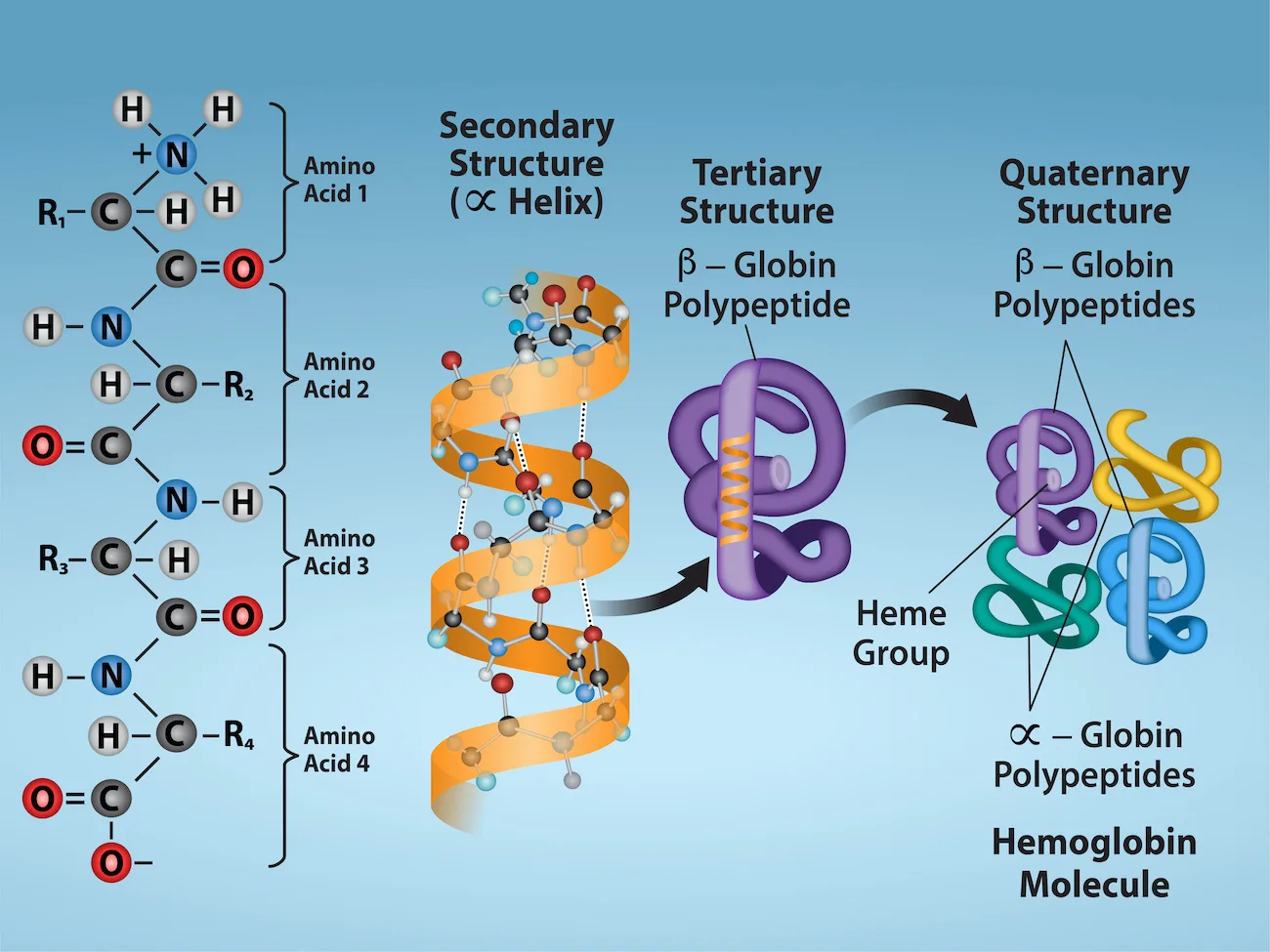
Extended description
Four panels, left to right, tracing one protein from its sequence to its final assembly. Primary structure: four generic amino acids, labeled Amino Acid 1 through 4, each drawn as its own backbone unit with an R group, joined in a vertical chain by peptide bonds. Secondary structure: the same backbone drawn as a coiled alpha-helix ribbon with ball-and-stick atoms. Tertiary structure: the helix folds into a single compact beta-globin polypeptide, drawn as a folded purple ribbon. Quaternary structure: four folded polypeptides — two beta-globin chains, one purple and one green, and two alpha-globin chains, one yellow and one light blue — assemble around a central heme group to form the complete hemoglobin molecule.
Denaturation and Protein Folding
Each protein has its own unique sequence and shape that chemical interactions hold together. If the protein is subject to changes in temperature, pH, or exposure to chemicals, the protein structure may change, losing its shape without losing its primary sequence in what scientists call denaturation. Denaturation is often reversible because the polypeptide’s primary structure is conserved in the process if the denaturing agent is removed, allowing the protein to resume its function. Sometimes denaturation is irreversible, leading to loss of function. One example of irreversible protein denaturation is frying an egg. The albumin protein in the liquid egg white denatures when placed in a hot pan. Not all proteins denature at high temperatures. For instance, bacteria that survive in hot springs have proteins that function at temperatures close to boiling. The stomach is also very acidic, has a low pH, and denatures proteins as part of the digestion process; however, the stomach’s digestive enzymes retain their activity under these conditions.
Protein folding is critical to its function. Scientists originally thought that the proteins themselves were responsible for the folding process. Only recently researchers discovered that often they receive assistance in the folding process from protein helpers, or chaperones (or chaperonins) that associate with the target protein during the folding process. They act by preventing polypeptide aggregation that comprise the complete protein structure, and they disassociate from the protein once the target protein is folded.
Summary
Proteins are a class of macromolecules that perform a diverse range of functions for the cell. They help in metabolism by acting as enzymes, carriers, or hormones, and provide structural support. The building blocks of proteins (monomers) are amino acids. Each amino acid has a central carbon that bonds to an amino group, a carboxyl group, a hydrogen atom, and an R group or side chain. There are 20 commonly occurring amino acids, each of which differs in the R group. A peptide bond links each amino acid to its neighbors. A long amino acid chain is a polypeptide.
Proteins are organized at four levels: primary, secondary, tertiary, and (optional) quaternary. The primary structure is the amino acids’ unique sequence. The polypeptide’s local folding to form structures such as the α-helix and β-pleated sheet constitutes the secondary structure. The overall three-dimensional structure is the tertiary structure. When two or more polypeptides combine to form the complete protein structure, the configuration is the protein’s quaternary structure. Protein shape and function are intricately linked. Any change in shape caused by changes in temperature or pH may lead to protein denaturation and a loss in function.
Key terms
- alpha-helix structure (α-helix) — type of secondary protein structure formed by folding the polypeptide into a helix shape with hydrogen bonds stabilizing the structure
- amino acid — a protein’s monomer; has a central carbon or alpha carbon to which an amino group, a carboxyl group, a hydrogen, and an R group or side chain is attached; the R group is different for all 20 common amino acids
- beta-pleated sheet (β-pleated) — secondary structure in proteins in which hydrogen bonding forms “pleats” between atoms on the polypeptide chain’s backbone
- chaperone — (also, chaperonin) protein that helps nascent protein in the folding process
- denaturation — loss of shape in a protein as a result of changes in temperature, pH, or chemical exposure
- enzyme — catalyst in a biochemical reaction that is usually a complex or conjugated protein
- hormone — chemical signaling molecule, usually protein or steroid, secreted by endocrine cells that act to control or regulate specific physiological processes
- peptide bond — bond formed between two amino acids by a dehydration reaction
- polypeptide — long chain of amino acids that peptide bonds link
- primary structure — linear sequence of amino acids in a protein
- protein — biological macromolecule comprised of one or more amino acid chains
- quaternary structure — association of discrete polypeptide subunits in a protein
- secondary structure — regular structure that proteins form by intramolecular hydrogen bonding between the oxygen atom of one amino acid residue and the hydrogen attached to the nitrogen atom of another amino acid residue
- tertiary structure — a protein’s three-dimensional conformation, including interactions between secondary structural elements; formed from interactions between amino acid side chains
Practice
Describe the functions proteins perform in the cell and in tissues
A catalyst in a biochemical reaction, usually a complex or conjugated protein, is called a(n) ________.
It speeds up a reaction, such as digestion, without itself being consumed.A chemical-signaling molecule, usually a small protein or steroid, secreted by endocrine cells to control or regulate a specific physiological process is called a(n) ________.
Insulin, which helps regulate the blood glucose level, is an example.Proteins help in metabolism by acting as enzymes, carriers, or ________, and provide structural support.
Insulin, a chemical-signaling molecule that regulates blood glucose, is an example of this kind of protein.Discuss the relationship between amino acids and proteins
The monomers that make up proteins are called ________.
These molecules link together by peptide bonds to build a polypeptide chain.The monomer of a protein — a central carbon bonded to an amino group, a carboxyl group, a hydrogen atom, and a variable side chain — is called a(n) ________.
Twenty common types of this monomer, each with a different side chain, link together to build every protein.A long chain of amino acids joined together by peptide bonds is called a ________.
The term is often used loosely to mean the same thing as “protein,” though the two are technically distinct.Explain the four levels of protein organization
The α-helix and the β-pleated sheet are part of which protein structure?
This level is the local folding pattern held together by hydrogen bonds along the peptide backbone — one step up from the raw amino acid sequence.Mad cow disease is an infectious disease where one misfolded protein causes all other copies of the protein to begin misfolding. This is an example of a disease impacting ____ structure.
Ask which level of structure describes a single polypeptide’s own three-dimensional shape, as opposed to its sequence or its assembly with other chains.Describe the differences in the four protein structures.
Show model answer
Did your answer mention:
The level of protein organization formed when two or more separate polypeptide subunits associate together is its ________ structure.
Hemoglobin’s four separate globin chains combine at this level.Describe the ways in which protein shape and function are linked
Explain what happens if even one amino acid is substituted for another in a polypeptide chain. Provide a specific example.
Show model answer
Did your answer mention:
Aquaporins are proteins embedded in the plasma membrane that allow water molecules to move between the extracellular matrix and the intracellular space. Based on its function and location, describe the key features of the protein’s shape and the chemical characteristics of its amino acids.
Show model answer
Did your answer mention:
The loss of a protein’s shape — without loss of its underlying amino acid sequence — as a result of changes in temperature, pH, or chemical exposure is called ________.
Frying an egg does this irreversibly to the albumin protein in the egg white.This section is adapted from Biology 2e, Section 3.4: Proteins by Mary Ann Clark, Jung Choi, Matthew Douglas, and OpenStax, © OpenStax, licensed under CC BY-NC-SA 4.0. Access the original for free at openstax.org. Changes: figures re-encoded as WebP, with seven of the nine re-kinded from the manifest’s file-extension guess of “photo” to “diagram” after inspection (every one is a molecular illustration, not a photograph — only the sickle-cell blood smear is a true photo) and a longer extended description added for the six figures that are labeled diagrams whose full reading is not carried by their captions (the twenty-amino-acid chart, the insulin chain, the hemoglobin/sickle-cell comparison, the α-helix/β-pleated-sheet illustration, the tertiary-structure interaction diagram, and the four-levels-of-structure diagram); the sickle-cell blood-smear alt corrected from the source’s “electron micrograph” to the bright-field light micrograph its own caption names; the twenty-amino-acid figure’s alt shortened from over 600 characters to what the image shows, with its category walk-through moved into the longdesc; inline references to figures and the table changed from the source’s print numbers (“Table 3.1,” “Figure 3.22,” “(Figure 3.27)” inside a caption) to descriptive phrases (“the table below,” “below,” “above”) since figures and tables are not numbered here; feature boxes (two Link to Learning notes and one Evolution Connection) rendered as callouts with their bold names; the Visual Connection question kept in the body immediately after its figure and rendered as a self-check, since the source keys it with a prose solution rather than a lettered option, with the solution’s garbled parenthetical “nonpolar (e.g., amino acid side chains)” read as “nonpolar amino acid side chains”; the end-of-section Review Questions and Critical Thinking Questions adapted into the closing interactive Practice block (multiple choice and self-check respectively); and six key-term recall items (enzyme, hormone, amino acid, polypeptide, quaternary structure, denaturation) added from the glossary to round out every objective group with an auto-graded item; rubric checkpoints added to each self-check, decomposing its model answer (the source solution) into check-off clauses with no new claims; and one summary-derived select-the-term multiple choice added (protein functions, naming hormones from the summary’s “enzymes, carriers, or hormones” list, with distractors drawn from the module’s own terms) since the first objective’s two glossary recall items were both textin and left that group short of three exercises. Three claims are corrected with visible Source notes: cysteine is replaced by lysine in the essential-amino-acid example (this book’s own nutrition table makes cysteine non-essential), the human–yeast cytochrome c comparison reads 44 differing positions rather than “the 44th position,” and the hemoglobin β-chain is 146 residues rather than 147 (errata 385–387).