Cell Structure
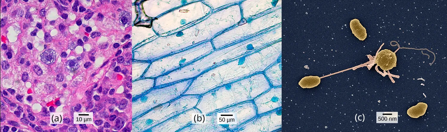
Close your eyes and picture a brick wall. What is the wall’s basic building block? It is a single brick. Like a brick wall, cells are the building blocks that make up your body.
Your body has many kinds of cells, each specialized for a specific purpose. Just as we use a variety of materials to build a home, the human body is constructed from many cell types. For example, epithelial cells protect the body’s surface and cover the organs and body cavities within. Bone cells help to support and protect the body. Immune system cells fight invading bacteria. Additionally, blood and blood cells carry nutrients and oxygen throughout the body while removing carbon dioxide. Each of these cell types plays a vital role during the body’s growth, development, and day-to-day maintenance. In spite of their enormous variety, however, cells from all organisms—even ones as diverse as bacteria, onion, and human—share certain fundamental characteristics.
Sections
- Studying Cells — the role cells play in every organism, how light microscopy and electron microscopy differ, and the three tenets of cell theory.
- Prokaryotic Cells — examples of prokaryotic and eukaryotic organisms, how their cells compare, the relative sizes of different cells, and why a cell must stay small.
- Eukaryotic Cells — the structure of eukaryotic cells, how animal and plant cells compare, the plasma membrane’s role, and the functions of the major cell organelles.
- The Endomembrane System and Proteins — the components of the endomembrane system and how they relate to its functions.
- The Cytoskeleton — the cytoskeleton and the roles of microfilaments, intermediate filaments, and microtubules; how cilia and flagella compare; and how prokaryotic, animal, and plant cells differ.
- Connections between Cells and Cellular Activities — the extracellular matrix, how plant and animal cells communicate with neighboring cells, and the roles of tight junctions, desmosomes, gap junctions, and plasmodesmata.
This chapter is adapted from Biology 2e, Chapter 4: Cell Structure by Mary Ann Clark, Jung Choi, Matthew Douglas, and OpenStax, © OpenStax, licensed under CC BY-NC-SA 4.0. Access the original for free at openstax.org. Each section page records its own changes from the source. Changes: the chapter-opening micrographs are the source’s, re-encoded for the web with the alt text rewritten to describe what each panel shows rather than repeat the caption’s identification of panel (a), which the source’s own alt and caption name inconsistently (alt: “cheek cells”; caption: “Nasal sinus cells”) — logged as a possible source defect rather than resolved locally.