Studying Cells
By the end of this section, you will be able to:
- Describe the role of cells in organisms
- Compare and contrast light microscopy and electron microscopy
- Summarize cell theory
A cell is the smallest unit of a living thing. Whether comprised of one cell (like bacteria) or many cells (like a human), we call it an organism. Thus, cells are the basic building blocks of all organisms.
Several cells of one kind that interconnect with each other and perform a shared function form tissues. These tissues combine to form an organ (your stomach, heart, or brain), and several organs comprise an organ system (such as the digestive system, circulatory system, or nervous system). Several systems that function together form an organism (like a human being). Here, we will examine the structure and function of cells.
There are many types of cells, which scientists group into one of two broad categories: prokaryotic and eukaryotic. For example, we classify both animal and plant cells as eukaryotic cells; whereas, we classify bacterial cells as prokaryotic. Before discussing the criteria for determining whether a cell is prokaryotic or eukaryotic, we will first examine how biologists study cells.
Microscopy
Cells vary in size. With few exceptions, we cannot see individual cells with the naked eye, so scientists use microscopes (micro- = “small”; -scope = “to look at”) to study them. A microscope is an instrument that magnifies an object. We photograph most cells with a microscope, so we can call these images micrographs.
The optics of a microscope’s lenses change the image orientation that the user sees. A specimen that is right-side up and facing right on the microscope slide will appear upside-down and facing left when one views through a microscope, and vice versa. Similarly, if one moves the slide left while looking through the microscope, it will appear to move right, and if one moves it down, it will seem to move up. This occurs because microscopes use two sets of lenses to magnify the image. Because of the manner by which light travels through the lenses, this two lens system produces an inverted image (binocular, or dissecting microscopes, work in a similar manner, but include an additional magnification system that makes the final image appear to be upright).
Light Microscopes
To give you a sense of cell size, a typical human red blood cell is about eight millionths of a meter or eight micrometers (abbreviated as eight µm) in diameter. A pin head is about two thousandths of a meter (two mm) in diameter. That means about 250 red blood cells could fit on a pinhead.
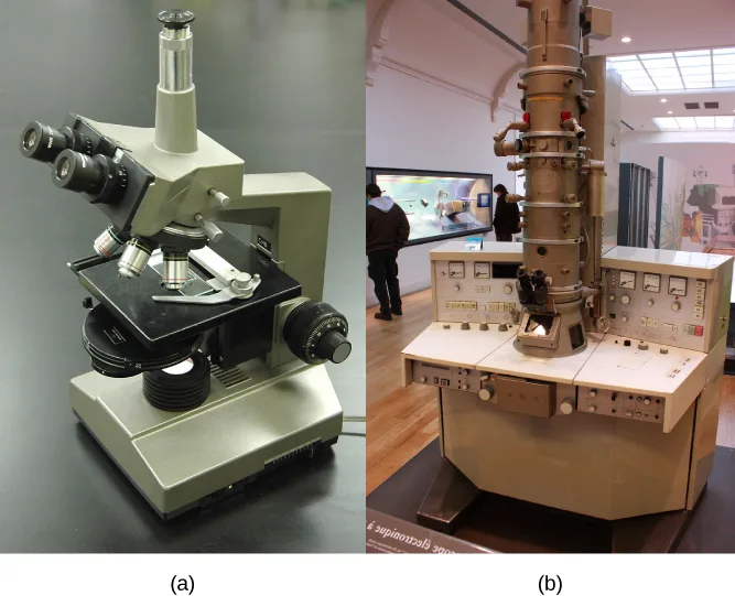
Most student microscopes are light microscopes (below). Visible light passes and bends through the lens system to enable the user to see the specimen. Light microscopes are advantageous for viewing living organisms, but since individual cells are generally transparent, their components are not distinguishable unless they are colored with special stains. Staining, however, usually kills the cells.
Light microscopes that undergraduates commonly use in the laboratory magnify up to approximately 400 times. Two parameters that are important in microscopy are magnification and resolving power. Magnification is the process of enlarging an object in appearance. Resolving power is the microscope’s ability to distinguish two adjacent structures as separate: the higher the resolution, the better the image’s clarity and detail. When one uses oil immersion lenses to study small objects, magnification usually increases to 1,000 times. In order to gain a better understanding of cellular structure and function, scientists typically use electron microscopes.
Electron Microscopes
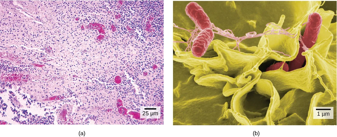
In contrast to light microscopes, electron microscopes (above) use a beam of electrons instead of a beam of light. Not only does this allow for higher magnification and, thus, more detail (below), it also provides higher resolving power. The method to prepare the specimen for viewing with an electron microscope kills the specimen. Electrons have short wavelengths (shorter than photons) that move best in a vacuum, so we cannot view living cells with an electron microscope.
In a scanning electron microscope, a beam of electrons moves back and forth across a cell’s surface, creating details of cell surface characteristics. In a transmission electron microscope, the electron beam penetrates the cell and provides details of a cell’s internal structures. As you might imagine, electron microscopes are significantly more bulky and expensive than light microscopes.
Cell Theory
The microscopes we use today are far more complex than those that Dutch shopkeeper Antony van Leeuwenhoek, used in the 1600s. Skilled in crafting lenses, van Leeuwenhoek observed the movements of single-celled organisms, which he collectively termed “animalcules.”
In the 1665 publication Micrographia, experimental scientist Robert Hooke coined the term “cell” for the box-like structures he observed when viewing cork tissue through a lens. In the 1670s, van Leeuwenhoek discovered bacteria and protozoa. Later advances in lenses, microscope construction, and staining techniques enabled other scientists to see some components inside cells.
By the late 1830s, botanist Matthias Schleiden and zoologist Theodor Schwann were studying tissues and proposed the unified cell theory, which states that one or more cells comprise all living things, the cell is the basic unit of life, and new cells arise from existing cells. Rudolf Virchow later made important contributions to this theory.
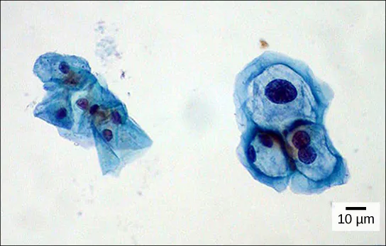
Career Connection. Cytotechnologist. Have you ever heard of a medical test called a Pap smear (below)? In this test, a doctor takes a small sample of cells from the patient’s uterine cervix and sends it to a medical lab where a cytotechnologist stains the cells and examines them for any changes that could indicate cervical cancer or a microbial infection.
Cytotechnologists (cyto- = “cell”) are professionals who study cells via microscopic examinations and other laboratory tests. They are trained to determine which cellular changes are within normal limits and which are abnormal. Their focus is not limited to cervical cells. They study cellular specimens that come from all organs. When they notice abnormalities, they consult a pathologist, a medical doctor who interprets and diagnoses changes that disease in body tissue and fluids cause.
Cytotechnologists play a vital role in saving people’s lives. When doctors discover abnormalities early, a patient’s treatment can begin sooner, which usually increases the chances of a successful outcome.
Summary
A cell is the smallest unit of life. Most cells are so tiny that we cannot see them with the naked eye. Therefore, scientists use microscopes to study cells. Electron microscopes provide higher magnification, higher resolution, and more detail than light microscopes. The unified cell theory states that one or more cells comprise all organisms, the cell is the basic unit of life, and new cells arise from existing cells.
Key terms
- cell theory — see unified cell theory
- electron microscope — an instrument that magnifies an object using an electron beam that passes and bends through a lens system to visualize a specimen
- light microscope — an instrument that magnifies an object using a beam of visible light that passes and bends through a lens system to visualize a specimen
- microscope — an instrument that magnifies an object
- unified cell theory — a biological concept that states that one or more cells comprise all organisms; the cell is the basic unit of life; and new cells arise from existing cells
Practice
Describe the role of cells in organisms
The ________ is the basic unit of life.
Unified cell theory names this as the smallest living unit — tissues, organs, and organisms all build up from it.Trace how cells build up to form a whole organism, naming each level in between.
Show model answer
Did your answer mention:
A cell is the ________ unit of life.
This is the section’s opening sentence, distinct from the cell theory’s own “basic unit of life” wording.Compare and contrast light microscopy and electron microscopy
When viewing a specimen through a light microscope, scientists use ________ to distinguish the individual components of cells.
Staining adds color to otherwise transparent cell components — though it usually kills the cells in the process.An instrument that magnifies an object using a beam of visible light that passes and bends through a lens system to visualize a specimen is called a(n) ________.
Its light beam does not kill the specimen, unlike the electron-based alternative.An instrument that magnifies an object using an electron beam that passes and bends through a lens system to visualize a specimen is called a(n) ________.
It requires a vacuum for its electron beam, so it cannot image a living specimen.In your everyday life, you have probably noticed that certain instruments are ideal for certain situations. For example, you would use a spoon rather than a fork to eat soup because a spoon is shaped for scooping, while soup would slip between the tines of a fork. The use of ideal instruments also applies in science. In what situation(s) would the use of a light microscope be ideal, and why?
Show model answer
Did your answer mention:
In what situation(s) would the use of a scanning electron microscope be ideal, and why?
Show model answer
Did your answer mention:
In what situation(s) would a transmission electron microscope be ideal, and why?
Show model answer
Did your answer mention:
What are the advantages and disadvantages of each of these types of microscopes?
Show model answer
Did your answer mention:
Summarize cell theory
The biological concept stating that one or more cells comprise all organisms, the cell is the basic unit of life, and new cells arise from existing cells is called the ________.
Schleiden and Schwann proposed it in the late 1830s; Virchow later made important contributions to it.According to the unified cell theory, new cells arise from ________.
This is the theory’s third tenet — think about where a new cell’s material ultimately comes from.Explain how the formation of an adult human follows the cell theory.
Show model answer
Did your answer mention:
This section is adapted from Biology 2e, Section 4.1: Studying Cells by Mary Ann Clark, Jung Choi, Matthew Douglas, and OpenStax, © OpenStax, licensed under CC BY-NC-SA 4.0. Access the original for free at openstax.org. Changes: figures re-encoded as WebP (all three photographic — two micrographs and one photo of physical instruments — matching the manifest’s guess, so none were re-kinded); inline references to figures changed from the source’s print numbers (“Figure 4.2a,” “Figure 4.3,” “Figure 4.4”) to “above”/“below” since figures are not numbered here; the two Link to Learning/interactive notes and the Career Connection feature box rendered as callouts with their bold names, the Career Connection’s own title set in italics beside the bold name, and its figure kept inside the same callout as in the source; the end-of-section Review Questions and Critical Thinking Questions adapted into the closing interactive Practice block (multiple choice and self-check respectively); three key-term recall items (light microscope, electron microscope, unified cell theory) added from the glossary to round out two objective groups with an auto-graded item; because no end-of-section exercise or glossary term addresses the “role of cells in organisms” objective, its group holds the source’s “basic unit of life” review question plus one self-check written directly from the section’s own cell-tissue-organ-organism paragraph (no new claims), disclosed here since it is not itself a keyed source item; rubric checkpoints added to each self-check, decomposing its model answer (the source solution) into check-off clauses with no new claims; and two summary-derived recall items added, one under each objective the summary sentences reach but the source’s own exercises did not fully round out to three items apiece (“smallest unit of life” under the first objective, “new cells arise from existing cells” as a multiple choice under the third).