Prokaryotic Cells
By the end of this section, you will be able to:
- Name examples of prokaryotic and eukaryotic organisms
- Compare and contrast prokaryotic and eukaryotic cells
- Describe the relative sizes of different cells
- Explain why cells must be small
Cells fall into one of two broad categories: prokaryotic and eukaryotic. We classify only the predominantly single-celled organisms Bacteria and Archaea as prokaryotes (pro- = “before”; -kary- = “nucleus”). Animal cells, plants, fungi, and protists are all eukaryotes (eu- = “true”).
Components of Prokaryotic Cells
All cells share four common components: 1) a plasma membrane, an outer covering that separates the cell’s interior from its surrounding environment; 2) cytoplasm, consisting of a jelly-like cytosol within the cell in which there are other cellular components; 3) DNA, the cell’s genetic material; and 4) ribosomes, which synthesize proteins. However, prokaryotes differ from eukaryotic cells in several ways.
A prokaryote is a simple, mostly single-celled (unicellular) organism that lacks a nucleus, or any other membrane-bound organelle. We will shortly come to see that this is significantly different in eukaryotes. Prokaryotic DNA is in the cell’s central part: the nucleoid.
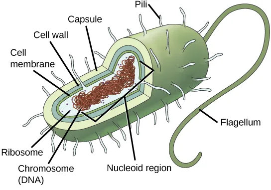
Extended description
A cutaway oval bacterium is drawn with its interior visible, and labels point to eight parts. Capsule: the outermost layer surrounding the cell. Cell wall: the layer just inside the capsule. Cell membrane: the innermost boundary layer, just inside the cell wall. Ribosomes: small dots scattered through the cytoplasm. Chromosome (DNA): a tangled coil filling the cell’s center. Nucleoid region: the area of cytoplasm where the chromosome is concentrated. Pili: short hair-like projections covering the cell’s surface. Flagellum: a single long, whip-like filament extending from one narrow end of the cell.
Most bacteria have a peptidoglycan cell wall and many have a polysaccharide capsule. The cell wall acts as an extra layer of protection, helps the cell maintain its shape, and prevents dehydration. The capsule enables the cell to attach to surfaces in its environment. Some prokaryotes have flagella, pili, or fimbriae. Flagella are used for locomotion. Pili exchange genetic material during conjugation, the process by which one bacterium transfers genetic material to another through direct contact. Bacteria use fimbriae to attach to a host cell.
Career Connection. Microbiologist. The most effective action anyone can take to prevent the spread of contagious illnesses is to wash their hands. Why? Because microbes (organisms so tiny that they can only be seen with microscopes) are ubiquitous. They live on doorknobs, money, your hands, and many other surfaces. If someone sneezes into his hand and touches a doorknob, and afterwards you touch that same doorknob, the microbes from the sneezer’s mucus are now on your hands. If you touch your hands to your mouth, nose, or eyes, those microbes can enter your body and could make you sick.
However, not all microbes (also called microorganisms) cause disease; most are actually beneficial. You have microbes in your gut that make vitamin K. Other microorganisms are used to ferment beer and wine.
Microbiologists are scientists who study microbes. Microbiologists can pursue a number of careers. Not only do they work in the food industry, they are also employed in the veterinary and medical fields. They can work in the pharmaceutical sector, serving key roles in research and development by identifying new antibiotic sources that can treat bacterial infections.
Environmental microbiologists may look for new ways to use specially selected or genetically engineered microbes to remove pollutants from soil or groundwater, as well as hazardous elements from contaminated sites. We call using these microbes bioremediation technologies. Microbiologists can also work in the bioinformatics field, providing specialized knowledge and insight for designing, developing, and optimizing specificity of computer models of, for example, bacterial epidemics.
Cell Size
At 0.1 to 5.0 µm in diameter, prokaryotic cells are significantly smaller than eukaryotic cells, which have diameters ranging from 10 to 100 µm. The prokaryotes’ small size allows ions and organic molecules that enter them to quickly diffuse to other parts of the cell. Similarly, any wastes produced within a prokaryotic cell can quickly diffuse. This is not the case in eukaryotic cells, which have developed different structural adaptations to enhance intracellular transport.
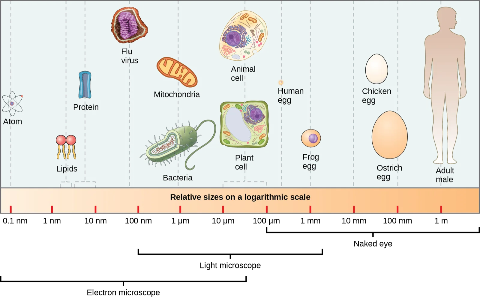
Extended description
A horizontal logarithmic scale runs from 0.1 nm to 1 m, marked at each power-of-ten tick: 0.1 nm, 1 nm, 10 nm, 100 nm, 1 µm, 10 µm, 100 µm, 1 mm, 10 mm, 100 mm, and 1 m. Above the scale, objects are placed at their approximate size, smallest to largest: an atom, about 0.1 nm; lipids and proteins, 1–10 nm; a flu virus, just under 100 nm; bacteria and a mitochondrion, about 1 µm; a plant cell and an animal cell, 10–100 µm; a human egg, 100 µm–1 mm; a frog egg, about 1 mm; a chicken egg and a larger ostrich egg, 10–100 mm; and an adult human, about 1 m. Below the scale, three overlapping brackets mark which instrument can resolve each size range: an electron microscope, from below 0.1 nm to almost 100 µm; a light microscope, from 100 nm to just over 1 mm; and the naked eye, from 100 µm upward.
Small size, in general, is necessary for all cells, whether prokaryotic or eukaryotic. Let’s examine why that is so. First, we’ll consider the area and volume of a typical cell. Not all cells are spherical in shape, but most tend to approximate a sphere. You may remember from your high school geometry course that the formula for the surface area of a sphere is , while the formula for its volume is . Thus, as the radius of a cell increases, its surface area increases as the square of its radius, but its volume increases as the cube of its radius (much more rapidly). Therefore, as a cell increases in size, its surface area-to-volume ratio decreases. This same principle would apply if the cell had a cube shape. If the cell grows too large, the plasma membrane will not have sufficient surface area to support the rate of diffusion required for the increased volume. In other words, as a cell grows, it becomes less efficient. One way to become more efficient is to divide. Other ways are to increase surface area by foldings of the cell membrane, become flat or thin and elongated, or develop organelles that perform specific tasks. These adaptations lead to developing more sophisticated cells, which we call eukaryotic cells.

What advantages might small cell size confer on a cell?
Show model answer
Did your answer mention:
Summary
Prokaryotes are single-celled organisms of the domains Bacteria and Archaea. All prokaryotes have plasma membranes, cytoplasm, ribosomes, and DNA that is not membrane-bound. Most have peptidoglycan cell walls and many have polysaccharide capsules. Prokaryotic cells range in diameter from 0.1 to 5.0 µm.
As a cell increases in size, its surface area-to-volume ratio decreases. If the cell grows too large, the plasma membrane will not have sufficient surface area to support the rate of diffusion required for the increased volume.
Key terms
- nucleoid — the central region of a prokaryotic cell where the chromosome is located
- prokaryote — unicellular organism that lacks a nucleus or any other membrane-bound organelle
Practice
Name examples of prokaryotic and eukaryotic organisms
Which of the following organisms is a prokaryote?
Prokaryotes are simple, unicellular organisms with no nucleus — the other three options are a eukaryotic protist, a virus (not a cell at all), and a eukaryotic alga.Prokaryotes are single-celled organisms of the domains ________.
The section’s summary names the two domains that make up all prokaryotes.Explain why not all microbes are harmful.
Show model answer
Did your answer mention:
Compare and contrast prokaryotic and eukaryotic cells
Bacteria that lack fimbriae are less likely to ________.
Fimbriae are the short, hair-like structures a bacterium uses to attach to a surface or a host cell.All prokaryotes have plasma membranes, cytoplasm, ribosomes, and DNA that is ________.
This is the key structural feature that separates prokaryotic DNA from a eukaryotic cell’s DNA.Antibiotics are medicines that are used to fight bacterial infections. These medicines kill prokaryotic cells without harming human cells. What part or parts of the bacterial cell do you think antibiotics target? Why?
Show model answer
Did your answer mention:
Describe the relative sizes of different cells
A unicellular organism that lacks a nucleus or any other membrane-bound organelle is called a(n) ________.
Bacteria and Archaea are the two domains classified this way, and both are far smaller than a typical eukaryotic cell.The central region of a prokaryotic cell where its chromosome is located is called the ________.
Unlike a eukaryotic nucleus, this region has no surrounding membrane.Prokaryotic cells range in diameter from 0.1 to ________.
The section’s summary gives this as the upper end of the prokaryotic size range.Explain why cells must be small
Prokaryotes depend on ________ to obtain some materials and to get rid of wastes.
Their small size means a molecule needs only a short distance to travel between the plasma membrane and any point in the cytoplasm.As a cell increases in size, its surface area-to-volume ratio ________.
Surface area grows with the square of the radius while volume grows with the cube, so one falls behind the other as a cell gets bigger.What advantages might large cell size have?
Show model answer
Did your answer mention:
This section is adapted from Biology 2e, Section 4.2: Prokaryotic Cells by Mary Ann Clark, Jung Choi, Matthew Douglas, and OpenStax, © OpenStax, licensed under CC BY-NC-SA 4.0. Access the original for free at openstax.org. Changes: figures re-encoded as WebP, with the generalized prokaryotic-cell diagram re-kinded from the manifest’s file-extension guess of “photo” to “diagram” (it is a labeled line drawing, not a photograph), and given a longer extended description walking its eight labels, since a labeled diagram’s full reading is not carried by its caption; the size-comparison scale figure’s over-long source alt was shortened to what the image shows, with its full object-by-object and instrument-bracket walk-through moved into an extended description; the cube-and-sphere comparison figure’s alt corrected — the source says the same sphere is shown in both boxes, but the image draws a visibly larger sphere in the larger box, matching the cell-growth point the caption and prose make — and reported as a source defect; the surface-area and volume formulas set in KaTeX rather than as printed running text; the Visual Connection question and its two-part answer split across its two questions — “what advantages might small cell size confer” kept in the body immediately after its figure as a self-check, and “what advantages might large cell size have” placed in the closing Practice block under the objective it argues, since the source pairs one compound solution with two distinct questions; feature boxes (one Career Connection) rendered as a callout with its bold name; the end-of-section Review Questions and Critical Thinking Questions adapted into the closing interactive Practice block (multiple choice and self-check respectively); the “nucleoid” glossary definition’s duplicated “central part … central part” phrasing cleaned to “the central region … where the chromosome is located,” reported as a source defect; and two key-term recall items (prokaryote, nucleoid) added from the glossary, since the module’s own exercise sets do not otherwise cover the relative-sizes objective; rubric checkpoints added to each self-check (the body self-check on small cell size included), decomposing its model answer (the source solution) into check-off clauses with no new claims; and four summary-derived recall items added, one per objective, to raise every group to the raised floor (“Bacteria and Archaea,” “not membrane-bound,” “5.0 µm,” and “decreases,” each a cloze or multiple choice on a summary sentence the module’s own exercises did not otherwise cover).