Eukaryotic Cells
By the end of this section, you will be able to:
- Describe the structure of eukaryotic cells
- Compare animal cells with plant cells
- State the role of the plasma membrane
- Summarize the functions of the major cell organelles
Have you ever heard the phrase “form follows function?” It’s a philosophy that many industries follow. In architecture, this means that buildings should be constructed to support the activities that will be carried out inside them. For example, a skyscraper should include several elevator banks. A hospital should have its emergency room easily accessible.
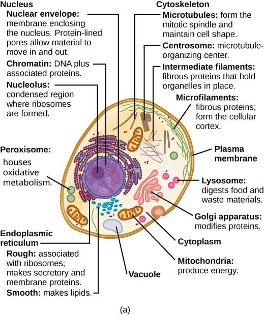
Our natural world also utilizes the principle of form following function, especially in cell biology, and this will become clear as we explore eukaryotic cells (below). Unlike prokaryotic cells, eukaryotic cells have: 1) a membrane-bound nucleus; 2) numerous membrane-bound organelles such as the endoplasmic reticulum, Golgi apparatus, chloroplasts, mitochondria, and others; and 3) several, rod-shaped chromosomes. Because a membrane surrounds a eukaryotic cell’s nucleus, it has a “true nucleus.” The word “organelle” means “little organ,” and, as we already mentioned, organelles have specialized cellular functions, just as your body’s organs have specialized functions.
At this point, it should be clear to you that eukaryotic cells have a more complex structure than prokaryotic cells. Organelles allow different functions to be compartmentalized in different areas of the cell. Before turning to organelles, let’s first examine two important components of the cell: the plasma membrane and the cytoplasm.
Extended description
Panel (a), a typical animal cell: an egg-shaped outline encloses the cytoplasm. A large central nucleus holds tangled chromatin threads and a smaller, darker nucleolus, all inside a double-membraned, pore-studded nuclear envelope. Around the nucleus, ribosome-studded rough endoplasmic reticulum gives way to smooth endoplasmic reticulum; nearby sits a stacked Golgi apparatus. Oval mitochondria, round peroxisomes and lysosomes, and a vacuole float in the cytoplasm alongside free ribosomes drawn as small dots. At the cell’s edge, microfilaments line the inner face of the plasma membrane, thread-like intermediate filaments anchor nearby organelles, microtubules radiate through the cytoplasm, and a centrosome of two perpendicular rod-shaped centrioles sits near the nucleus.
Extended description
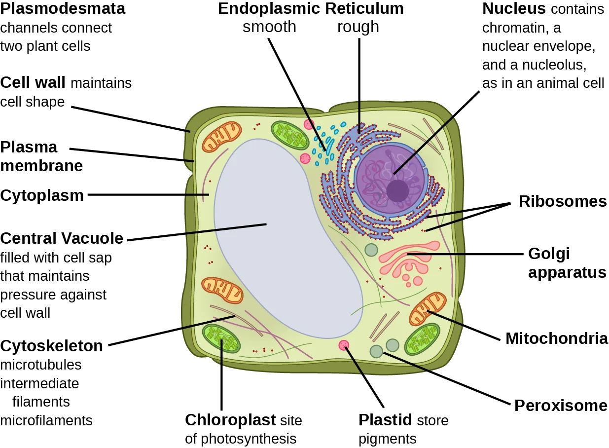
Panel (b), a typical plant cell: the same nucleus, endoplasmic reticulum, Golgi apparatus, mitochondria, peroxisomes, ribosomes, and cytoskeleton (microtubules, intermediate filaments, and microfilaments, but no centrosome) appear, now enclosed by an added rigid cell wall outside the plasma membrane. Oval chloroplasts and smaller plastids are scattered through the cytoplasm, and one enormous central vacuole fills most of the cell’s interior, pressing the cytoplasm into a thin layer against the cell wall.
If the nucleolus were not able to carry out its function, what other cellular organelles would be affected?
Show model answer
Did your answer mention:
The Plasma Membrane
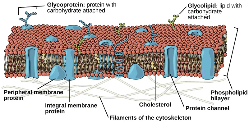
Like prokaryotes, eukaryotic cells have a plasma membrane (below), a phospholipid bilayer with embedded proteins that separates the internal contents of the cell from its surrounding environment. A phospholipid is a lipid molecule with two fatty acid chains and a phosphate-containing group. The plasma membrane controls the passage of organic molecules, ions, water, and oxygen into and out of the cell. Wastes (such as carbon dioxide and ammonia) also leave the cell by passing through the plasma membrane.
Extended description
A cutaway block of the phospholipid bilayer, drawn as two rows of red round phosphate heads with wavy tan tails pointing inward toward each other. Embedded blue shapes span the bilayer: several are labeled integral membrane protein, and one forms a protein channel with a visible central pore. A smaller blue shape resting against the inner (cytoplasmic) row, beside the cytoskeletal filaments, is labeled peripheral membrane protein. Short branched chains of blue beads labeled glycoprotein and green beads labeled glycolipid extend outward from the outer face. Small yellow ovals labeled cholesterol sit within the bilayer near two of the channels. Along the inner face, pale wavy lines labeled filaments of the cytoskeleton run beneath the membrane.
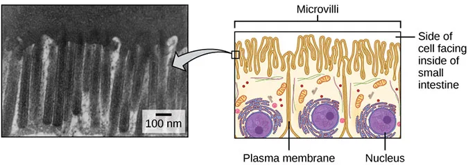
The plasma membranes of cells that specialize in absorption fold into fingerlike projections that we call microvilli (singular = microvillus) (below). Such cells typically line the small intestine, the organ that absorbs nutrients from digested food. This is an excellent example of form following function. People with celiac disease have an immune response to gluten, which is a protein in wheat, barley, and rye. The immune response damages microvilli, and thus, afflicted individuals cannot absorb nutrients. This leads to malnutrition, cramping, and diarrhea. Patients suffering from celiac disease must follow a gluten-free diet.
Extended description
On the left, a black-and-white electron micrograph shows a row of tall, narrow, finger-like microvilli projecting upward from the plasma membrane, with a 100 nm scale bar. An arrow connects a small boxed region of the micrograph to a colored diagram on the right, which shows three adjacent intestinal lining cells, each topped with a fringe of microvilli labeled at the top of the panel; the plasma membrane surrounding each cell and the round purple nucleus inside each cell are labeled, and a bracket labels the microvilli-covered surface as the side of the cell facing the interior of the small intestine.
The Cytoplasm
The cytoplasm is the cell’s entire region between the plasma membrane and the nuclear envelope (a structure we will discuss shortly). It is comprised of organelles suspended in the gel-like cytosol, the cytoskeleton, and various chemicals (above). Even though the cytoplasm consists of 70 to 80 percent water, it has a semi-solid consistency, which comes from the proteins within it. However, proteins are not the only organic molecules in the cytoplasm. Glucose and other simple sugars, polysaccharides, amino acids, nucleic acids, fatty acids, and derivatives of glycerol are also there. Ions of sodium, potassium, calcium, and many other elements also dissolve in the cytoplasm. Many metabolic reactions, including protein synthesis, take place in the cytoplasm.
The Nucleus
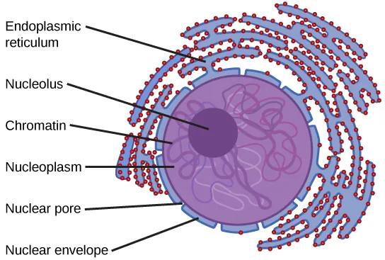
Typically, the nucleus is the most prominent organelle in a cell (above). The nucleus (plural = nuclei) houses the cell’s DNA and directs the synthesis of ribosomes and proteins. Let’s look at it in more detail (below).
Extended description
A large circle representing the nucleus sits at the center, its outer edge scalloped with small pores to represent the double-membraned, pored nuclear envelope. Inside, tangled pink and purple loops labeled chromatin fill most of the circle around a smaller, solid dark-purple circle labeled nucleolus, positioned left of center. Curved ribbons studded with small red dots, labeled endoplasmic reticulum, wrap around the outside of the nuclear envelope on the right side of the figure, fanning outward from it.
The Nuclear Envelope
The nuclear envelope is a double-membrane structure that constitutes the nucleus’ outermost portion (above). Both the nuclear envelope’s inner and outer membranes are phospholipid bilayers.
The nuclear envelope is punctuated with pores that control the passage of ions, molecules, and RNA between the nucleoplasm and cytoplasm. The nucleoplasm is the semi-solid fluid inside the nucleus, where we find the chromatin and the nucleolus.
Chromatin and Chromosomes
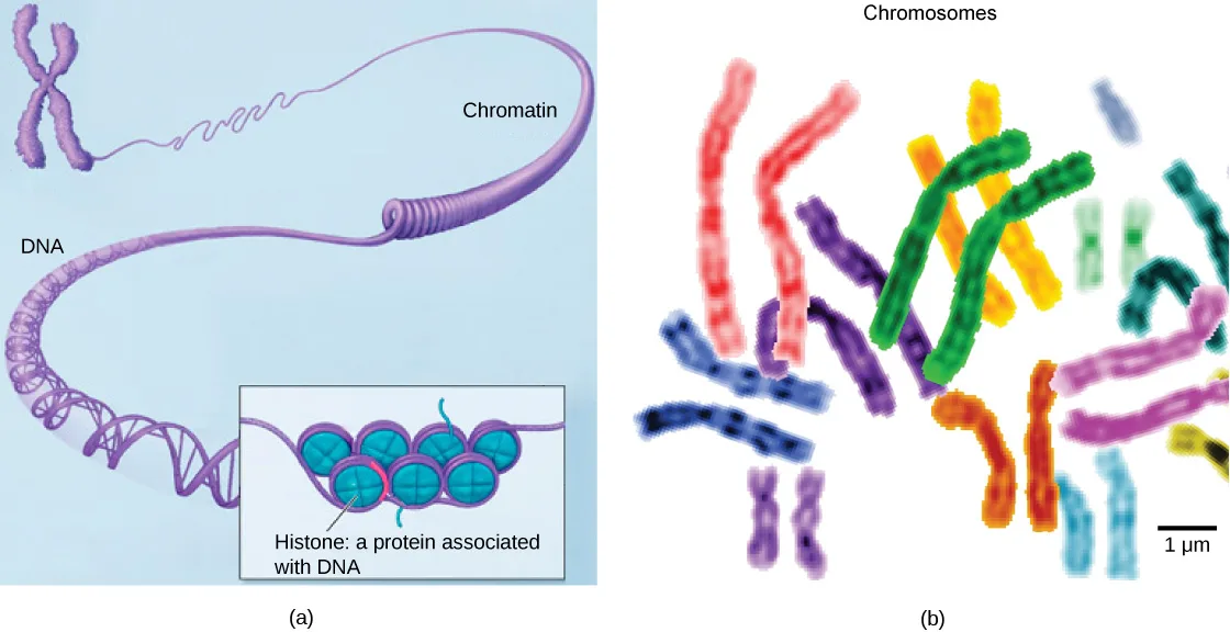
To understand chromatin, it is helpful to first explore chromosomes, structures within the nucleus that are made up of DNA, the hereditary material. You may remember that in prokaryotes, DNA is organized into a single circular chromosome. In eukaryotes, chromosomes are linear structures. Every eukaryotic species has a specific number of chromosomes in the nucleus of each cell. For example, in humans, the chromosome number is 46, while in fruit flies, it is eight. Chromosomes are only visible and distinguishable from one another when the cell is getting ready to divide. When the cell is in the growth and maintenance phases of its life cycle, proteins attach to chromosomes, and they resemble an unwound, jumbled bunch of threads. We call these unwound protein-chromosome complexes chromatin (below). Chromatin describes the material that makes up the chromosomes both when condensed and decondensed.
Extended description
Panel (a): starting at upper left, a small X-shaped, tightly coiled purple chromosome unwinds rightward into a loosely coiled thread labeled chromatin, then further unwinds into the double-helix DNA labeled at lower left. An inset box shows a short stretch of the double helix wrapped around a cluster of round teal beads labeled histone: a protein associated with DNA. Panel (b), labeled Chromosomes: roughly twenty X- and rod-shaped chromosomes of varying lengths, each dyed a distinct color (red, purple, orange, yellow, green, blue, teal, pink) so that the two copies of a chromosome pair share a color, are scattered loosely across a white background, with a 1 µm scale bar at lower right.
The Nucleolus
We already know that the nucleus directs the synthesis of ribosomes, but how does it do this? Some chromosomes have sections of DNA that encode ribosomal RNA. A darkly staining area within the nucleus called the nucleolus (plural = nucleoli) aggregates the ribosomal RNA with associated proteins to assemble the ribosomal subunits that are then transported out through the pores in the nuclear envelope to the cytoplasm.
Ribosomes
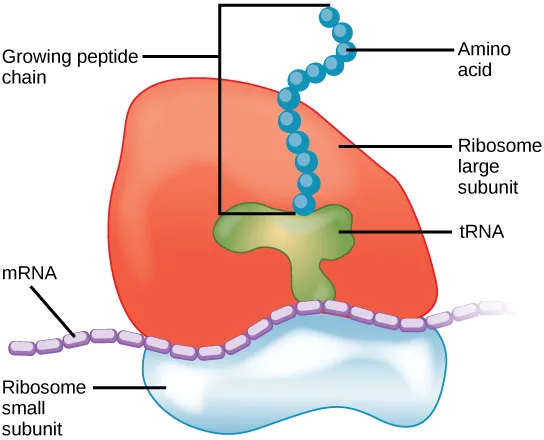
Ribosomes are the cellular structures responsible for protein synthesis. When we view them through an electron microscope, ribosomes appear either as clusters (polyribosomes) or single, tiny dots that float freely in the cytoplasm. They may be attached to the plasma membrane’s cytoplasmic side or the endoplasmic reticulum’s cytoplasmic side and the nuclear envelope’s outer membrane (above). Electron microscopy shows us that ribosomes, which are large protein and RNA complexes, consist of two subunits, large and small (below). Ribosomes receive their “orders” for protein synthesis from the nucleus where the DNA transcribes into messenger RNA (mRNA). The mRNA travels to the ribosomes, which translate the code provided by the sequence of the nitrogenous bases in the mRNA into a specific order of amino acids in a protein. Amino acids are the building blocks of proteins.
Extended description
A large, rounded orange-red shape (labeled ribosome large subunit) rests on top of a smaller, paler blue-white shape (labeled ribosome small subunit). A beaded purple strand labeled mRNA runs along the groove between the two subunits. A green Y-shaped molecule labeled tRNA sits in a notch where the subunits meet, and a chain of blue beads labeled amino acid climbs upward from the tRNA’s position, out of the top of the large subunit, forming the labeled growing peptide chain.
Because protein synthesis is an essential function of all cells (including enzymes, hormones, antibodies, pigments, structural components, and surface receptors), there are ribosomes in practically every cell. Ribosomes are particularly abundant in cells that synthesize large amounts of protein. For example, the pancreas is responsible for creating several digestive enzymes and the cells that produce these enzymes contain many ribosomes. Thus, we see another example of form following function.
Mitochondria
Scientists often call mitochondria (singular = mitochondrion) “powerhouses” or “energy factories” of both plant and animal cells because they are responsible for making adenosine triphosphate (ATP), the cell’s main energy-carrying molecule. ATP represents the cell’s short-term stored energy. Cellular respiration is the process of making ATP using the chemical energy in glucose and other nutrients. In mitochondria, this process uses oxygen and produces carbon dioxide as a waste product. In fact, the carbon dioxide that you exhale with every breath comes from the cellular reactions that produce carbon dioxide as a byproduct.
In keeping with our theme of form following function, it is important to point out that muscle cells have a very high concentration of mitochondria that produce ATP. Your muscle cells need considerable energy to keep your body moving. When your cells don’t get enough oxygen, they do not make much ATP. Instead, producing lactic acid accompanies the small amount of ATP they make in the absence of oxygen.
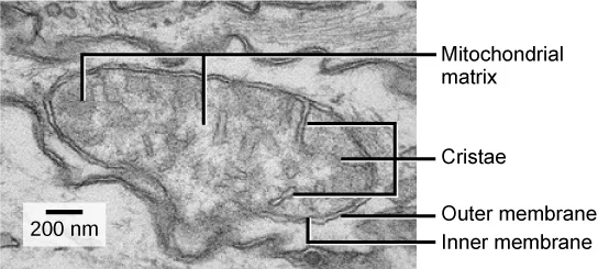
Mitochondria are oval-shaped, double membrane organelles (below) that have their own ribosomes and DNA. Each membrane is a phospholipid bilayer embedded with proteins. The inner layer has folds called cristae. We call the area surrounded by the folds the mitochondrial matrix. The cristae and the matrix have different roles in cellular respiration.
Peroxisomes
Peroxisomes are small, round organelles enclosed by single membranes. They carry out oxidation reactions that break down fatty acids and amino acids. They also detoxify many poisons that may enter the body. (Many of these oxidation reactions release hydrogen peroxide, H₂O₂, which would be damaging to cells; however, when these reactions are confined to peroxisomes, enzymes safely break down the H₂O₂ into oxygen and water.) For example, peroxisomes in liver cells detoxify alcohol. Glyoxysomes, which are specialized peroxisomes in plants, are responsible for converting stored fats into sugars. Plant cells contain many different types of peroxisomes that play a role in metabolism, pathogen defense, and stress response, to mention a few.
Vesicles and Vacuoles
Vesicles and vacuoles are membrane-bound sacs that function in storage and transport. Other than the fact that vacuoles are somewhat larger than vesicles, there is a very subtle distinction between them. Vesicle membranes can fuse with either the plasma membrane or other membrane systems within the cell. Additionally, some agents such as enzymes within plant vacuoles break down macromolecules. The vacuole’s membrane does not fuse with the membranes of other cellular components.
Animal Cells versus Plant Cells
At this point, you know that each eukaryotic cell has a plasma membrane, cytoplasm, a nucleus, ribosomes, mitochondria, peroxisomes, and in some, vacuoles, but there are some striking differences between animal and plant cells. While both animal and plant cells have microtubule organizing centers (MTOCs), animal cells also have centrioles associated with the MTOC: a complex we call the centrosome. Animal cells each have a centrosome and lysosomes; whereas, most plant cells do not. Plant cells have a cell wall, chloroplasts and other specialized plastids, and a large central vacuole; whereas, animal cells do not.
The Centrosome
The centrosome is a microtubule-organizing center found near the nuclei of animal cells. It contains a pair of centrioles, two structures that lie perpendicular to each other (below). Each centriole is a cylinder of nine triplets of microtubules.
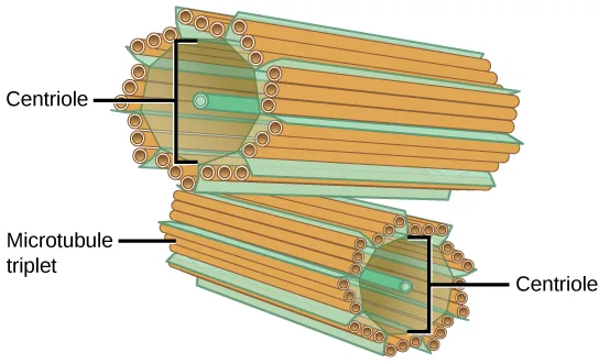
The centrosome (the organelle where all microtubules originate) replicates itself before a cell divides, and the centrioles appear to have some role in pulling the duplicated chromosomes to opposite ends of the dividing cell. However, the centriole’s exact function in cell division isn’t clear, because cells that have had the centrosome removed can still divide, and plant cells, which lack centrosomes, are capable of cell division.
Lysosomes
Animal cells have another set of organelles that most plant cells do not: lysosomes. The lysosomes are the cell’s “garbage disposal.” In plant cells, the digestive processes take place in vacuoles. Enzymes within the lysosomes aid in breaking down proteins, polysaccharides, lipids, nucleic acids, and even worn-out organelles. These enzymes are active at a much lower pH than the cytoplasm’s. Therefore, the pH within lysosomes is more acidic than the cytoplasm’s pH. Many reactions that take place in the cytoplasm could not occur at a low pH, so again, the advantage of compartmentalizing the eukaryotic cell into organelles is apparent.
The Cell Wall
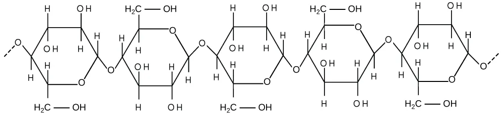
If you examine the plant-cell panel of the diagram above, you will see a structure external to the plasma membrane. This is the cell wall, a rigid covering that protects the cell, provides structural support, and gives shape to the cell. Fungal and some protistan cells also have cell walls. While the prokaryotic cell walls’ chief component is peptidoglycan, the major organic molecule in the plant (and some protists’) cell wall is cellulose (below), a polysaccharide comprised of glucose units. Have you ever noticed that when you bite into a raw vegetable, like celery, it crunches? That’s because you are tearing the celery cells’ rigid cell walls with your teeth.
Chloroplasts
Like the mitochondria, chloroplasts have their own DNA and ribosomes, but chloroplasts have an entirely different function. Chloroplasts are plant cell organelles that carry out photosynthesis. Photosynthesis is the series of reactions that use carbon dioxide, water, and light energy to make glucose and oxygen. This is a major difference between plants and animals. Plants (autotrophs) are able to make their own food, like sugars used in cellular respiration to provide ATP energy generated in the plant mitochondria. Animals (heterotrophs) must ingest their food.
Like mitochondria, chloroplasts have outer and inner membranes, but within the space enclosed by a chloroplast’s inner membrane is a set of interconnected and stacked fluid-filled membrane sacs we call thylakoids (below). Each thylakoid stack is a granum (plural = grana). We call the fluid enclosed by the inner membrane that surrounds the grana the stroma.
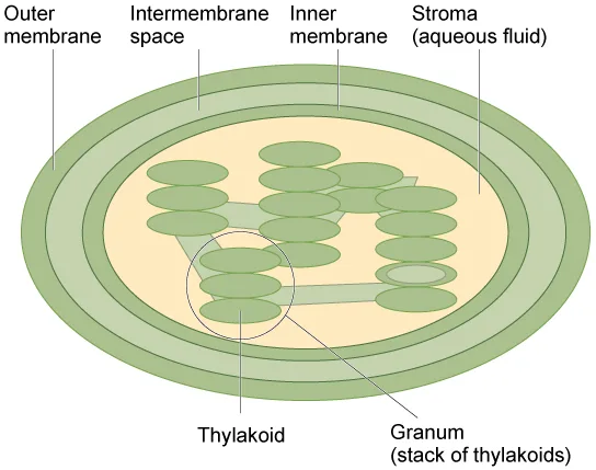
The chloroplasts contain a green pigment, chlorophyll, which captures the light energy that drives the reactions of photosynthesis. Like plant cells, photosynthetic protists also have chloroplasts. Some bacteria perform photosynthesis, but their chlorophyll is not relegated to an organelle.
Evolution Connection. Endosymbiosis. We have mentioned that both mitochondria and chloroplasts contain DNA and ribosomes. Have you wondered why? Strong evidence points to endosymbiosis as the explanation.
Symbiosis is any type of close and long term relationship between two organisms from different species that depend on each other for their survival. In some cases both organisms benefit from the relationship. Endosymbiosis (endo- = “within”) is a mutually beneficial relationship in which one organism lives inside the other. Endosymbiotic relationships abound in nature. We have already mentioned that microbes that produce vitamin K live inside the human gut. This relationship is beneficial for us because we are unable to synthesize vitamin K. It is also beneficial for the microbes because they are protected from other organisms and from drying out, and they receive abundant food from the environment of the large intestine.
Scientists have long noticed that bacteria, mitochondria, and chloroplasts are similar in size. We also know that bacteria have DNA and ribosomes, just like mitochondria and chloroplasts. Scientists believe that host cells and bacteria formed an endosymbiotic relationship when the host cells ingested both aerobic and autotrophic bacteria (cyanobacteria) but did not destroy them. Through many millions of years of evolution, these ingested bacteria became more specialized in their functions, with the aerobic bacteria becoming mitochondria and the autotrophic bacteria becoming chloroplasts.
The Central Vacuole
Previously, we mentioned vacuoles as essential components of plant cells. If you look at the plant-cell panel of the diagram above, you will see that plant cells each have a large central vacuole that occupies most of the cell’s volume. The central vacuole plays a key role in regulating the cell’s concentration of water in changing environmental conditions. Have you ever noticed that if you forget to water a plant for a few days, it wilts? That’s because as the water concentration in the soil becomes lower than the water concentration in the plant, water moves out of the central vacuoles and cytoplasm. As the central vacuole shrinks, it leaves the cell wall unsupported. This loss of support to the plant’s cell walls results in the wilted appearance.
The central vacuole also supports the cell’s expansion. When the central vacuole holds more water, the cell becomes larger without having to invest considerable energy in synthesizing new cytoplasm.
Summary
Like a prokaryotic cell, a eukaryotic cell has a plasma membrane, cytoplasm, and ribosomes, but a eukaryotic cell is typically larger than a prokaryotic cell, has a true nucleus (meaning a membrane surrounds its DNA), and has other membrane-bound organelles that allow for compartmentalizing functions. The plasma membrane is a phospholipid bilayer embedded with proteins. The nucleus’s nucleolus is the site of ribosome assembly. We find ribosomes either in the cytoplasm or attached to the cytoplasmic side of the plasma membrane or endoplasmic reticulum. They perform protein synthesis. Mitochondria participate in cellular respiration. They are responsible for the majority of ATP produced in the cell. Peroxisomes hydrolyze fatty acids, amino acids, and some toxins. Vesicles and vacuoles are storage and transport compartments. In plant cells, vacuoles also help break down macromolecules.
Animal cells also have a centrosome and lysosomes. The centrosome has two bodies perpendicular to each other, the centrioles, and has an unknown purpose in cell division. Lysosomes are the digestive organelles of animal cells.
Plant cells and plant-like cells each have a cell wall, chloroplasts, and a central vacuole. The plant cell wall, whose primary component is cellulose, protects the cell, provides structural support, and gives the cell shape. Photosynthesis takes place in chloroplasts. The central vacuole can expand without having to produce more cytoplasm.
Key terms
- cell wall — rigid cell covering comprised of various molecules that protects the cell, provides structural support, and gives shape to the cell
- central vacuole — large plant cell organelle that regulates the cell’s storage compartment, holds water, and plays a significant role in cell growth as the site of macromolecule degradation
- centrosome — region in animal cells made of two centrioles that serves as an organizing center for microtubules
- chlorophyll — green pigment that captures the light energy that drives the light reactions of photosynthesis
- chloroplast — plant cell organelle that carries out photosynthesis
- chromatin — protein-DNA complex that serves as the chromosomes’ building material
- chromosome — structure within the nucleus that comprises chromatin that contains DNA, the hereditary material
- cytoplasm — entire region between the plasma membrane and the nuclear envelope, consisting of organelles suspended in the gel-like cytosol, the cytoskeleton, and various chemicals
- cytosol — the cytoplasm’s gel-like material in which cell structures are suspended
- eukaryotic cell — cell that has a membrane-bound nucleus and several other membrane-bound compartments or sacs
- lysosome — organelle in an animal cell that functions as the cell’s digestive component; it breaks down proteins, polysaccharides, lipids, nucleic acids, and even worn-out organelles
- mitochondria — (singular = mitochondrion) cellular organelles responsible for carrying out cellular respiration, resulting in producing ATP, the cell’s main energy-carrying molecule
- nuclear envelope — double-membrane structure that constitutes the nucleus’ outermost portion
- nucleolus — darkly staining body within the nucleus that is responsible for assembling ribosome subunits
- nucleoplasm — semi-solid fluid inside the nucleus that contains the chromatin and nucleolus
- nucleus — cell organelle that houses the cell’s DNA and directs ribosome and protein synthesis
- organelle — compartment or sac within a cell
- peroxisome — small, round organelle that contains hydrogen peroxide, oxidizes fatty acids and amino acids, and detoxifies many poisons
- plasma membrane — phospholipid bilayer with embedded (integral) or attached (peripheral) proteins, and separates the cell’s internal content from its surrounding environment
- ribosome — cellular structure that carries out protein synthesis
- vacuole — membrane-bound sac, somewhat larger than a vesicle, which functions in cellular storage and transport
- vesicle — small, membrane-bound sac that functions in cellular storage and transport; its membrane is capable of fusing with the plasma membrane and the membranes of the endoplasmic reticulum and Golgi apparatus
Practice
Describe the structure of eukaryotic cells
Which of the following is surrounded by two phospholipid bilayers?
The nuclear envelope, which encloses this fluid, is itself made of two separate phospholipid bilayers — an inner and an outer nuclear membrane.Which of the following is found in both eukaryotic and prokaryotic cells?
Ask which of these is not membrane-bound — prokaryotic cells have no membrane-bound organelles at all.Unlike a prokaryotic cell, a eukaryotic cell has a ________ (meaning a membrane surrounds its DNA).
The section’s summary uses this exact phrase for the membrane-bound nucleus that gives eukaryotic cells their name.Compare animal cells with plant cells
In plant cells, the function of the lysosomes is carried out by ________.
Plant cells lack lysosomes altogether; the digestive processes instead take place in a different, larger organelle.A rigid covering outside the plasma membrane that protects a plant cell, provides structural support, and gives it shape is called the ________.
Fungal and some protistan cells have this structure too, but animal cells do not.Plant cells and plant-like cells each have a cell wall, chloroplasts, and a ________.
The section’s summary names this large, water-storing organelle as the third structure plant cells have that animal cells lack.State the role of the plasma membrane
Why are plasma membranes arranged as a bilayer rather than a monolayer?
Show model answer
Did your answer mention:
A phospholipid bilayer with embedded or attached proteins that separates a cell’s internal contents from its surrounding environment is called the ________.
Integral and peripheral proteins sit in and on this structure, along with embedded cholesterol.The plasma membrane is a ________ embedded with proteins.
The section’s summary opens its plasma-membrane sentence with this two-word structural term.Summarize the functions of the major cell organelles
Peroxisomes got their name because hydrogen peroxide is:
Look at what the oxidation reactions peroxisomes carry out actually release as a byproduct.Tay-Sachs disease is a genetic disorder that results in the destruction of neurons due to a buildup of sphingolipids in the cells. Which organelle is malfunctioning in Tay-Sachs?
Sphingolipids are complex lipids that would normally be broken down by this organelle’s digestive enzymes.You already know that ribosomes are abundant in red blood cells. In what other cells of the body would you find them in great abundance? Why?
Show model answer
Did your answer mention:
What are the structural and functional similarities and differences between mitochondria and chloroplasts?
Show model answer
Did your answer mention:
This section is adapted from Biology 2e, Section 4.3: Eukaryotic Cells by Mary Ann Clark, Jung Choi, Matthew Douglas, and OpenStax, © OpenStax, licensed under CC BY-NC-SA 4.0. Access the original for free at openstax.org. Changes: figures re-encoded as WebP, with ten of the eleven re-kinded from the manifest’s file-extension guess of “photo” to “diagram” after inspection (every one is a drawn illustration, or a diagram paired with a micrograph whose labels carry the teaching — only the mitochondrion electron micrograph is a photograph, and its four baked-in labels are all named in its caption); an extended description added for the six figures that are labeled diagrams whose full reading is not carried by their captions (the animal-cell and plant-cell diagrams, which the source prints as panels (a) and (b) of one figure and this page renders as two consecutive figures — the second carrying a short caption written here — because each panel is its own image; the plasma membrane, the microvilli micrograph-and-diagram pair, the nucleus, the chromatin-and-chromosomes pair, and the ribosome); inline references to figures changed from the source’s print-figure links ("(see below)", plain parenthetical links) to descriptive phrases (“above,” “below”) since figures are not numbered here, and the source’s lettered cross-reference “if you look at b” resolved to “the plant-cell panel of the diagram above”; the evolution feature box rendered as a callout with its bold name and the source’s own subheading (“Endosymbiosis”) kept as an italic clause; the visual-connection question kept in the body immediately after its figure and rendered as a self-check, since the source keys it with a prose solution rather than a lettered option; a typo in the source prose (“pathogene defense”) corrected to “pathogen defense”, and a missing “that” supplied in the Evolution Connection’s sentence on symbiosis; the end-of-section Review Questions and Critical Thinking Questions adapted into the closing interactive Practice block (multiple choice and self-check respectively); and two key-term recall items (cell wall, plasma membrane) added from the glossary to round out every objective group with an auto-graded item; rubric checkpoints added to each self-check (the body self-check on the nucleolus included), decomposing its model answer (the source solution) into check-off clauses with no new claims; and three summary-derived recall items added, one per objective still short of the raised floor (“true nucleus” under the first objective, “central vacuole” as a multiple choice under the second, and “phospholipid bilayer” under the third).