The Endomembrane System and Proteins
By the end of this section, you will be able to:
- List the components of the endomembrane system
- Recognize the relationship between the endomembrane system and its functions
The endomembrane system (endo = “within”) is a group of membranes and organelles in eukaryotic cells that works together to modify, package, and transport lipids and proteins. It includes the nuclear envelope, lysosomes, and vesicles, which we have already mentioned, and the endoplasmic reticulum and Golgi apparatus, which we will cover shortly. Although not technically within the cell, the plasma membrane is included in the endomembrane system because, as you will see, it interacts with the other endomembranous organelles. The endomembrane system does not include either mitochondria or chloroplast membranes.
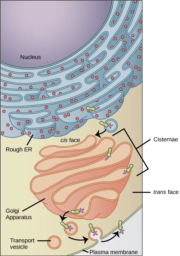
Extended description
A vertical cross-section, top to bottom. At top, a rounded purple nucleus sits beside the rough endoplasmic reticulum, drawn as stacked, folded blue membrane sheets studded with small red dots (ribosomes). A green integral membrane protein, with a purple carbohydrate attached on the side facing into the membrane sac, sits within the ER membrane. An arrow shows a small circular vesicle carrying the same green protein and purple carbohydrate budding from the ER and moving down to the Golgi apparatus, drawn as a stack of curved orange-and-pink membrane layers labeled cis face (top, nearest the ER) and trans face (bottom). As the protein passes through the Golgi’s stacked cisternae, arrows show it gaining additional purple carbohydrate. A vesicle then buds from the Golgi’s trans face and travels down to the plasma membrane at the bottom of the figure, where it fuses; the carbohydrate, originally facing the inside of the ER membrane, now faces outward from the cell surface, and a transport vesicle icon at lower left labels the vesicle stage.
If a peripheral membrane protein were synthesized in the lumen (inside) of the ER, would it end up on the inside or outside of the plasma membrane?
Show model answer
Did your answer mention:
The Endoplasmic Reticulum
The endoplasmic reticulum (ER) is a series of interconnected membranous sacs and tubules that collectively modifies proteins and synthesizes lipids. However, these two functions take place in separate areas of the ER: the rough ER and the smooth ER, respectively.
We call the ER tubules’ hollow portion the lumen or cisternal space. The ER’s membrane, which is a phospholipid bilayer embedded with proteins, is continuous with the nuclear envelope.
Rough ER
Scientists have named the rough endoplasmic reticulum (RER) as such because the ribosomes attached to its cytoplasmic surface give it a studded appearance when viewing it through an electron microscope (below).
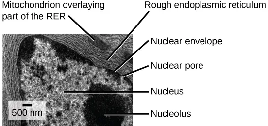
Extended description
Labels point from the image to six features. ‘Mitochondrion overlaying part of the RER’ and ‘Rough endoplasmic reticulum’ point to a striated, layered membrane band curving across the upper portion of the image. ‘Nuclear envelope’ and ‘Nuclear pore’ point to the boundary of the large dark circular body that fills most of the frame. ‘Nucleus’ points to that dark circular body, and ‘Nucleolus’ points to a smaller, denser dark patch within it. A scale bar in the lower left reads 500 nm.
Ribosomes transfer their newly synthesized proteins into the RER’s lumen where they undergo structural modifications, such as folding or acquiring side chains. These modified proteins incorporate into cellular membranes—the ER or the ER’s or other organelles’ membranes. The proteins can also secrete from the cell (such as protein hormones, enzymes). The RER also makes phospholipids for cellular membranes.
If the phospholipids or modified proteins are not destined to stay in the RER, they will reach their destinations via transport vesicles that bud from the RER’s membrane (above).
Since the RER is engaged in modifying proteins (such as enzymes, for example) that secrete from the cell, you would be correct in assuming that the RER is abundant in cells that secrete proteins. This is the case with liver cells, for example.
Smooth ER
The smooth endoplasmic reticulum (SER) is continuous with the RER but has few or no ribosomes on its cytoplasmic surface (above). SER functions include synthesis of carbohydrates, lipids, and steroid hormones; detoxification of medications and poisons; and storing calcium ions.
In muscle cells, a specialized SER, the sarcoplasmic reticulum, is responsible for storing calcium ions that are needed to trigger the muscle cells’ coordinated contractions.
Career Connection. Cardiologist. Heart disease is the leading cause of death in the United States. This is primarily due to our sedentary lifestyle and our high trans-fat diets.
Heart failure is just one of many disabling heart conditions. Heart failure does not mean that the heart has stopped working. Rather, it means that the heart can’t pump with sufficient force to transport oxygenated blood to all the vital organs. Left untreated, heart failure can lead to kidney failure and other organ failure.
Cardiac muscle tissue comprises the heart’s wall. Heart failure occurs when cardiac muscle cells’ endoplasmic reticula do not function properly. As a result, an insufficient number of calcium ions are available to trigger a sufficient contractile force.
Cardiologists (cardi- = “heart”; -ologist = “one who studies”) are doctors who specialize in treating heart diseases, including heart failure. Cardiologists can diagnose heart failure via a physical examination, results from an electrocardiogram (ECG, a test that measures the heart’s electrical activity), a chest X-ray to see whether the heart is enlarged, and other tests. If the cardiologist diagnoses heart failure, they will typically prescribe appropriate medications and recommend a reduced table salt intake and a supervised exercise program.
The Golgi Apparatus
We have already mentioned that vesicles can bud from the ER and transport their contents elsewhere, but where do the vesicles go? Before reaching their final destination, the lipids or proteins within the transport vesicles still need sorting, packaging, and tagging so that they end up in the right place. Sorting, tagging, packaging, and distributing lipids and proteins takes place in the Golgi apparatus (also called the Golgi body), a series of flattened membranous sacs (below).
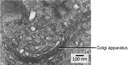
The side of the Golgi apparatus that is closer to the ER is called the cis face. The opposite side is the trans face. The transport vesicles that formed from the ER travel to the cis face, fuse with it, and empty their contents into the Golgi apparatus’ lumen. As the proteins and lipids travel through the Golgi, they undergo further modifications that allow them to be sorted. The most frequent modification is adding short sugar molecule chains. These newly modified proteins and lipids then tag with phosphate groups or other small molecules in order to travel to their proper destinations.
Finally, the modified and tagged proteins are packaged into secretory vesicles that bud from the Golgi’s trans face. While some of these vesicles deposit their contents into other cell parts where they will be used, other secretory vesicles fuse with the plasma membrane and release their contents outside the cell.
In another example of form following function, cells that engage in a great deal of secretory activity (such as salivary gland cells that secrete digestive enzymes or immune system cells that secrete antibodies) have an abundance of Golgi.
In plant cells, the Golgi apparatus has the additional role of synthesizing polysaccharides, some of which are incorporated into the cell wall and some of which other cell parts use.
Career Connection. Geneticist. Many diseases arise from genetic mutations that prevent synthesizing critical proteins. One such disease is Lowe disease (or oculocerebrorenal syndrome, because it affects the eyes, brain, and kidneys). In Lowe disease, there is a deficiency in an enzyme localized to the Golgi apparatus. Children with Lowe disease are born with cataracts, typically develop kidney disease after the first year of life, and may have intellectual disabilities.
A mutation on the X chromosome causes Lowe disease. The X chromosome is one of the two human sex chromosomes, as these chromosomes determine a person’s sex. Females possess two X chromosomes while males possess one X and one Y chromosome. In females, the genes on only one of the two X chromosomes are expressed. Females who carry the Lowe disease gene on one of their X chromosomes are carriers and do not show symptoms of the disease. However, males only have one X chromosome and the genes on this chromosome are always expressed. Therefore, males will always have Lowe disease if their X chromosome carries the Lowe disease gene. Geneticists have identified the mutated gene’s location, as well as many other mutation locations that cause genetic diseases. Through prenatal testing, a pregnant person can find out if the fetus they are carrying may be afflicted with one of several genetic diseases.
Geneticists analyze prenatal genetic test results and may counsel pregnant people on available options. They may also conduct genetic research that leads to new drugs or foods, or perform DNA analyses for forensic investigations.
Lysosomes
In addition to their role as the digestive component and organelle-recycling facility of animal cells, lysosomes are part of the endomembrane system. Lysosomes also use their hydrolytic enzymes to destroy pathogens (disease-causing organisms) that might enter the cell. A good example of this occurs in macrophages, a group of white blood cells which are part of your body’s immune system. In a process that scientists call phagocytosis or endocytosis, a section of the macrophage’s plasma membrane invaginates (folds in) and engulfs a pathogen. The invaginated section, with the pathogen inside, then pinches itself off from the plasma membrane and becomes a vesicle. The vesicle fuses with a lysosome. The lysosome’s hydrolytic enzymes then destroy the pathogen (below).
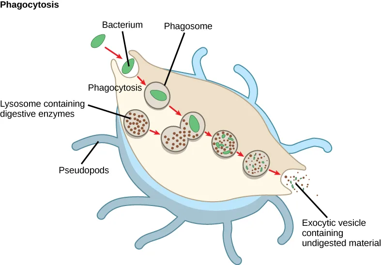
Extended description
A large tan cell body fringed with thin, branching blue surface projections labeled Pseudopods. A small green oval labeled Bacterium sits just outside the cell at upper left. A red arrow points from it to a shallow dent in the cell’s edge labeled Phagocytosis, where the membrane is folding around the bacterium. A second red arrow leads to the first of four circles arranged in a row across the cell’s interior: the first circle, labeled Phagosome, contains only the green bacterium; a nearby cluster of small brown dots is labeled Lysosome containing digestive enzymes. Red arrows connect the four circles in sequence, showing the brown dots merging into each circle while the green shape shrinks and fragments — intact in the first circle, partly broken up in the second and third, and reduced to a few small green flecks among the brown dots in the fourth. A final red arrow leads outside the cell to a small scatter of dots labeled Exocytic vesicle containing undigested material.
Summary
The endomembrane system includes the nuclear envelope, lysosomes, vesicles, the ER, and Golgi apparatus, as well as the plasma membrane. These cellular components work together to modify, package, tag, and transport proteins and lipids that form the membranes.
The RER modifies proteins and synthesizes phospholipids in cell membranes. The SER synthesizes carbohydrates, lipids, and steroid hormones; engages in the detoxification of medications and poisons; and stores calcium ions. Sorting, tagging, packaging, and distributing lipids and proteins take place in the Golgi apparatus. Budding RER and Golgi membranes create lysosomes. Lysosomes digest macromolecules, recycle worn-out organelles, and destroy pathogens.
Key terms
- endomembrane system — group of organelles and membranes in eukaryotic cells that work together modifying, packaging, and transporting lipids and proteins
- endoplasmic reticulum (ER) — series of interconnected membranous structures within eukaryotic cells that collectively modify proteins and synthesize lipids
- Golgi apparatus — eukaryotic organelle comprised of a series of stacked membranes that sorts, tags, and packages lipids and proteins for distribution
- rough endoplasmic reticulum (RER) — region of the endoplasmic reticulum that is studded with ribosomes and engages in protein modification and phospholipid synthesis
- smooth endoplasmic reticulum (SER) — region of the endoplasmic reticulum that has few or no ribosomes on its cytoplasmic surface and synthesizes carbohydrates, lipids, and steroid hormones; detoxifies certain chemicals (like pesticides, preservatives, medications, and environmental pollutants), and stores calcium ions
Practice
List the components of the endomembrane system
Which of the following is not a component of the endomembrane system?
Three of these four are named in the section as endomembrane-system components; the fourth has its own separate set of membranes, outside the system.Congenital disorders of glycosylation are a growing class of rare diseases. Which organelle would be most commonly involved in the glycoprotein disorder portion of the group?
Glycosylation means attaching sugar chains to a protein — recall which organelle performs that particular modification once the RER has handed the protein off.A group of organelles and membranes in eukaryotic cells that work together to modify, package, and transport lipids and proteins is called the ________.
It is the name for the whole group — the nuclear envelope, ER, Golgi apparatus, lysosomes, vesicles, and plasma membrane all belong to it.In your opinion, is the nuclear membrane part of the endomembrane system? Why or why not? Defend your answer.
Show model answer
Did your answer mention:
Recognize the relationship between the endomembrane system and its functions
The process by which a cell engulfs a foreign particle is known as:
This is the term the macrophage example uses for a cell folding its membrane around a pathogen and engulfing it.Which of the following is most likely to have the greatest concentration of smooth endoplasmic reticulum?
Match the organelle to one of the SER’s specific functions — protein secretion and pathogen destruction point to different organelles instead.Which of the following sequences correctly lists in order the steps involved in the incorporation of a proteinaceous molecule within a cell?
Trace the path in the order this section describes it — ribosome, then ER, then Golgi, then vesicle — and rule out any option that gives the ribosome or the vesicle the wrong job.A series of interconnected membranous sacs and tubules within eukaryotic cells that collectively modifies proteins and synthesizes lipids is called the ________.
It has two functionally distinct regions — one studded with ribosomes, one without.In the context of cell biology, what do we mean by form follows function? What are at least two examples of this concept?
Show model answer
Did your answer mention:
This section is adapted from Biology 2e, Section 4.4: The Endomembrane System and Proteins by Mary Ann Clark, Jung Choi, Matthew Douglas, and OpenStax, © OpenStax, licensed under CC BY-NC-SA 4.0. Access the original for free at openstax.org. Changes: figures re-encoded as WebP, with one re-kinded from the manifest’s file-extension guess of “photo” to “diagram” after inspection (the phagocytosis illustration is a drawn cartoon, not a photograph, despite its JPEG source); a longer extended description added for the three figures that are labeled diagrams or micrographs whose full reading is not carried by their captions (the ER-to-Golgi-to-membrane flow diagram, the labeled rough-ER micrograph, and the phagocytosis flow diagram); the first figure’s alt shortened from over 600 characters to what the image shows, with its step-by-step walk-through moved into the longdesc; inline references to figures changed from the source’s parenthetical figure links to descriptive phrases (“above,” “below”) since figures are not numbered here; the Link to Learning note’s bare “here” replaced with descriptive link text; two Career Connection notes rendered as callouts with their bold names and italicized job titles; the Visual Connection question kept in the body immediately after its figure and rendered as a self-check, since the source keys it with a prose solution rather than a lettered option; the end-of-section Review Questions and Critical Thinking Questions adapted into the closing interactive Practice block (multiple choice and self-check respectively); and two key-term recall items (endomembrane system, endoplasmic reticulum) added from the glossary to round out both objective groups with an auto-graded item; and rubric checkpoints added to each self-check (the body self-check on the peripheral membrane protein included), decomposing its model answer (the source solution) into check-off clauses with no new claims — no new Practice items were needed, since both objective groups already carried the module’s full Review Question and Critical Thinking Question sets at or above the raised per-objective and per-section floors.