The Cytoskeleton
By the end of this section, you will be able to:
- Describe the cytoskeleton
- Compare the roles of microfilaments, intermediate filaments, and microtubules
- Compare and contrast cilia and flagella
- Summarize the differences among the components of prokaryotic cells, animal cells, and plant cells
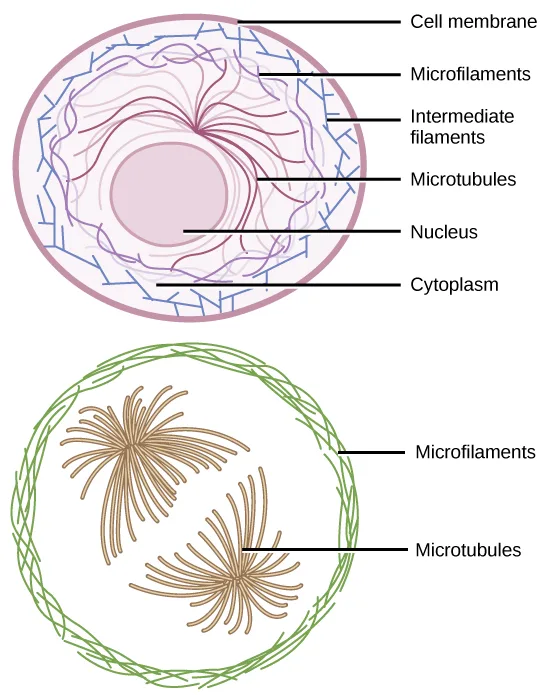
If you were to remove all the organelles from a cell, would the plasma membrane and the cytoplasm be the only components left? No. Within the cytoplasm, there would still be ions and organic molecules, plus a network of protein fibers that help maintain the cell’s shape, secure some organelles in specific positions, allow cytoplasm and vesicles to move within the cell, and enable cells within multicellular organisms to move. Collectively, scientists call this network of protein fibers the cytoskeleton. There are three types of fibers within the cytoskeleton: microfilaments, intermediate filaments, and microtubules (below). Here, we will examine each.
Extended description
Top panel: a cross-section of a generalized cell labeled, from outside in, Cell membrane (the outer boundary), Microfilaments (thin blue zigzag lines just inside the membrane and scattered through the cytoplasm), Intermediate filaments (dark red strands radiating outward from around the nucleus), Microtubules (lighter purple strands also radiating from near the nucleus), Nucleus (the central circle), and Cytoplasm (the space between the nucleus and the membrane). Bottom panel: a single cell outlined by a coiled green ring labeled Microfilaments, containing two brown, star-shaped clusters of radiating fibers — one nearer the upper left, one nearer the lower right — labeled Microtubules.
Microfilaments
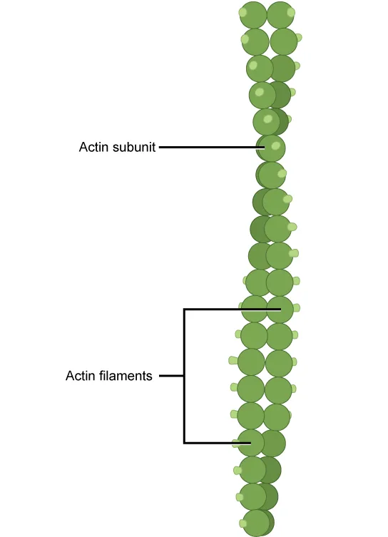
Of the three types of protein fibers in the cytoskeleton, microfilaments are the narrowest. They function in cellular movement, have a diameter of about 7 nm, and are comprised of two globular protein intertwined strands, which we call actin (below). For this reason, we also call microfilaments actin filaments.
Extended description
A vertical chain of two intertwined strands of ball-shaped actin subunits, one strand slightly offset from the other in a twisted double-strand pattern. A leader line labeled ‘Actin subunit’ points to a single ball partway down the chain. A bracket labeled ‘Actin filaments’ spans a short stretch of the twisted double strand near the middle of the chain.
ATP powers actin to assemble its filamentous form, which serves as a track for the movement of a motor protein we call myosin. This enables actin to engage in cellular events requiring motion, such as cell division in eukaryotic cells and cytoplasmic streaming, which is the cell cytoplasm’s circular movement in plant cells. Actin and myosin are plentiful in muscle cells. When your actin and myosin filaments slide past each other, your muscles contract.
Microfilaments also provide some rigidity and shape to the cell. They can depolymerize (disassemble) and reform quickly, thus enabling a cell to change its shape and move. White blood cells (your body’s infection-fighting cells) make good use of this ability. They can move to an infection site and phagocytize the pathogen.
Intermediate Filaments
Several strands of fibrous proteins that are wound together comprise intermediate filaments (below). Cytoskeleton elements get their name from the fact that their diameter, 8 to 10 nm, is between those of microfilaments and microtubules.
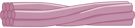
Intermediate filaments have no role in cell movement. Their function is purely structural. They bear tension, thus maintaining the cell’s shape, and anchor the nucleus and other organelles in place. The figure above shows how intermediate filaments create a supportive scaffolding inside the cell.
The intermediate filaments are the most diverse group of cytoskeletal elements. Several fibrous protein types are in the intermediate filaments. You are probably most familiar with keratin, the fibrous protein that strengthens your hair, nails, and the skin’s epidermis.
Microtubules
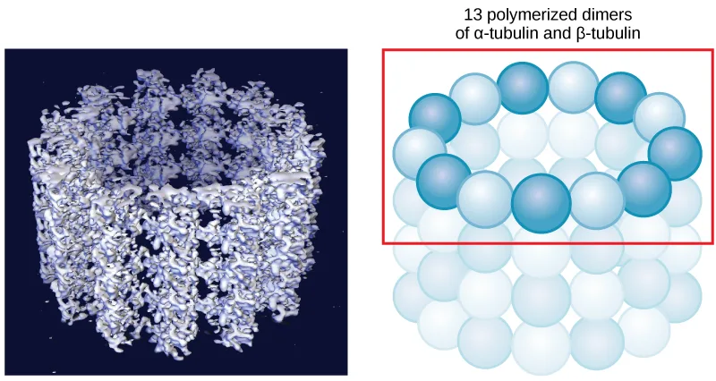
As their name implies, microtubules are small hollow tubes. Polymerized dimers of α-tubulin and β-tubulin, two globular proteins, comprise the microtubule’s walls (below). With a diameter of about 25 nm, microtubules are cytoskeletons’ widest components. They help the cell resist compression, provide a track along which vesicles move through the cell, and pull replicated chromosomes to opposite ends of a dividing cell. Like microfilaments, microtubules can disassemble and reform quickly.
Extended description
Left panel: a dense, textured three-dimensional rendering of a hollow, roughly cylindrical microtubule wall reconstructed from molecular structure data, its surface a mesh of interwoven ridges, set against a dark background. Right panel: a red-outlined box highlights one ring of 13 spheres, alternating darker and lighter blue, arranged in a circle and labeled ‘13 polymerized dimers of α-tubulin and β-tubulin’; below the highlighted ring, additional rows of the same spheres continue in a repeating grid, suggesting the tube’s wall extending along its length.
Microtubules are also the structural elements of flagella, cilia, and centrioles (the latter are the centrosome’s two perpendicular bodies). In animal cells, the centrosome is the microtubule-organizing center. In eukaryotic cells, flagella and cilia are quite different structurally from their counterparts in prokaryotes, as we discuss below.
Flagella and Cilia
The flagella (singular = flagellum) are long, hair-like structures that extend from the plasma membrane and enable an entire cell to move (for example, sperm, Euglena, and some prokaryotes). When present, the cell has just one flagellum or a few flagella. However, when cilia (singular = cilium) are present, many of them extend along the plasma membrane’s entire surface. They are short, hair-like structures that move entire cells (such as paramecia) or substances along the cell’s outer surface (for example, the cilia of cells lining the Fallopian tubes that move the ovum toward the uterus, or cilia lining the cells of the respiratory tract that trap particulate matter and move it toward your nostrils.)
Despite their differences in length and number, flagella and cilia share a common structural arrangement of microtubules called a “9 + 2 array.” This is an appropriate name because a single flagellum or cilium is made of a ring of nine microtubule doublets, surrounding a single microtubule doublet in the center (below).
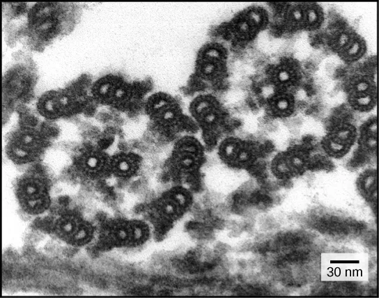
You have now completed a broad survey of prokaryotic and eukaryotic cell components. For a summary of cellular components in prokaryotic and eukaryotic cells, see the table below.
Components of Prokaryotic and Eukaryotic Cells
| Cell Component | Function | Present in Prokaryotes? | Present in Animal Cells? | Present in Plant Cells? |
|---|---|---|---|---|
| Plasma membrane | Separates cell from external environment; controls passage of organic molecules, ions, water, oxygen, and wastes into and out of cell | Yes | Yes | Yes |
| Cytoplasm | Provides turgor pressure to plant cells as fluid inside the central vacuole; site of many metabolic reactions; medium in which organelles are found | Yes | Yes | Yes |
| Nucleolus | Darkened area within the nucleus where ribosomal subunits are synthesized. | No | Yes | Yes |
| Nucleus | Cell organelle that houses DNA and directs synthesis of ribosomes and proteins | No | Yes | Yes |
| Ribosomes | Protein synthesis | Yes | Yes | Yes |
| Mitochondria | ATP production/cellular respiration | No | Yes | Yes |
| Peroxisomes | Oxidize and thus break down fatty acids and amino acids, and detoxify poisons | No | Yes | Yes |
| Vesicles and vacuoles | Storage and transport; digestive function in plant cells | No | Yes | Yes |
| Centrosome | Unspecified role in cell division in animal cells; microtubule source in animal cells | No | Yes | No |
| Lysosomes | Digestion of macromolecules; recycling of worn-out organelles | No | Yes | Some |
| Cell wall | Protection, structural support, and maintenance of cell shape | Yes, primarily peptidoglycan | No | Yes, primarily cellulose |
| Chloroplasts | Photosynthesis | No | No | Yes |
| Endoplasmic reticulum | Modifies proteins and synthesizes lipids | No | Yes | Yes |
| Golgi apparatus | Modifies, sorts, tags, packages, and distributes lipids and proteins | No | Yes | Yes |
| Cytoskeleton | Maintains cell’s shape, secures organelles in specific positions, allows cytoplasm and vesicles to move within cell, and enables unicellular organisms to move independently | Yes | Yes | Yes |
| Flagella | Cellular locomotion | Some | Some | No, except for some plant sperm cells |
| Cilia | Cellular locomotion, movement of particles along plasma membrane’s extracellular surface, and filtration | Some | Some | No |
Summary
The cytoskeleton has three different protein element types. From narrowest to widest, they are the microfilaments (actin filaments), intermediate filaments, and microtubules. Biologists often associate microfilaments with myosin. They provide rigidity and shape to the cell and facilitate cellular movements. Intermediate filaments bear tension and anchor the nucleus and other organelles in place. Microtubules help the cell resist compression, serve as tracks for motor proteins that move vesicles through the cell, and pull replicated chromosomes to opposite ends of a dividing cell. They are also the structural element of centrioles, flagella, and cilia.
Key terms
- cilium — (plural = cilia) short, hair-like structure that extends from the plasma membrane in large numbers and functions to move an entire cell or move substances along the cell’s outer surface
- cytoskeleton — protein fiber network that collectively maintains the cell’s shape, secures some organelles in specific positions, allows cytoplasm and vesicles to move within the cell, and enables unicellular organisms to move independently
- flagellum — (plural = flagella) long, hair-like structure that extends from the plasma membrane and moves the cell
- intermediate filament — cytoskeletal component, comprised of several fibrous protein intertwined strands, that bears tension, supports cell-cell junctions, and anchors cells to extracellular structures
- microfilament — the cytoskeleton system’s narrowest element; it provides rigidity and shape to the cell and enables cellular movements
- microtubule — the cytoskeleton system’s widest element; it helps the cell resist compression, provides a track along which vesicles move through the cell, pulls replicated chromosomes to opposite ends of a dividing cell, and is the structural element of centrioles, flagella, and cilia
Practice
Describe the cytoskeleton
A network of protein fibers that maintains a cell’s shape, secures organelles in specific positions, and allows cytoplasm, vesicles, and the whole cell to move is called the ________.
It is not a single organelle but a network of protein fibers running throughout the cytoplasm.Which of the following have the ability to disassemble and reform quickly?
Both of these fiber types can quickly disassemble and reform, which is what lets a cell rapidly change shape.From narrowest to widest, they are the microfilaments (actin filaments), intermediate filaments, and ________.
The section’s summary lists the three cytoskeletal fiber types in size order, ending with the widest.Compare the roles of microfilaments, intermediate filaments, and microtubules
Which of the following do not play a role in intracellular movement?
Which of the three fiber types is described as purely structural, with no role in cell movement?Describe how microfilaments and microtubules are involved in the phagocytosis and destruction of a pathogen by a macrophage.
Show model answer
Did your answer mention:
The narrowest of the three cytoskeletal fiber types, composed of two intertwined strands of the protein actin, is called a(n) ________.
Its diameter is about 7 nm — the narrowest of the three — and it drives muscle contraction and the crawling of cells.A cytoskeletal component made of several intertwined fibrous protein strands that bears tension, supports cell-cell junctions, and anchors cells to extracellular structures is called a(n) ________.
Its diameter, 8 to 10 nm, falls between the other two fiber types, which is how it got its name.The widest of the three cytoskeletal fiber types, which resists compression, serves as a track for organelle movement, and pulls replicated chromosomes apart during cell division, is called a(n) ________.
At about 25 nm across, it is the widest of the three, and it forms the core of centrioles, cilia, and flagella.Compare and contrast cilia and flagella
In humans, _____ are used to move a cell within its environment while _____ are used to move the environment relative to the cell.
One of these structures moves the whole cell; the other moves material past a stationary cell.What are the similarities and differences between the structures of centrioles and flagella?
Show model answer
Did your answer mention:
How do cilia and flagella differ?
Show model answer
Did your answer mention:
A long, hair-like structure that extends from the plasma membrane and moves the entire cell is called a(n) ________.
A cell that has one of these — such as a sperm cell — has only one or a few.A short, hair-like structure that extends from the plasma membrane in large numbers and moves either the entire cell or substances along the cell’s outer surface is called a(n) ________.
A cell covered in many of these short structures uses them to move itself or to move substances past its surface.Summarize the differences among the components of prokaryotic cells, animal cells, and plant cells
Compare and contrast the boundaries that plant, animal, and bacteria cells use to separate themselves from their surrounding environment.
Show model answer
Did your answer mention:
According to the table above, which cell structure is present in prokaryotic cells and in plant cells, but absent in animal cells?
Look for the row where the answer is “Yes” under both Prokaryotes and Plant Cells, but “No” under Animal Cells.According to the table above, which structure is present in animal and plant cells but absent in prokaryotic cells?
Check each option’s own row: only one is “No” under Prokaryotes but “Yes” under both Animal Cells and Plant Cells.This section is adapted from Biology 2e, Section 4.5: The Cytoskeleton by Mary Ann Clark, Jung Choi, Matthew Douglas, and OpenStax, © OpenStax, licensed under CC BY-NC-SA 4.0. Access the original for free at openstax.org. Changes: figures re-encoded as WebP, with all five re-kinded from the manifest’s file-extension guess of “photo” — every source file is a JPEG — to their actual kind after inspection: four are diagrams (the two labeled cell schematics, the actin-subunit illustration, the intermediate-filament bundle illustration, and the paired microtubule molecular-model/schematic figure, whose teaching point is its labeled schematic panel) and only the flagella cross-section is a true photograph (a transmission electron micrograph); a longer extended description added for the three figures whose full labeling is not carried by their captions (the two-panel cell diagram, the labeled actin-subunit illustration, and the paired microtubule model/schematic); the flagella micrograph’s alt corrected from the source’s singular “a cross section” to the two cross sections the image and its own caption both show; inline references to the figures and the table changed from the source’s print numbers (“Figure 4.22,” “Table 4.1”) to descriptive phrases (“below,” “the table below”) since figures and tables are not numbered here; the one Link to Learning feature box rendered as a callout with its bold name and its embedded video’s real URL kept; the end-of-section Review Questions and Critical Thinking Questions adapted into the closing interactive Practice block (multiple choice and self-check respectively); six key-term recall items (cytoskeleton, microfilament, intermediate filament, microtubule, flagellum, cilium) added from the glossary so every objective group has at least one; one multiple-choice item written directly from the section’s own comparison table (asking which structure is present in prokaryotic and plant cells but absent in animal cells) because neither the Review Questions nor the Critical Thinking Questions test the fourth objective with an auto-gradable item; a second table-derived multiple choice added under the same objective to reach the raised per-objective floor (asking which structure is present in animal and plant cells but absent in prokaryotic cells), disclosed here as locally written since it draws only on the page’s own table with no new claim; a comparison-table sortbins was evaluated for this section’s prokaryote/animal/plant table and rejected — the table’s overlapping “Yes/Yes/No” and “Some” values leave only three cells (centrosome, cell wall, chloroplasts) that distinguish a single category without ambiguity, short of the format’s four-item minimum; a summary-derived cloze item added under the first objective (“microtubules,” completing the fiber-size ordering the summary states); rubric checkpoints added to each self-check, decomposing its model answer (the source solution) into check-off clauses with no new claims. One source solution is corrected with a visible Source note: the lipopolysaccharide layer of gram-negative bacteria is their outer membrane, not a capsule, as this book’s own prokaryote-structure section describes it (erratum 388).