Connections between Cells and Cellular Activities
By the end of this section, you will be able to:
- Describe the extracellular matrix
- List examples of the ways that plant cells and animal cells communicate with adjacent cells
- Summarize the roles of tight junctions, desmosomes, gap junctions, and plasmodesmata
You already know that tissue is a group of similar cells working together. As you might expect, if cells are to work together, they must communicate with each other, just as you need to communicate with others if you work on a group project. Let’s take a look at how cells communicate with each other.
Extracellular Matrix of Animal Cells
While cells in most multicellular organisms release materials into the extracellular space, animal cells will be discussed as an example. The primary components of these materials are proteins, and the most abundant protein is collagen. Collagen fibers are interwoven with proteoglycans, which are carbohydrate-containing protein molecules. Collectively, we call these materials the extracellular matrix (below). Not only does the extracellular matrix hold the cells together to form a tissue, but it also allows the cells within the tissue to communicate with each other. How can this happen?
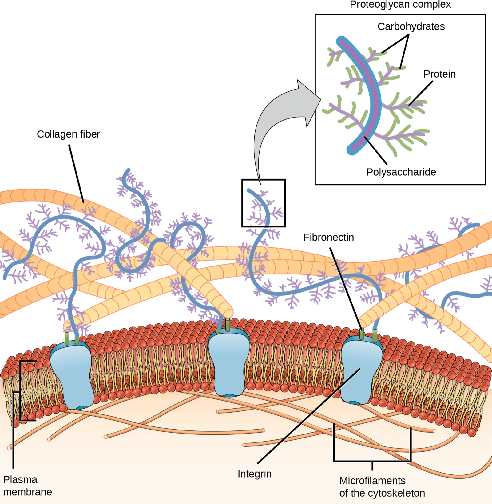
Extended description
A stretch of plasma membrane is drawn as a lipid bilayer, with microfilaments of the cytoskeleton lining its inner, cytoplasmic face. Three golden collagen fibers, each entwined with thinner purple-and-green proteoglycan strands, run across the top of the illustration above the membrane. Three blue, goblet-shaped integrin proteins span the membrane, each connected upward by a short fibronectin protein to one of the collagen fibers. A boxed inset, linked by an arrow to one proteoglycan strand, magnifies a single proteoglycan complex: a central polysaccharide core, with protein chains branching from the core and carbohydrate chains branching in turn from the proteins.
Cells have protein receptors on their plasma membranes’ extracellular surfaces. When a molecule within the matrix binds to the receptor, it changes the receptor’s molecular structure. The receptor, in turn, changes the microfilaments’ conformation positioned just inside the plasma membrane. These conformational changes induce chemical signals inside the cell that reach the nucleus and turn “on” or “off” the transcription of specific DNA sections, which affects the associated protein production, thus changing the activities within the cell.
Blood clotting provides an example of the extracellular matrix’s role in cell communication. When the cells lining a blood vessel are damaged, they display a protein receptor, which we call tissue factor. When tissue factor binds with another factor in the extracellular matrix, it causes platelets to adhere to the damaged blood vessel’s wall, stimulates the adjacent smooth muscle cells in the blood vessel to contract (thus constricting the blood vessel), and initiates a series of steps that stimulate the platelets to produce clotting factors.
Intercellular Junctions
Cells can also communicate with each other via direct contact, or intercellular junctions. There are differences in the ways that plant and animal and fungal cells communicate. Plasmodesmata are junctions between plant cells; whereas, animal cell contacts include tight junctions, gap junctions, and desmosomes.
Plasmodesmata
In general, long stretches of the plasma membranes of neighboring plant cells cannot touch one another because the cell wall that surrounds each cell separates them. How then, can a plant transfer water and other soil nutrients from its roots, through its stems, and to its leaves? Such transport uses the vascular tissues (xylem and phloem) primarily. There also exist structural modifications, which we call plasmodesmata (singular = plasmodesma). Numerous channels that pass between adjacent plant cells’ cell walls connect their cytoplasm, and enable transport of materials from cell to cell, and thus throughout the plant (below).
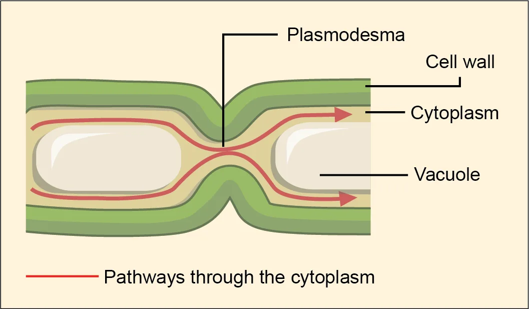
Extended description
Two plant cells sit side by side, each bounded by a thick green cell wall on the outside, with a thin cytoplasm layer just inside it and a large central vacuole filling most of the cell. Where the two cells meet, both cell walls pinch inward to form a single narrow channel — the plasmodesma — connecting the two cytoplasms. Red arrows, labeled “Pathways through the cytoplasm,” run through the cytoplasm of each cell and cross through the plasmodesma, showing the route materials take from one cell to the other.
Tight Junctions
A tight junction is a watertight seal between two adjacent animal cells (below). Proteins (predominantly two proteins called claudins and occludins) tightly hold the cells against each other.
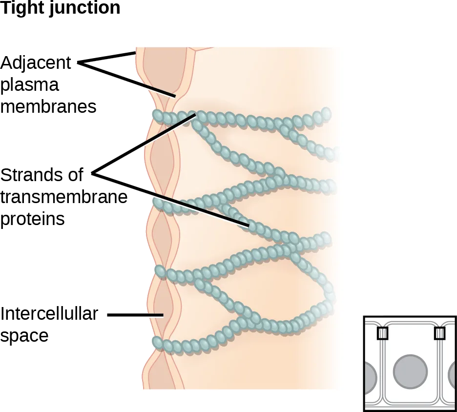
Extended description
Two parallel, pink-shaded plasma membranes, labeled “Adjacent plasma membranes,” run vertically, with the narrow intercellular space between them labeled at the bottom. Between the membranes, beaded strands, labeled “Strands of transmembrane proteins,” weave back and forth in an interlocking zigzag pattern, sealing the space between the two membranes at multiple points. A small inset in the lower right shows the same junction in a top-down cutaway view of a row of cells, with the tight junction drawn as a continuous belt encircling the apical end of each cell.
This tight adherence prevents materials from leaking between the cells; tight junctions are typically found in epithelial tissues that line internal organs and cavities, and comprise most of the skin. For example, the tight junctions of the epithelial cells lining your urinary bladder prevent urine from leaking out into the extracellular space.
Desmosomes
Also only in animal cells are desmosomes, which act like spot welds between adjacent epithelial cells (below). Cadherins, short proteins in the plasma membrane connect to intermediate filaments to create desmosomes. The cadherins connect two adjacent cells and maintain the cells in a sheet-like formation in organs and tissues that stretch, like the skin, heart, and muscles.
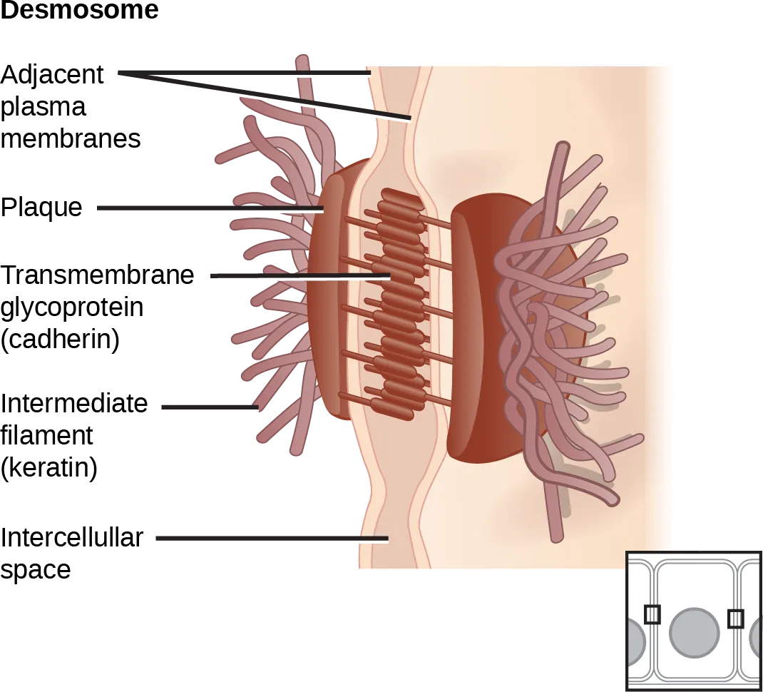
Extended description
Two plasma membranes, labeled “Adjacent plasma membranes,” run vertically with a narrow intercellular space between them. A dense plaque sits against the inner face of each membrane; the two plaques are bridged across the intercellular space by parallel rows of rod-shaped transmembrane glycoprotein (cadherin) filaments. From each plaque, a bundle of curling intermediate filament (keratin) strands radiates outward into the cell’s cytoplasm. A small inset in the lower right shows the same junction in a top-down cutaway view of a row of cells, with the desmosome drawn as a small square spot connecting two adjacent cells, unlike the continuous belt of the tight-junction inset.
Gap Junctions
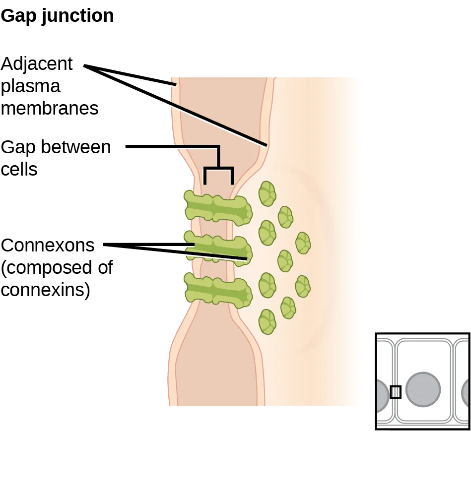
Gap junctions in animal cells are like plasmodesmata in plant cells in that they are channels between adjacent cells that allow for transporting ions, nutrients, and other substances that enable cells to communicate (below). Structurally, however, gap junctions and plasmodesmata differ.
Extended description
Two plasma membranes, labeled “Adjacent plasma membranes,” run vertically, narrowing toward each other to leave a thin gap between cells, labeled “Gap between cells.” Three barrel-shaped connexons, each made of six connexin subunits and labeled “Connexons (composed of connexins),” span the gap, aligning end to end between the two membranes to form continuous channels. On the right side of the membrane, small circles representing water and small molecules cluster beside the channels, illustrating substances passing from one cell into the other. A small inset in the lower right shows the same junction in a top-down cutaway view of a row of cells, with the gap junction drawn as a small pore connecting two adjacent cells.
Gap junctions develop when a set of six proteins (connexins) in the plasma membrane arrange themselves in an elongated donut-like configuration - a connexon. When the connexon’s pores (“doughnut holes”) in adjacent animal cells align, a channel between the two cells forms. Gap junctions are particularly important in cardiac muscle. The electrical signal for the muscle to contract passes efficiently through gap junctions, allowing the heart muscle cells to contract in tandem.
Summary
Animal cells communicate via their extracellular matrices and are connected to each other via tight junctions, desmosomes, and gap junctions. Plant cells are connected and communicate with each other via plasmodesmata.
When protein receptors on the plasma membrane’s surface of an animal cell bind to a substance in the extracellular matrix, a chain of reactions begins that changes activities taking place within the cell. Plasmodesmata are channels between adjacent plant cells, while gap junctions are channels between adjacent animal cells. However, their structures are quite different. A tight junction is a watertight seal between two adjacent cells, while a desmosome acts like a spot weld.
Key terms
- desmosome — linkages between adjacent epithelial cells that form when cadherins in the plasma membrane attach to intermediate filaments
- extracellular matrix — material secreted from animal or fungal cells that provides mechanical protection and anchoring for the cells in the tissue
- gap junction — channel between two adjacent animal cells that allows ions, nutrients, and low molecular weight substances to pass between cells, enabling the cells to communicate
- plasmodesma — (plural = plasmodesmata) channel that passes between adjacent plant cells’ cell walls, connects their cytoplasm, and allows transporting of materials from cell to cell
- tight junction — protein adherence that creates a firm seal between two adjacent animal cells
Practice
Describe the extracellular matrix
Explain how the extracellular matrix functions.
Show model answer
Did your answer mention:
Material secreted from animal or fungal cells that provides mechanical protection and anchoring for the cells in a tissue is called the ________.
Collagen fibers interwoven with proteoglycans are its primary components.When protein receptors on the plasma membrane’s surface of an animal cell bind to a substance in the ________, a chain of reactions begins that changes activities taking place within the cell.
The section’s summary describes this binding as the trigger for the cell’s internal chain of reactions.List examples of the ways that plant cells and animal cells communicate with adjacent cells
Which of the following are only in plant cells?
Only one of these four connection types crosses a rigid cell wall rather than joining two plasma membranes directly.How does the structure of a plasmodesma differ from that of a gap junction?
Show model answer
Did your answer mention:
A channel that passes between the cell walls of adjacent plant cells, connecting their cytoplasm and allowing materials to move from cell to cell, is called a ________.
Its plural form describes many such channels connecting a plant’s cells throughout its tissues.Summarize the roles of tight junctions, desmosomes, gap junctions, and plasmodesmata
The key components of desmosomes are cadherins and __________.
The text names two protein components of a desmosome: short membrane proteins called cadherins, and a cytoskeletal filament type they connect to.Diseased animal cells may produce molecules that activate death cascades to kill the cells in a controlled manner. Why would neighboring healthy cells also die?
Only one of these four options names a channel that lets small molecules pass directly between the cytoplasm of two animal cells.Pathogenic E. coli have recently been shown to degrade tight junction proteins during infection. How would this provide an advantage to the bacteria?
Show model answer
Did your answer mention:
A protein adherence that seals two adjacent animal cells together so that no material can leak between them is called a ________.
Claudins and occludins are the two main proteins that create it.A channel between two adjacent animal cells that allows ions, nutrients, and low molecular weight substances to pass between them, enabling the cells to communicate, is called a ________.
Six connexin proteins in each cell’s membrane arrange into a donut-shaped connexon that lines this channel.A linkage between adjacent epithelial cells that forms when cadherins in the plasma membrane attach to intermediate filaments is called a ________.
It acts like a spot weld holding two cells together in tissues that stretch, such as skin, heart, and muscle.This section is adapted from Biology 2e, Section 4.6: Connections between Cells and Cellular Activities by Mary Ann Clark, Jung Choi, Matthew Douglas, and OpenStax, © OpenStax, licensed under CC BY-NC-SA 4.0. Access the original for free at openstax.org. Changes: figures re-encoded as WebP, all five already correctly guessed as diagrams; each figure’s alt shortened to what it shows and teaches, with the full label-by-label walk-through moved into a longdesc, since none of the five captions carries the diagram’s labeled detail; the inline print figure reference to a plant-cell-wall figure in a different section (4.3) dropped, since that figure is not on this page, and the remaining same-page figure references changed from the source’s blank cross-reference markers to “below”; the Link to Learning note rendered as a callout with its bold name; the end-of-section Review Questions and Critical Thinking Questions adapted into the closing interactive Practice block (multiple choice and self-check respectively); and five key-term recall items (extracellular matrix, plasmodesma, tight junction, gap junction, desmosome) added from the glossary to give every objective group an auto-graded item; rubric checkpoints added to each self-check, decomposing its model answer (the source solution) into check-off clauses with no new claims; and one summary-derived cloze item added under the first objective (“extracellular matrix,” completing the summary’s sentence on what receptor binding triggers) to reach the raised per-objective floor.