Bulk Transport
By the end of this section, you will be able to:
- Describe endocytosis, including phagocytosis, pinocytosis, and receptor-mediated endocytosis
- Understand the process of exocytosis
In addition to moving small ions and molecules through the membrane, cells also need to remove and take in larger molecules and particles (see the table below for examples). Some cells are even capable of engulfing entire unicellular microorganisms. You might have correctly hypothesized that when a cell uptakes and releases large particles, it requires energy. A large particle, however, cannot pass through the membrane, even with energy that the cell supplies.
Endocytosis
Endocytosis is a type of active transport that moves particles, such as large molecules, parts of cells, and even whole cells, into a cell. There are different endocytosis variations, but all share a common characteristic: the cell’s plasma membrane invaginates, forming a pocket around the target particle. The pocket pinches off, resulting in the particle containing itself in a newly created intracellular vesicle formed from the plasma membrane.
Phagocytosis
Phagocytosis (the condition of “cell eating”) is the process by which a cell takes in large particles, such as other cells or relatively large particles. For example, when microorganisms invade the human body, a type of white blood cell, a neutrophil, will remove the invaders through this process, surrounding and engulfing the microorganism, which the neutrophil then destroys (below).
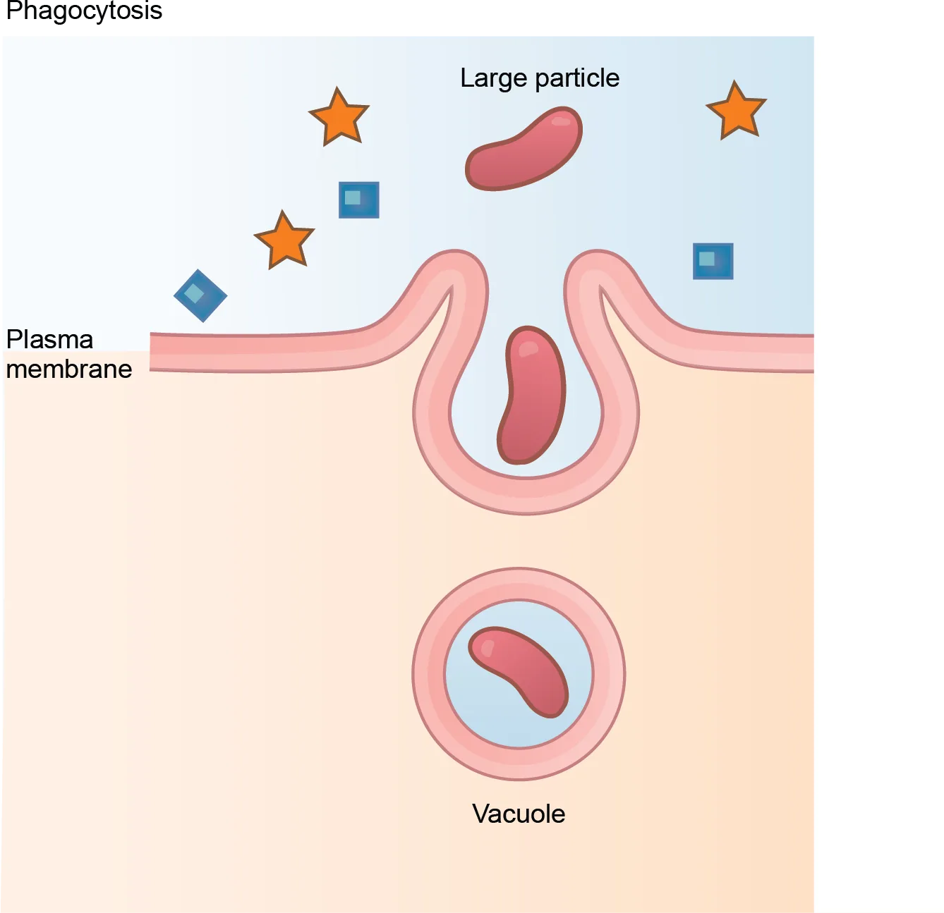
Extended description
The diagram is captioned “Phagocytosis.” At the top, in the extracellular fluid, a large rounded particle labeled “Large particle” sits among several smaller orange stars and blue diamonds and squares. Below, the plasma membrane, labeled, curves upward on both sides around the large particle, forming a pocket that nearly encloses it. At the bottom, a separate circular vacuole, labeled, sits fully enclosed in the cytoplasm with the same particle inside it.
In preparation for phagocytosis, a portion of the plasma membrane’s inward-facing surface becomes coated with the protein clathrin, which stabilizes this membrane’s section. The membrane’s coated portion then extends from the cell’s body and surrounds the particle, eventually enclosing it. Once the vesicle containing the particle is enclosed within the cell, the clathrin disengages from the membrane and the vesicle merges with a lysosome for breaking down the material in the newly formed compartment (endosome). When accessible nutrients from the vesicular contents’ degradation have been extracted, the newly formed endosome merges with the plasma membrane and releases its contents into the extracellular fluid. The endosomal membrane again becomes part of the plasma membrane.
Pinocytosis
A variation of endocytosis is pinocytosis. This literally means “cell drinking”. Discovered by Warren Lewis in 1929, this American embryologist and cell biologist described a process whereby he assumed that the cell was purposefully taking in extracellular fluid. In reality, this is a process that takes in molecules, including water, which the cell needs from the extracellular fluid. Pinocytosis results in a much smaller vesicle than does phagocytosis, and the vesicle does not need to merge with a lysosome (below).
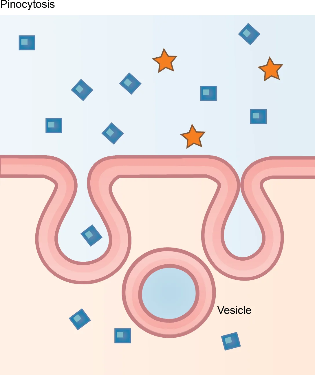
Extended description
The diagram is captioned “Pinocytosis.” At the top, in the extracellular fluid, small orange stars and blue diamonds and squares are scattered above the plasma membrane. The membrane folds inward at two neighboring places, forming two invaginations; one still holds a diamond shape inside its fold. Below, a small circular vesicle, labeled, has pinched free in the cytoplasm, drawn empty, with diamond shapes floating loose in the cytoplasm around it.
A variation of pinocytosis is potocytosis. This process uses a coating protein, caveolin, on the plasma membrane’s cytoplasmic side, which performs a similar function to clathrin. The cavities in the plasma membrane that form the vacuoles have membrane receptors and lipid rafts in addition to caveolin. The vacuoles or vesicles formed in caveolae (singular caveola) are smaller than those in pinocytosis. Potocytosis brings small molecules into the cell and transports them through the cell for their release on the other side, a process we call transcytosis. In some cases, the caveolae deliver their cargo to membranous organelles like the ER.
Receptor-mediated Endocytosis
A targeted variation of endocytosis employs receptor proteins in the plasma membrane that have a specific binding affinity for certain substances (below).
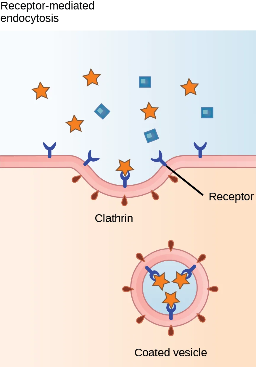
Extended description
The diagram is captioned “Receptor-mediated endocytosis.” Y-shaped receptors, labeled with a leader line, are embedded in the plasma membrane, some with an orange star bound to their tips; unbound orange stars and blue squares and diamonds float in the extracellular fluid above. On the cytoplasmic side, short red rod shapes labeled “Clathrin” coat the membrane where it curves inward around the bound receptors. Below, a completed circular “Coated vesicle,” labeled, shows the same red coat on its outside and two or three orange stars still bound to receptors inside it.
In receptor-mediated endocytosis, as in phagocytosis, clathrin attaches to the plasma membrane’s cytoplasmic side. If a compound’s uptake is dependent on receptor-mediated endocytosis and the process is ineffective, the material will not be removed from the tissue fluids or blood. Instead, it will stay in those fluids and increase in concentration. The failure of receptor-mediated endocytosis causes some human diseases. For example, receptor mediated endocytosis removes low density lipoprotein or LDL (or “bad” cholesterol) from the blood. In the human genetic disease familial hypercholesterolemia, the LDL receptors are defective or missing entirely. People with this condition have life-threatening levels of cholesterol in their blood, because their cells cannot clear LDL particles.
Although receptor-mediated endocytosis is designed to bring specific substances that are normally in the extracellular fluid into the cell, other substances may gain entry into the cell at the same site. Flu viruses, diphtheria, and cholera toxin all have sites that cross-react with normal receptor-binding sites and gain entry into cells.
Link to Learning
See receptor-mediated endocytosis in action, and click on different parts of an interactive endocytosis animation for a focused animation.
Exocytosis
The reverse process of moving material into a cell is the process of exocytosis. Exocytosis is the opposite of the processes we discussed above in that its purpose is to expel material from the cell into the extracellular fluid. Waste material is enveloped in a membrane and fuses with the plasma membrane’s interior. This fusion opens the membranous envelope on the cell’s exterior, and the waste material expels into the extracellular space (below). Other examples of cells releasing molecules via exocytosis include extracellular matrix protein secretion and neurotransmitter secretion into the synaptic cleft by synaptic vesicles.
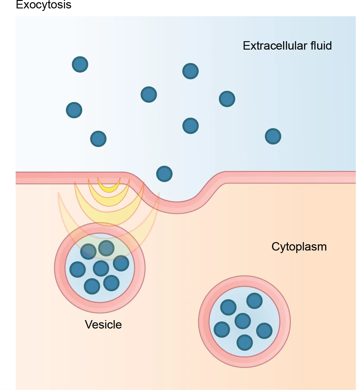
Extended description
The diagram is captioned “Exocytosis.” Small dark blue dots are scattered through the extracellular fluid at the top. The plasma membrane bulges outward where a vesicle from the cytoplasm has fused with it, opening to release its blue dots into the extracellular fluid above. A second, still-intact vesicle, labeled “Vesicle,” sits nearby in the cytoplasm, also filled with blue dots and not yet fused with the membrane.
Methods of Transport, Energy Requirements, and Types of Transported Material
| Transport Method | Active/Passive | Material Transported |
|---|---|---|
| Diffusion | Passive | Small-molecular weight material |
| Osmosis | Passive | Water |
| Facilitated transport/diffusion | Passive | Sodium, potassium, calcium, glucose |
| Primary active transport | Active | Sodium, potassium, calcium |
| Secondary active transport | Active | Amino acids, lactose |
| Phagocytosis | Active | Large macromolecules, whole cells, or cellular structures |
| Pinocytosis and potocytosis | Active | Small molecules (liquids/water) |
| Receptor-mediated endocytosis | Active | Large quantities of macromolecules |
Summary
Active transport methods require directly using ATP to fuel the transport. In a process scientists call phagocytosis, other cells can engulf large particles, such as macromolecules, cell parts, or whole cells. In phagocytosis, a portion of the membrane invaginates and flows around the particle, eventually pinching off and leaving the particle entirely enclosed by a plasma membrane’s envelope. The cell breaks down vesicle contents, with the particles either used as food or dispatched. Pinocytosis is a similar process on a smaller scale. The plasma membrane invaginates and pinches off, producing a small envelope of fluid from outside the cell. Pinocytosis imports substances that the cell needs from the extracellular fluid. The cell expels waste in a similar but reverse manner. It pushes a membranous vacuole to the plasma membrane, allowing the vacuole to fuse with the membrane and incorporate itself into the membrane structure, releasing its contents to the exterior.
Key terms
- caveolin — protein that coats the plasma membrane’s cytoplasmic side and participates in the liquid uptake process by potocytosis
- clathrin — protein that coats the plasma membrane’s inward-facing surface and assists in forming specialized structures, like coated pits, for phagocytosis
- endocytosis — type of active transport that moves substances, including fluids and particles, into a cell
- exocytosis — process of passing bulk material out of a cell
- pinocytosis — a variation of endocytosis that imports small molecules, including water, that the cell needs from the extracellular fluid (Source note: the source glossary says “macromolecules”; this section’s own text calls pinocytosis “cell drinking” and its transport table lists small molecules (liquids/water) as its cargo.)
- potocytosis — variation of pinocytosis that uses a different coating protein (caveolin) on the plasma membrane’s cytoplasmic side
- receptor-mediated endocytosis — variation of endocytosis that involves using specific binding proteins in the plasma membrane for specific molecules or particles, and clathrin-coated pits that become clathrin-coated vesicles
Practice
Describe endocytosis, including phagocytosis, pinocytosis, and receptor-mediated endocytosis
Which transport mechanism can bring whole cells into a cell?
Which process was compared to a neutrophil engulfing an entire invading microorganism?In what important way does receptor-mediated endocytosis differ from phagocytosis?
Phagocytosis engulfs whatever large particle is nearby; think about what makes the receptor pathway selective.Many viruses enter host cells through receptor-mediated endocytosis. What is an advantage of this entry strategy?
A compound’s uptake by this pathway depends on binding a receptor that only certain cells display.Why is it important that there are different types of proteins in plasma membranes for the transport of materials into and out of a cell?
Show model answer
Did your answer mention:
The protein that coats the plasma membrane’s inward-facing surface and assists in forming specialized structures, like coated pits, for phagocytosis is called ________.
This protein stabilizes the pocket that forms around a particle being engulfed during phagocytosis, and it also coats the pits used in receptor-mediated endocytosis.Understand the process of exocytosis
What happens to the membrane of a vesicle after exocytosis?
The vesicle’s envelope opens on the cell’s exterior; think about where that membrane ends up.Which of the following organelles relies on exocytosis to complete its function?
Which organelle packages materials into vesicles for release from the cell?Imagine a cell can perform exocytosis, but only minimal endocytosis. What would happen to the cell?
Exocytosis adds membrane to the cell surface each time a vesicle fuses with it; endocytosis is what normally removes an equivalent amount.The process of passing bulk material out of a cell is called ________.
This is the reverse of endocytosis: material inside the cell is released to the extracellular fluid.This section is adapted from Biology 2e, Section 5.4: Bulk Transport by Mary Ann Clark, Jung Choi, Matthew Douglas, and OpenStax, © OpenStax, licensed under CC BY-NC-SA 4.0. Access the original for free at openstax.org. Changes: figures re-encoded as WebP; all four figures kept as “diagram” (matching the manifest’s guess) after inspection, since each is a labeled line illustration rather than a photograph, and an extended description was added to each since its labels (large particle/vacuole, vesicle, receptor/clathrin/coated vesicle, vesicle/cytoplasm) are not repeated in its caption; the table’s merged three-column title row, which Markdown cannot represent, was rendered as a bold line above the table with the column headers as the table’s own header row; inline references to the figures and table changed from the source’s print numbers to descriptive phrases (“below”) since figures and tables are not numbered here; the Link to Learning note’s link text was expanded from the source’s bare “parts” to “different parts of an interactive endocytosis animation” so the link describes its destination; the end-of-section Review Questions and one of the two Critical Thinking Questions were adapted into the closing interactive Practice block (multiple choice and self-check respectively) — the second Critical Thinking Question, about why ions have difficulty crossing membranes via channels and carrier proteins, was omitted because it tests passive-transport material this section does not carry; and two key-term recall items (clathrin, exocytosis) were added from the glossary so each objective group carries an auto-graded item; and rubric checkpoints added to the self-check, decomposing its model answer (the source solution) into check-off clauses with no new claims. One key-term definition is corrected with a visible Source note: pinocytosis imports small molecules and water, as this section’s own text and table say, not macromolecules (erratum 389).