Energy in Living Systems
By the end of this section, you will be able to:
- Discuss the importance of electrons in the transfer of energy in living systems
- Explain how ATP is used by cells as an energy source
Energy conversion within a cell involves many coordinated chemical pathways. Most of these pathways are combinations of oxidation and reduction reactions, which occur at the same time. An oxidation reaction strips an electron from an atom in a compound, and the addition of this electron to another compound is a reduction reaction. Because oxidation and reduction usually occur together, these pairs of reactions are called oxidation reduction reactions, or redox reactions.
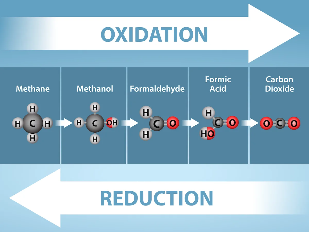
Extended description
A horizontal strip of five labeled panels, methane through carbon dioxide, with a wide white arrow above reading Oxidation pointing right across all five and a wide white arrow below reading Reduction pointing left across all five. Panel 1, Methane: a central black carbon bonded to four white hydrogens. Panel 2, Methanol: the same carbon now bonded to three hydrogens and one red oxygen carrying its own white hydrogen (OH). Panel 3, Formaldehyde: the carbon bonded to two hydrogens and double-bonded to one oxygen. Panel 4, Formic Acid: the carbon bonded to one hydrogen, double-bonded to one oxygen, and single-bonded to a hydroxyl (OH) group. Panel 5, Carbon Dioxide: the carbon double-bonded to two oxygens, with no hydrogens remaining. Moving left to right, hydrogens are progressively replaced by oxygens (oxidation); moving right to left, the reverse occurs (reduction).
Electrons and Energy
The removal of an electron from a molecule (oxidizing it), results in a decrease in potential energy in the oxidized compound. However, the electron (sometimes as part of a hydrogen atom) does not remain unbonded in the cytoplasm of a cell. Rather, the electron is shifted to a second compound, reducing the second compound. The shift of an electron from one compound to another removes some potential energy from the first compound (the oxidized compound) and increases the potential energy of the second compound (the reduced compound). The transfer of electrons between molecules is important because most of the energy stored in atoms and used to fuel cell functions is in the form of high-energy electrons. The transfer of energy in the form of high-energy electrons allows the cell to transfer and use energy in an incremental fashion—in small packages rather than in a single, destructive burst. This chapter focuses on the extraction of energy from food; you will see that as you track the path of the transfers, you are tracking the path of electrons moving through metabolic pathways.
Electron Carriers
In living systems, a small class of compounds functions as electron shuttles: they bind and carry high-energy electrons between compounds in biochemical pathways. The principal electron carriers we will consider are derived from the B vitamin group and are derivatives of nucleotides. These compounds can be easily reduced (that is, they accept electrons) or oxidized (they lose electrons). Nicotinamide adenine dinucleotide (NAD) (below) is derived from vitamin B₃, niacin. NAD⁺ is the oxidized form of the molecule; NADH is the reduced form of the molecule after it has accepted two electrons and a proton (which together are the equivalent of a hydrogen atom with an extra electron). Note that if a compound has an “H” on it, it is generally reduced (e.g., NADH is the reduced form of NAD).
NAD⁺ can accept electrons from an organic molecule according to the general equation:
RH (Reducing agent) + NAD⁺ (Oxidizing agent) → NADH (Reduced) + R (Oxidized)
When electrons are added to a compound, it is reduced. A compound that reduces another is called a reducing agent. In the above equation, RH is a reducing agent, and NAD⁺ is reduced to NADH. When electrons are removed from a compound, it is oxidized. A compound that oxidizes another is called an oxidizing agent. In the above equation, NAD⁺ is an oxidizing agent, and RH is oxidized to R.
Similarly, flavin adenine dinucleotide (FAD) is derived from vitamin B₂, also called riboflavin. Its reduced form is FADH₂. A second variation of NAD, NADP, contains an extra phosphate group. Both NAD⁺ and FAD are extensively used in energy extraction from sugars, and NADP plays an important role in anabolic reactions and photosynthesis in plants.
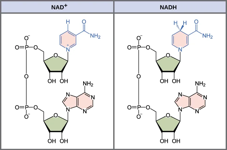
Extended description
Two labeled panels, NAD⁺ on the left and NADH on the right, each showing an identical dinucleotide skeleton: a nicotinamide ring at top linked through a phosphate-sugar backbone (two phosphate groups bridging two five-membered ribose rings) down to an adenine ring at bottom. In the NAD⁺ panel, the nicotinamide ring carries a single hydrogen at its top position and a positive charge on its ring nitrogen. In the NADH panel, the same ring instead carries two hydrogens at that top position (drawn with a wedge and a dash to show both add to the same carbon) and no charge on the nitrogen — the two added electrons and the added hydrogen are the only difference between the two structures.
ATP in Living Systems
A living cell cannot store significant amounts of free energy. Excess free energy would result in an increase of heat in the cell, which would result in excessive thermal motion that could damage and then destroy the cell. Rather, a cell must be able to handle that energy in a way that enables the cell to store energy safely and release it for use only as needed. Living cells accomplish this by using the compound adenosine triphosphate (ATP). ATP is often called the “energy currency” of the cell, and, like currency, this versatile compound can be used to fill any energy need of the cell. How? It functions similarly to a rechargeable battery.
When ATP is broken down, usually by the removal of its terminal phosphate group, energy is released. The energy is used to do work by the cell, usually when the released phosphate binds to another molecule, thereby activating it. For example, in the mechanical work of muscle contraction, ATP supplies the energy to move the contractile muscle proteins. Recall the active transport work of the sodium-potassium pump in cell membranes. ATP alters the structure of the integral protein that functions as the pump, changing its affinity for sodium and potassium. In this way, the cell performs work, pumping ions against their electrochemical gradients.
ATP Structure and Function
At the heart of ATP is a molecule of adenosine monophosphate (AMP), which is composed of an adenine molecule bonded to a ribose molecule and to a single phosphate group (below). Ribose is a five-carbon sugar found in RNA, and AMP is one of the nucleotides in RNA. The addition of a second phosphate group to this core molecule results in the formation of adenosine diphosphate (ADP); the addition of a third phosphate group forms adenosine triphosphate (ATP).
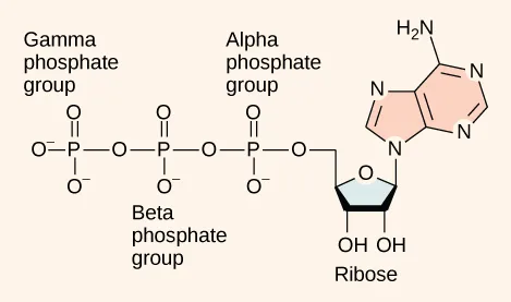
Extended description
An adenine ring (fused five- and six-membered rings with an NH₂ group and three ring nitrogens) at upper right, bonded to a five-membered ribose sugar bearing two OH groups at lower right, labeled Ribose. The ribose is bonded on its left to a chain of three phosphate groups in a row, each drawn as a phosphorus atom double-bonded to one oxygen and single-bonded to two negatively charged oxygens, with the chain linked oxygen-to-phosphorus. The three phosphates are labeled, nearest the ribose outward, Alpha phosphate group, Beta phosphate group, and Gamma phosphate group.
The addition of a phosphate group to a molecule requires energy. Phosphate groups are negatively charged and thus repel one another when they are arranged in series, as they are in ADP and ATP. This repulsion makes the ADP and ATP molecules inherently unstable. The release of one or two phosphate groups from ATP, a process called dephosphorylation, releases energy.
Energy from ATP
Hydrolysis is the process of breaking complex macromolecules apart. During hydrolysis, water is split, or lysed, and the resulting hydrogen atom (H⁺) and a hydroxyl group (OH⁻), or hydroxide, are added to the larger molecule. The hydrolysis of ATP produces ADP, together with an inorganic phosphate ion (Pi), and the release of free energy. To carry out life processes, ATP is continuously broken down into ADP, and like a rechargeable battery, ADP is continuously regenerated into ATP by the reattachment of a third phosphate group. Water, which was broken down into its hydrogen atom and hydroxyl group (hydroxide) during ATP hydrolysis, is regenerated when a third phosphate is added to the ADP molecule, reforming ATP.
Obviously, energy must be infused into the system to regenerate ATP. Where does this energy come from? In nearly every living thing on Earth, the energy comes from the metabolism of glucose, fructose, or galactose, all isomers with the chemical formula C₆H₁₂O₆ but different molecular configurations. In this way, ATP is a direct link between the limited set of exergonic pathways of glucose catabolism and the multitude of endergonic pathways that power living cells.
Phosphorylation
Recall that, in some chemical reactions, enzymes may bind to several substrates that react with each other on the enzyme, forming an intermediate complex. An intermediate complex is a temporary structure, and it allows one of the substrates (such as ATP) and reactants to more readily react with each other; in reactions involving ATP, ATP is one of the substrates and ADP is a product. During an endergonic chemical reaction, ATP forms an intermediate complex with the substrate and enzyme in the reaction. This intermediate complex allows the ATP to transfer its third phosphate group, with its energy, to the substrate, a process called phosphorylation. Phosphorylation refers to the addition of the phosphate (~P). This is illustrated by the following generic reaction, in which A and B represent two different substrates:
A + enzyme + ATP → [A − enzyme − ~P] → B + enzyme + ADP + phosphate ion
When the intermediate complex breaks apart, the energy is used to modify the substrate and convert it into a product of the reaction. The ADP molecule and a free phosphate ion are released into the medium and are available for recycling through cell metabolism.
Substrate Phosphorylation
ATP is generated through two mechanisms during the breakdown of glucose. A few ATP molecules are generated (that is, regenerated from ADP) as a direct result of the chemical reactions that occur in the catabolic pathways. A phosphate group is removed from an intermediate reactant in the pathway, and the free energy of the reaction is used to add the third phosphate to an available ADP molecule, producing ATP (below). This very direct method of phosphorylation is called substrate-level phosphorylation.
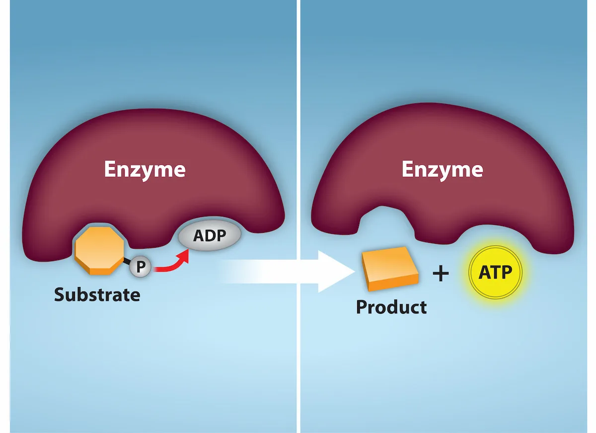
Extended description
Two panels labeled left to right showing an enzyme, drawn as a large maroon crescent, before and after a reaction. Left panel: a small orange octagon labeled Substrate sits in one pocket of the enzyme, connected by a black bond to a phosphate group labeled P, which sits beside a gray oval labeled ADP in a neighboring pocket; a red arrow curves from the phosphate toward the ADP. Right panel, after the white arrow: the enzyme now has two empty pockets; below it sit an orange rectangular block labeled Product and, separately, a yellow circle labeled ATP.
Oxidative Phosphorylation
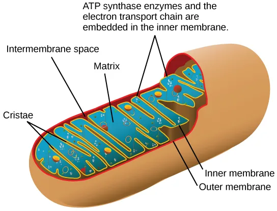
Most of the ATP generated during glucose catabolism, however, is derived from a much more complex process, chemiosmosis, which takes place in mitochondria (below) within a eukaryotic cell or the plasma membrane of a prokaryotic cell. Chemiosmosis, a process of ATP production in cellular metabolism, is used to generate 90 percent of the ATP made during glucose catabolism and is also the method used in the light reactions of photosynthesis to harness the energy of sunlight. The production of ATP using the process of chemiosmosis is called oxidative phosphorylation because of the involvement of oxygen in the process.
Extended description
A bean-shaped mitochondrion drawn in cross-section. A tan outer membrane surrounds a red inner membrane, which folds inward repeatedly to form finger-like ridges labeled Cristae. The narrow tan-colored gap between the outer and inner membranes is labeled Intermembrane space. The blue-green interior enclosed by the folded inner membrane is labeled Matrix, and contains scattered small colored shapes representing enzymes and metabolites. Along the inner membrane, small embedded protein shapes are labeled ATP synthase enzymes and the electron transport chain are embedded in the inner membrane, with leader lines pointing to several of the ridge surfaces.
Summary
ATP functions as the energy currency for cells. It allows the cell to store energy briefly and transport it within the cell to support endergonic chemical reactions. The structure of ATP is that of an RNA nucleotide with three phosphates attached. As ATP is used for energy, a phosphate group or two are detached, and either ADP or AMP is produced. Energy derived from glucose catabolism is used to convert ADP into ATP. When ATP is used in a reaction, the third phosphate is temporarily attached to a substrate in a process called phosphorylation. The two processes of ATP regeneration that are used in conjunction with glucose catabolism are substrate-level phosphorylation and oxidative phosphorylation through the process of chemiosmosis.
Key terms
- chemiosmosis — process in which there is a production of adenosine triphosphate (ATP) in cellular metabolism by the involvement of a proton gradient across a membrane
- dephosphorylation — removal of a phosphate group from a molecule
- oxidative phosphorylation — production of ATP using the process of chemiosmosis in the presence of oxygen
- phosphorylation — addition of a high-energy phosphate to a compound, usually a metabolic intermediate, a protein, or ADP
- redox reaction — chemical reaction that consists of the coupling of an oxidation reaction and a reduction reaction
- substrate-level phosphorylation — production of ATP from ADP using the excess energy from a chemical reaction and a phosphate group from a reactant
Practice
Discuss the importance of electrons in the transfer of energy in living systems
A reducing chemical reaction ________.
Reduction is what happens to the compound that gains the electron a reducing agent gives up.A chemical reaction that consists of the coupling of an oxidation reaction and a reduction reaction is called a(n) ________ reaction.
The name blends the two halves of the coupled reaction: reduction and oxidation.The high-energy electron carrier derived from vitamin B₃ (niacin), whose oxidized and reduced forms recur throughout the pathways that extract energy from food, is abbreviated ________.
Its reduced form, after accepting two electrons and a proton, is NADH.Explain how ATP is used by cells as an energy source
The energy currency used by cells is ________.
This molecule releases energy when its terminal phosphate group is removed.Production of ATP from ADP using the excess energy from a chemical reaction and a phosphate group from a reactant is called ________ phosphorylation.
This is the direct method of phosphorylation, as opposed to the process that runs through the electron transport chain.Production of ATP using chemiosmosis in the presence of oxygen is called ________ phosphorylation.
This process generates about 90 percent of the ATP made during glucose catabolism.Why is it beneficial for cells to use ATP rather than energy directly from the bonds of carbohydrates? What are the greatest drawbacks to harnessing energy directly from the bonds of several different compounds?
Show model answer
Did your answer mention:
The removal of a phosphate group from a molecule is called ________.
This is the process that releases the energy stored when one or two phosphate groups leave ATP.This section is adapted from Biology 2e, Section 7.1: Energy in Living Systems by Mary Ann Clark, Jung Choi, Matthew Douglas, and OpenStax, © OpenStax, licensed under CC BY-NC-SA 4.0. Access the original for free at openstax.org. Changes: figures re-encoded as WebP, with four of the five re-kinded from the manifest’s file-extension guess of “photo” (guessed from their JPEG source files) to “diagram” after inspection — every one of the five is a molecular or cellular illustration, not a photograph, and the fifth (a PNG) was already guessed correctly; a longdesc added for all five figures, since each is a labeled diagram whose full reading (structures, panels, or leader-line labels) is not carried by its caption; the source’s two display equations — the NAD⁺ reduction equation and the generic phosphorylation reaction, both chemical equations rather than mathematics — rendered as Unicode text lines instead of typeset math; inline references to figures changed from the source’s print numbers (“Figure 7.3,” “Figure 7.4,” etc.) to the descriptive phrase “below” since figures are not numbered here; the section’s one feature box (Career Connection) rendered as a callout with its bold name; the two Review Questions and the one Critical Thinking Question adapted into the closing interactive Practice block (multiple choice and self-check respectively); five recall textin items (redox reaction, NAD, substrate-level phosphorylation, oxidative phosphorylation, and dephosphorylation) added from the glossary and body text — NAD is defined in the body rather than the glossary — to round out both objective groups with an auto-graded item; and rubric checkpoints added to the section’s self-check, decomposing its model answer (the source solution) into check-off clauses with no new claims.