Oxidative Phosphorylation
By the end of this section, you will be able to:
- Describe how electrons move through the electron transport chain and explain what happens to their energy levels during this process
- Explain how a proton (H⁺) gradient is established and maintained by the electron transport chain
You have just read about two pathways in glucose catabolism—glycolysis and the citric acid cycle—that generate ATP. Most of the ATP generated during the aerobic catabolism of glucose, however, is not generated directly from these pathways. Instead, it is derived from a process that begins by moving electrons through a series of electron carriers that undergo redox reactions. This process causes hydrogen ions to accumulate within the intermembranous space. Therefore, a concentration gradient forms in which hydrogen ions diffuse out of the intermembranous space into the mitochondrial matrix by passing through ATP synthase. The current of hydrogen ions powers the catalytic action of ATP synthase, which phosphorylates ADP, producing ATP.
Electron Transport Chain
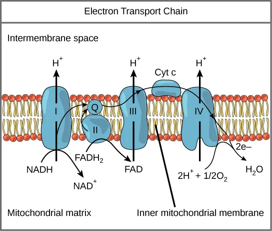
The electron transport chain (below) is the last component of aerobic respiration and is the only part of glucose metabolism that uses atmospheric oxygen. Oxygen continuously diffuses into plant tissues (typically through stomata), as well as into fungi and bacteria; however, in animals, oxygen enters the body through a variety of respiratory systems. Electron transport is a series of redox reactions that resembles a relay race or bucket brigade in that electrons are passed rapidly from one component to the next, to the endpoint of the chain where the electrons reduce molecular oxygen and, along with associated protons, produces water. There are four complexes composed of proteins, labeled I through IV in the diagram below, and the aggregation of these four complexes, together with associated mobile, accessory electron carriers, is called the electron transport chain. The electron transport chain is present with multiple copies in the inner mitochondrial membrane of eukaryotes and within the plasma membrane of prokaryotes.
Extended description
A diagram titled Electron Transport Chain. Intermembrane space is labeled at the top of the figure, above a horizontal phospholipid bilayer (round orange head groups on tan tails); Mitochondrial matrix is labeled at the bottom. Four blue protein shapes are embedded in the membrane, left to right: complex I; a small round complex II sitting beside a purple oval labeled Q; complex III; and complex IV, with a smaller blue dome labeled Cyt c resting on top of the membrane between complexes III and IV. A diagonal leader line labeled Inner mitochondrial membrane points up to the bilayer between complexes III and IV. Below the membrane, a black arrow labeled NADH points up into complex I, with a second arrow curving down and away to NAD⁺; a black arrow labeled FADH₂ points up into complex II, with a second arrow curving down and away to FAD. Curved black arrows trace electrons moving from complex I to Q, from complex II to Q, from Q to complex III, from complex III to Cyt c, and from Cyt c to complex IV. Three vertical arrows labeled H⁺, rising from complexes I, III, and IV, point up into the intermembrane space. At the right, an arrow labeled 2e⁻ leads into complex IV, where it joins an arrow labeled 2H⁺ + ½O₂ to produce an arrow labeled H₂O.
Complex I
First, two electrons are carried to the first complex via NADH. This complex, labeled I, is composed of flavin mononucleotide (FMN) and an iron-sulfur (Fe-S)-containing protein. FMN, which is derived from vitamin B₂ (also called riboflavin), is one of several prosthetic groups or cofactors in the electron transport chain. A prosthetic group is a nonprotein molecule required for the activity of a protein. Prosthetic groups are organic or inorganic, nonpeptide molecules bound to a protein that facilitate its function. Prosthetic groups include coenzymes, which are the prosthetic groups of enzymes. The enzyme in complex I is NADH dehydrogenase and is composed of 44 separate polypeptide chains. Complex I can pump four hydrogen ions across the membrane from the matrix into the intermembrane space, and it is in this way that the hydrogen ion gradient is established and maintained between the two compartments separated by the inner mitochondrial membrane.
Q and Complex II
Complex II directly receives FADH₂—which does not pass through complex I. The compound connecting the first and second complexes to the third is ubiquinone. The Q molecule is lipid soluble and freely moves through the hydrophobic core of the membrane. Once it is reduced (QH₂), ubiquinone delivers its electrons to the next complex in the electron transport chain. Q receives the electrons derived from NADH from complex I, and the electrons derived from FADH₂ from complex II. This enzyme and FADH₂ form a small complex that delivers electrons directly to the electron transport chain, bypassing the first complex. Since these electrons bypass and thus do not energize the proton pump in the first complex, fewer ATP molecules are made from the FADH₂ electrons. The number of ATP molecules ultimately obtained is directly proportional to the number of protons pumped across the inner mitochondrial membrane.
Complex III
The third complex is composed of cytochrome b—another Fe-S protein, a Rieske center (2Fe-2S center), and cytochrome c proteins. This complex is also called cytochrome oxidoreductase. Cytochrome proteins have a prosthetic group of heme. The heme molecule is similar to the heme in hemoglobin, but it carries electrons, not oxygen. As a result, the iron ion at its core is reduced and oxidized as it passes the electrons, fluctuating between different oxidation states: Fe⁺⁺ (reduced) and Fe⁺⁺⁺ (oxidized). The heme molecules in the cytochromes have slightly different characteristics due to the effects of the different proteins binding to them, giving slightly different characteristics to each complex. Complex III pumps protons through the membrane and passes its electrons to cytochrome c for transport to the fourth complex of proteins and enzymes. (Cytochrome c receives electrons from Q; however, whereas Q carries pairs of electrons, cytochrome c can accept only one at a time.)
Complex IV
The fourth complex is composed of cytochrome proteins c, a, and a₃. This complex contains two heme groups (one in each of the two cytochromes, a, and a₃) and three copper ions (a pair of CuA and one CuB in cytochrome a₃). The cytochromes hold an oxygen molecule very tightly between the iron and copper ions until the oxygen is completely reduced by the gain of two electrons. The reduced oxygen then picks up two hydrogen ions from the surrounding medium to make water (H₂O). The removal of the hydrogen ions from the system contributes to the ion gradient that forms the foundation for the process of chemiosmosis.
Chemiosmosis
In chemiosmosis, the free energy from the series of redox reactions just described is used to pump hydrogen ions (protons) across the mitochondrial membrane. The uneven distribution of H⁺ ions across the membrane establishes both concentration and electrical gradients (thus, an electrochemical gradient), owing to the hydrogen ions’ positive charge and their aggregation on one side of the membrane.
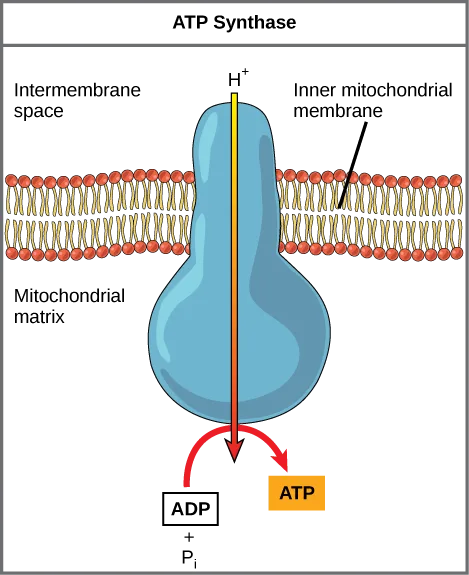
If the membrane were continuously open to simple diffusion by the hydrogen ions, the ions would tend to diffuse back across into the matrix, driven by the concentrations producing their electrochemical gradient. Recall that many ions cannot diffuse through the nonpolar regions of phospholipid membranes without the aid of ion channels. Similarly, hydrogen ions in the matrix space can only pass through the inner mitochondrial membrane by an integral membrane protein called ATP synthase (below). This complex protein acts as a tiny generator, turned by the force of the hydrogen ions diffusing through it, down their electrochemical gradient. The turning of parts of this molecular machine facilitates the addition of a phosphate to ADP, forming ATP, using the potential energy of the hydrogen ion gradient.
Extended description
A single light-blue, hourglass-shaped ATP synthase complex sits within a horizontal phospholipid bilayer (round orange head groups on tan tails). Above the membrane the region is labeled Intermembrane space; below it, Mitochondrial matrix. A leader line labeled Inner mitochondrial membrane points to the bilayer. A vertical arrow labeled H⁺, shaded from yellow at the top to red at the bottom, runs down through the middle of the complex from the intermembrane space into the matrix. At the bottom of the complex, a red curved arrow points from a box labeled ADP plus P subscript i to an orange box labeled ATP, showing the complex converting ADP and inorganic phosphate into ATP as the proton passes through.
Dinitrophenol (DNP) is an “uncoupler” that makes the inner mitochondrial membrane “leaky” to protons. It was used until 1938 as a weight-loss drug. What effect would you expect DNP to have on the change in pH across the inner mitochondrial membrane? Why do you think this might be an effective weight-loss drug?
Show model answer
Did your answer mention:
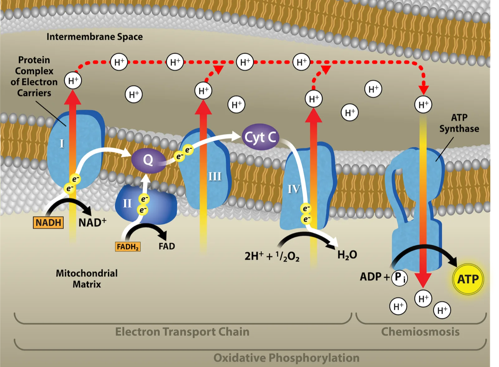
Chemiosmosis (below) is used to generate 90 percent of the ATP made during aerobic glucose catabolism; it is also the method used in the light reactions of photosynthesis to harness the energy of sunlight in the process of photophosphorylation. Recall that the production of ATP using the process of chemiosmosis in mitochondria is called oxidative phosphorylation. The overall result of these reactions is the production of ATP from the energy of the electrons removed from hydrogen atoms. These atoms were originally part of a glucose molecule. At the end of the pathway, the electrons are used to reduce an oxygen molecule to oxygen ions. The extra electrons on the oxygen attract hydrogen ions (protons) from the surrounding medium, and water is formed. Thus, oxygen is the final electron acceptor in the electron transport chain.
Extended description
A wide diagram spanning the same phospholipid bilayer as the electron transport chain figure, with complexes I through IV, Q, and Cyt C arranged the same way at the left under a label reading Protein Complex of Electron Carriers with a leader line to complex I, and a bulb-shaped ATP synthase added at the right under a label reading ATP Synthase with a leader line to it. Intermembrane Space is labeled at the top; Mitochondrial Matrix at the bottom. Several white circles labeled H⁺ float in the intermembrane space above the complexes; a dashed red arrow traces a path connecting them, arcing rightward and down into the top of ATP synthase. A vertical arrow shaded yellow to red, also labeled H⁺, runs down through ATP synthase into the matrix, where a black curved arrow shows ADP plus P subscript i combining, next to a highlighted circle labeled ATP, with a few more white circles labeled H⁺ nearby in the matrix. Below the whole membrane, a bracket labeled Electron Transport Chain spans complexes I through IV, a second bracket labeled Chemiosmosis spans ATP synthase, and a third, longer bracket beneath both is labeled Oxidative Phosphorylation.
Cyanide inhibits cytochrome c oxidase, a component of the electron transport chain. If cyanide poisoning occurs, would you expect the pH of the intermembrane space to increase or decrease? What effect would cyanide have on ATP synthesis?
Show model answer
Did your answer mention:
ATP Yield
The number of ATP molecules generated from the catabolism of glucose varies. For example, the number of hydrogen ions that the electron transport chain complexes can pump through the membrane varies between species. Another source of variance stems from the shuttle of electrons across the membranes of the mitochondria. (The NADH generated from glycolysis cannot easily enter mitochondria.) Thus, electrons are picked up on the inside of mitochondria by either NAD⁺ or FAD⁺. As you have learned earlier, these FAD⁺ molecules can transport fewer ions; consequently, fewer ATP molecules are generated when FAD⁺ acts as a carrier. NAD⁺ is used as the electron transporter in the liver and FAD⁺ acts in the brain.
Another factor that affects the yield of ATP molecules generated from glucose is the fact that intermediate compounds in these pathways are also used for other purposes. Glucose catabolism connects with the pathways that build or break down all other biochemical compounds in cells, and the result is somewhat messier than the ideal situations described thus far. For example, sugars other than glucose are fed into the glycolytic pathway for energy extraction. In addition, the five-carbon sugars that form nucleic acids are made from intermediates in glycolysis. Certain nonessential amino acids can be made from intermediates of both glycolysis and the citric acid cycle. Lipids, such as cholesterol and triglycerides, are also made from intermediates in these pathways, and both amino acids and triglycerides are broken down for energy through these pathways. Overall, in living systems, these pathways of glucose catabolism extract about 34 percent of the energy contained in glucose, with the remainder being released as heat.
Summary
The electron transport chain is the portion of aerobic respiration that uses free oxygen as the final electron acceptor of the electrons removed from the intermediate compounds in glucose catabolism. The electron transport chain is composed of four large, multiprotein complexes embedded in the inner mitochondrial membrane and two small diffusible electron carriers shuttling electrons between them. The electrons are passed through a series of redox reactions, with a small amount of free energy used at three points to transport hydrogen ions across a membrane. This process contributes to the gradient used in chemiosmosis. The electrons passing through the electron transport chain gradually lose energy. High-energy electrons donated to the chain by either NADH or FADH₂ complete the chain, as low-energy electrons reduce oxygen molecules and form water. The level of free energy of the electrons drops from about 60 kcal/mol in NADH or 45 kcal/mol in FADH₂ to about 0 kcal/mol in water. The end products of the electron transport chain are water and ATP. A number of intermediate compounds of the citric acid cycle can be diverted into the anabolism of other biochemical molecules, such as nonessential amino acids, sugars, and lipids. These same molecules can serve as energy sources for the glucose pathways.
Key terms
- ATP synthase — (also F1F0 ATP synthase) membrane-embedded protein complex that adds a phosphate to ADP with energy from protons diffusing through it
- prosthetic group — (also prosthetic cofactor) molecule bound to a protein that facilitates the function of the protein
- ubiquinone — soluble electron transporter in the electron transport chain that connects the first or second complex to the third
Practice
Describe how electrons move through the electron transport chain and explain what happens to their energy levels during this process
What compound receives electrons from NADH?
It is the first prosthetic group electrons reach, at complex I.How do the roles of ubiquinone and cytochrome c differ from the roles of the other components of the electron transport chain?
Show model answer
Did your answer mention:
What accounts for the different number of ATP molecules that are formed through cellular respiration?
Show model answer
Did your answer mention:
The soluble electron transporter in the electron transport chain that connects the first or second complex to the third is called ________.
The section abbreviates this molecule’s name with a single capital letter.The level of free energy of the electrons drops from about 60 kcal/mol in NADH or ________ kcal/mol in FADH₂ to about 0 kcal/mol in water.
The section’s summary gives the free-energy level of the electrons FADH₂ donates, between NADH’s 60 kcal/mol and water’s 0 kcal/mol.A nonprotein molecule bound to a protein that facilitates the protein’s function is called a ________.
FMN, in complex I, is one example of this kind of nonpeptide helper molecule.Explain how a proton (H⁺) gradient is established and maintained by the electron transport chain
Chemiosmosis involves ________.
It is the ions, driven through ATP synthase by their own gradient, that do the work — not the electrons or the atoms.The membrane-embedded protein complex that adds a phosphate to ADP using energy from protons diffusing through it is called ________.
Hydrogen ions turn this molecular machine as they flow down their electrochemical gradient into the matrix.The electrons are passed through a series of redox reactions, with a small amount of free energy used at ________ points to transport hydrogen ions across the membrane.
Complexes I, III, and IV are the points along the chain that pump protons.This section is adapted from Biology 2e, Section 7.4: Oxidative Phosphorylation by Mary Ann Clark, Jung Choi, Matthew Douglas, and OpenStax, © OpenStax, licensed under CC BY-NC-SA 4.0. Access the original for free at openstax.org. Changes: figures re-encoded as WebP, with one of three re-kinded from the manifest’s file-extension guess of “photo” to “diagram” after inspection (all three are line-drawn schematics of the transport chain and ATP synthase, not photographs); all three source alts, which were transcribed with disruptive letter-by-letter text-to-speech spacing (“A T P,” “N A D H”), rewritten from the images, and an extended description added to each since none is a photograph whose full content is carried by its caption; both Visual Connection questions (Dinitrophenol/pH, cyanide/pH) kept in the body immediately after their figures and rendered as self-checks, since the source keys both with a prose solution rather than a lettered option; ion charges (Fe⁺⁺, Fe⁺⁺⁺) and the H⁺, NAD⁺/FAD⁺ superscripts set as Unicode; the copper-ion subscripts CuA/CuB and inorganic phosphate Pi set as inline HTML since no Unicode subscript letter exists for them; the end-of-section Review Questions and Critical Thinking Questions adapted into the closing interactive Practice block (multiple choice and self-check respectively); and two key-term recall items (ubiquinone, prosthetic group) added from the glossary, plus a third (ATP synthase) to round out the second objective group with an auto-graded item, since this section’s own end-of-section sets leave that objective with only the one Review Question above; a stray “B.” printed after “ubiquinone” in the source’s “Q and Complex II” paragraph (also present in the PDF) is omitted as a source typesetting defect; rubric checkpoints added to all four self-checks, the two body Visual Connections included, decomposing each model answer (the source solution) into check-off clauses with no new claims; and two summary-derived cloze textin items added — the 45 kcal/mol free-energy level of FADH₂’s electrons under the first objective, and the three points along the chain that pump hydrogen ions under the second objective — to raise the second objective’s group and the section as a whole to the practice floor.