Signaling Molecules and Cellular Receptors
By the end of this section, you will be able to:
- Describe four types of signaling mechanisms found in multicellular organisms
- Compare internal receptors with cell-surface receptors
- Recognize the relationship between a ligand’s structure and its mechanism of action
There are two kinds of communication in the world of living cells. Communication between cells is called intercellular signaling, and communication within a cell is called intracellular signaling. An easy way to remember the distinction is by understanding the Latin origin of the prefixes: inter- means “between” (for example, intersecting lines are those that cross each other) and intra- means “inside” (as in intravenous).
Chemical signals are released by signaling cells in the form of small, usually volatile or soluble molecules called ligands. A ligand is a molecule that binds another specific molecule, in some cases, delivering a signal in the process. Ligands can thus be thought of as signaling molecules. Ligands interact with proteins in target cells, which are cells that are affected by chemical signals; these proteins are also called receptors. Ligands and receptors exist in several varieties; however, a specific ligand will have a specific receptor that typically binds only that ligand.
Forms of Signaling
There are four categories of chemical signaling found in multicellular organisms: paracrine signaling, endocrine signaling, autocrine signaling, and direct signaling across gap junctions (below). The main difference between the different categories of signaling is the distance that the signal travels through the organism to reach the target cell. We should note here that not all cells are affected by the same signals.
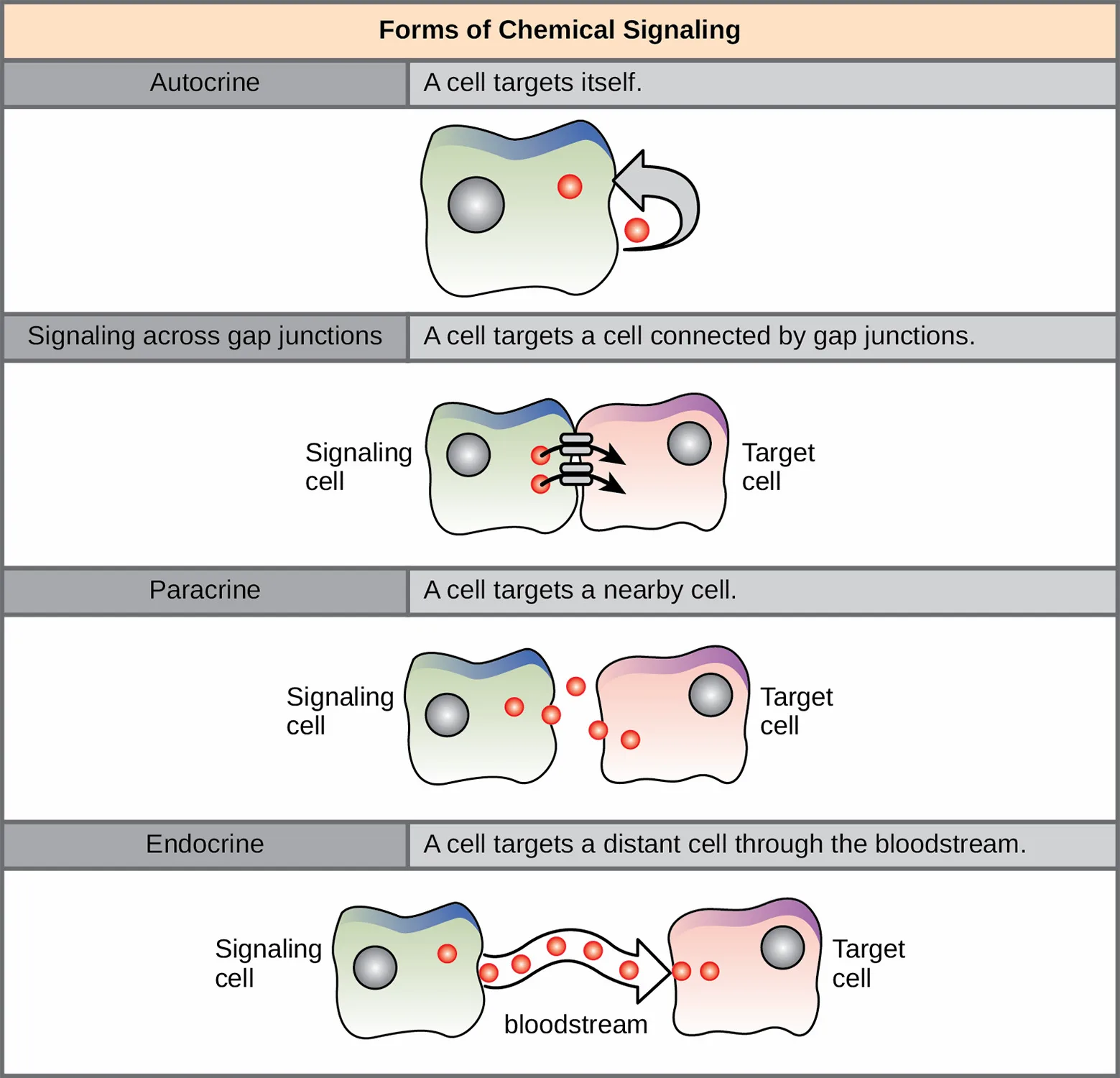
Paracrine Signaling
Signals that act locally between cells that are close together are called paracrine signals. Paracrine signals move by diffusion through the extracellular matrix. These types of signals usually elicit quick responses that last only a short period of time. In order to keep the response localized, paracrine ligand molecules are normally quickly degraded by enzymes or removed by neighboring cells. Removing the signals will reestablish the concentration gradient for the signal, allowing them to quickly diffuse through the intracellular space if released again.
One example of paracrine signaling is the transfer of signals across synapses between nerve cells. A nerve cell consists of a cell body, several short, branched extensions called dendrites that receive stimuli, and a long extension called an axon, which transmits signals to other nerve cells or muscle cells. The junction between nerve cells where signal transmission occurs is called a synapse. A synaptic signal is a chemical signal that travels between nerve cells. Signals within the nerve cells are propagated by fast-moving electrical impulses. When these impulses reach the end of the axon, the signal continues on to a dendrite of the next cell by the release of chemical ligands called neurotransmitters from the presynaptic cell (the cell emitting the signal). The neurotransmitters are transported across the very small distances (20–40 nanometers) between nerve cells, which are called chemical synapses (below). The small distance between nerve cells allows the signal to travel quickly; this enables an immediate response, such as, “Take your hand off the stove!”
When the neurotransmitter binds the receptor on the surface of the postsynaptic cell, the electrochemical potential of the target cell changes, and the next electrical impulse is launched. The neurotransmitters that are released into the chemical synapse are degraded quickly or get reabsorbed by the presynaptic cell so that the recipient nerve cell can recover quickly and be prepared to respond rapidly to the next synaptic signal.
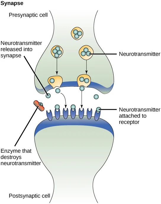
Extended description
Labeled top to bottom. ‘Presynaptic cell’ forms a bulbous terminal at the top, containing three round vesicles, with two more fusing with the membrane below them labeled ‘Neurotransmitter,’ each holding several small blue dots. Arrows lead from the vesicles down to the cell membrane, where a label reads ‘Neurotransmitter released into synapse’ as dots pass into the gap below. An orange capsule shape at the left edge of the gap is labeled ‘Enzyme that destroys neurotransmitter.’ Below the gap, the ‘Postsynaptic cell’ membrane is studded with a row of purple receptor proteins; one dot sits docked on a receptor, labeled ‘Neurotransmitter attached to receptor.’
Endocrine Signaling
Signals from distant cells are called endocrine signals, and they originate from endocrine cells. (In the body, many endocrine cells are located in endocrine glands, such as the thyroid gland, the hypothalamus, and the pituitary gland.) These types of signals usually produce a slower response but have a longer-lasting effect. The ligands released in endocrine signaling are called hormones, signaling molecules that are produced in one part of the body but affect other body regions some distance away.
Hormones travel the large distances between endocrine cells and their target cells via the bloodstream, which is a relatively slow way to move throughout the body. Because of their form of transport, hormones become diluted and are present in low concentrations when they act on their target cells. This is different from paracrine signaling, in which local concentrations of ligands can be very high.
Autocrine Signaling
Autocrine signals are produced by signaling cells that can also bind to the ligand that is released. This means the signaling cell and the target cell can be the same or a similar cell (the prefix auto- means self, a reminder that the signaling cell sends a signal to itself). This type of signaling often occurs during the early development of an organism to ensure that cells develop into the correct tissues and take on the proper function. Autocrine signaling also regulates pain sensation and inflammatory responses. Further, if a cell is infected with a virus, the cell can signal itself to undergo programmed cell death, killing the virus in the process. In some cases, neighboring cells of the same type are also influenced by the released ligand. In embryological development, this process of stimulating a group of neighboring cells may help to direct the differentiation of identical cells into the same cell type, thus ensuring the proper developmental outcome.
Direct Signaling Across Gap Junctions
Gap junctions in animals and plasmodesmata in plants are connections between the plasma membranes of neighboring cells. These fluid-filled channels allow small signaling molecules, called intracellular mediators, to diffuse between the two cells. Small molecules or ions, such as calcium ions (Ca²⁺), are able to move between cells, but large molecules like proteins and DNA cannot fit through the channels. The specificity of the channels ensures that the cells remain independent but can quickly and easily transmit signals. The transfer of signaling molecules communicates the current state of the cell that is directly next to the target cell; this allows a group of cells to coordinate their response to a signal that only one of them may have received. In plants, plasmodesmata are ubiquitous, making the entire plant into a giant communication network.
Types of Receptors
Receptors are protein molecules in the target cell or on its surface that bind ligand. There are two types of receptors, internal receptors and cell-surface receptors.
Internal receptors
Internal receptors, also known as intracellular or cytoplasmic receptors, are found in the cytoplasm of the cell and respond to hydrophobic ligand molecules that are able to travel across the plasma membrane. Once inside the cell, many of these molecules bind to proteins that act as regulators of mRNA synthesis (transcription) to mediate gene expression. Gene expression is the cellular process of transforming the information in a cell’s DNA into a sequence of amino acids, which ultimately forms a protein. When the ligand binds to the internal receptor, a conformational change is triggered that exposes a DNA-binding site on the protein. The ligand-receptor complex moves into the nucleus, then binds to specific regulatory regions of the chromosomal DNA and promotes the initiation of transcription (below). Transcription is the process of copying the information in a cell’s DNA into a special form of RNA called messenger RNA (mRNA); the cell uses information in the mRNA (which moves out into the cytoplasm and associates with ribosomes) to link specific amino acids in the correct order, producing a protein. Internal receptors can directly influence gene expression without having to pass the signal on to other receptors or messengers.
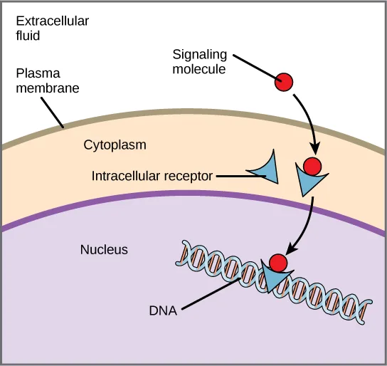
Extended description
Layered top to bottom: ‘Extracellular fluid’ above a tan band labeled ‘Plasma membrane’; below it, the ‘Cytoplasm’ contains a blue triangular shape labeled ‘Intracellular receptor.’ A red circle labeled ‘Signaling molecule’ arrives from the extracellular fluid, crosses the membrane, and an arrow shows it binding the receptor triangle. A second arrow carries the joined receptor-signaling molecule pair down through a purple region labeled ‘Nucleus’ to a coiled double helix labeled ‘DNA.’
Cell-Surface Receptors
Cell-surface receptors, also known as transmembrane receptors, are cell surface, membrane-anchored (integral) proteins that bind to external ligand molecules. This type of receptor spans the plasma membrane and performs signal transduction, through which an extracellular signal is converted into an intracellular signal. Ligands that interact with cell-surface receptors do not have to enter the cell that they affect. Cell-surface receptors are also called cell-specific proteins or markers because they are specific to individual cell types.
Because cell-surface receptor proteins are fundamental to normal cell functioning, it should come as no surprise that a malfunction in any one of these proteins could have severe consequences. Errors in the protein structures of certain receptor molecules have been shown to play a role in hypertension (high blood pressure), asthma, heart disease, and cancer.
Each cell-surface receptor has three main components: an external ligand-binding domain called the extracellular domain, a hydrophobic membrane-spanning region called a transmembrane domain, and an intracellular domain inside the cell. The size and extent of each of these domains vary widely, depending on the type of receptor.
Evolution Connection. How Viruses Recognize a Host. Unlike living cells, many viruses do not have a plasma membrane or any of the structures necessary to sustain metabolic life. Some viruses are simply composed of an inert protein shell enclosing DNA or RNA. To reproduce, viruses must invade a living cell, which serves as a host, and then take over the host’s cellular apparatus. But how does a virus recognize its host?
Viruses often bind to cell-surface receptors on the host cell. For example, the virus that causes human influenza (flu) binds specifically to receptors on membranes of cells of the respiratory system. Chemical differences in the cell-surface receptors among hosts mean that a virus that infects a specific species (for example, humans) often cannot infect another species (for example, chickens).
However, viruses have very small amounts of DNA or RNA compared to humans, and, as a result, viral reproduction can occur rapidly. Viral reproduction invariably produces errors that can lead to changes in newly produced viruses; these changes mean that the viral proteins that interact with cell-surface receptors may evolve in such a way that they can bind to receptors in a new host. Such changes happen randomly and quite often in the reproductive cycle of a virus, but the changes only matter if a virus with new binding properties comes into contact with a suitable host. In the case of influenza, this situation can occur in settings where animals and people are in close contact, such as poultry and swine farms. (A. B. Sigalov, The School of Nature. IV. Learning from Viruses, Self/Nonself 1, no. 4 (2010): 282–298; Y. Cao, X. Koh, L. Dong, X. Du, A. Wu, X. Ding, H. Deng, Y. Shu, J. Chen, T. Jiang, Rapid Estimation of Binding Activity of Influenza Virus Hemagglutinin to Human and Avian Receptors, PLoS One 6, no. 4 (2011): e18664.) Once a virus jumps the former “species barrier” to a new host, it can spread quickly. Scientists watch newly appearing viruses (called emerging viruses) closely in the hope that such monitoring can reduce the likelihood of global viral epidemics.
Cell-surface receptors are involved in most of the signaling in multicellular organisms. There are three general categories of cell-surface receptors: ion channel-linked receptors, G-protein-linked receptors, and enzyme-linked receptors.
Ion channel-linked receptors bind a ligand and open a channel through the membrane that allows specific ions to pass through. To form a channel, this type of cell-surface receptor has an extensive membrane-spanning region. In order to interact with the double layer of phospholipid fatty acid tails that form the center of the plasma membrane, many of the amino acids in the membrane-spanning region are hydrophobic in nature. Conversely, the amino acids that line the inside of the channel are hydrophilic to allow for the passage of water or ions. When a ligand binds to the extracellular region of the channel, there is a conformational change in the protein’s structure that allows ions such as sodium, calcium, magnesium, and hydrogen to pass through (below).
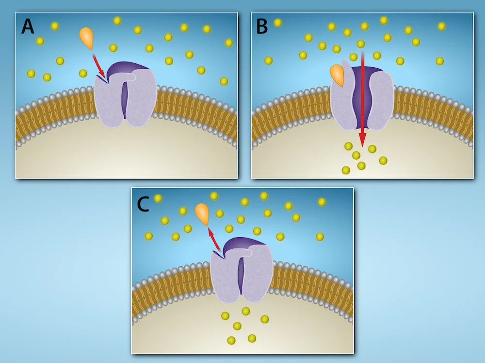
Extended description
Panel A: a closed channel protein spans the membrane; a teardrop-shaped signaling molecule approaches from above on a red arrow, while yellow ion dots are scattered on both sides of the membrane. Panel B: the signaling molecule has docked in the channel’s binding site, and a red arrow through the open channel shows yellow ions flowing from outside to inside. Panel C: the signaling molecule is shown detaching upward on a red arrow, and the channel has returned to its closed shape, with ions again scattered on both sides rather than flowing through.
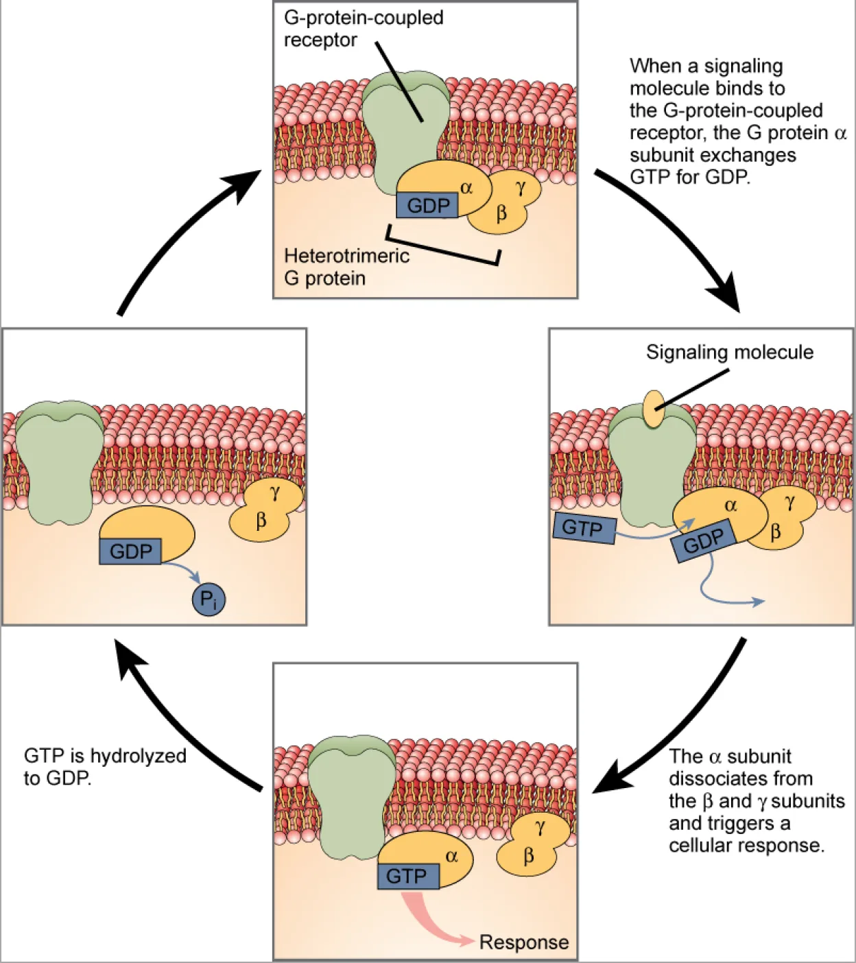
G-protein-linked receptors bind a ligand and activate a membrane protein called a G-protein. The activated G-protein then interacts with either an ion channel or an enzyme in the membrane (below). All G-protein-linked receptors have seven transmembrane domains, but each receptor has its own specific extracellular domain and G-protein-binding site.
Cell signaling using G-protein-linked receptors occurs as a cyclic series of events. Before the ligand binds, the inactive G-protein can bind to a site on the receptor specific for its binding. Once the ligand binds to the receptor, the resulting change in shape activates the G-protein, which releases guanosine diphosphate (GDP) and picks up guanosine triphosphate (GTP). (Source note: the source prints “guanosine 3-phosphate”; GTP is guanosine triphosphate, three phosphates on the ribose, as its abbreviation and its standard name (PubChem CID 135398633) say.) The subunits of the G-protein then split into the α subunit and the βγ subunit. One or both of these G-protein fragments may be able to activate other proteins as a result. After awhile, the GTP on the active α subunit of the G-protein is hydrolyzed to GDP and the βγ subunit is deactivated. The subunits reassociate to form the inactive G-protein and the cycle begins anew.
Extended description
Four panels arranged in a clockwise cycle and connected by curved arrows. Top panel: an inactive G-protein-coupled receptor spans the membrane, with a heterotrimeric G protein below it — the α subunit bound to GDP, alongside the β and γ subunits. An arrow to the right, labeled ‘When a signaling molecule binds to the G-protein-coupled receptor, the G protein α subunit exchanges GTP for GDP,’ leads to the right panel, where a signaling molecule has bound the receptor and GTP is replacing GDP on the α subunit. An arrow down, labeled ‘The α subunit dissociates from the β and γ subunits and triggers a cellular response,’ leads to the bottom panel, where the GTP-bound α subunit has separated from β and γ and points to a ‘Response’ arrow. An arrow left, labeled ‘GTP is hydrolyzed to GDP,’ leads to the left panel, where the lone α subunit releases a phosphate (Pᵢ) as its GTP converts to GDP, with β and γ nearby before the cycle returns to the top panel.
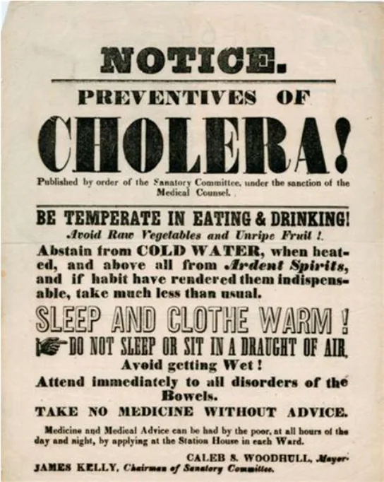
G-protein-linked receptors have been extensively studied and much has been learned about their roles in maintaining health. Bacteria that are pathogenic to humans can release poisons that interrupt specific G-protein-linked receptor function, leading to illnesses such as pertussis, botulism, and cholera. In cholera (below), for example, the water-borne bacterium Vibrio cholerae produces a toxin, choleragen, that binds to cells lining the small intestine. The toxin then enters these intestinal cells, where it modifies a G-protein that controls the opening of a chloride channel and causes it to remain continuously active, resulting in large losses of fluids from the body and potentially fatal dehydration as a result.
Enzyme-linked receptors are cell-surface receptors with intracellular domains that are associated with an enzyme. In some cases, the intracellular domain of the receptor itself is an enzyme. Other enzyme-linked receptors have a small intracellular domain that interacts directly with an enzyme. The enzyme-linked receptors normally have large extracellular and intracellular domains, but the membrane-spanning region consists of a single alpha-helical region of the peptide strand. When a ligand binds to the extracellular domain, a signal is transferred through the membrane, activating the enzyme. Activation of the enzyme sets off a chain of events within the cell that eventually leads to a response. One example of this type of enzyme-linked receptor is the tyrosine kinase receptor (below). A kinase is an enzyme that transfers phosphate groups from ATP to another protein. The tyrosine kinase receptor transfers phosphate groups to tyrosine molecules (tyrosine residues). First, signaling molecules bind to the extracellular domain of two nearby tyrosine kinase receptors. The two neighboring receptors then bond together, or dimerize. Phosphates are then added to tyrosine residues on the intracellular domain of the receptors (phosphorylation). The phosphorylated residues can then transmit the signal to the next messenger within the cytoplasm.
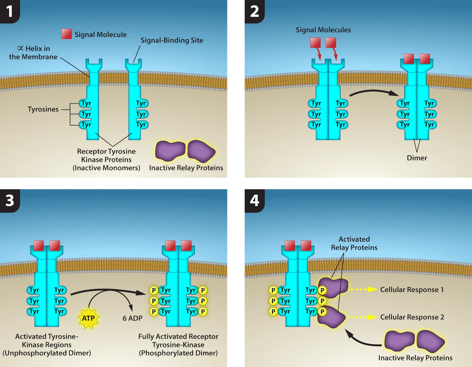
- A receptor tyrosine kinase is an enzyme-linked receptor with a single helical transmembrane region, and extracellular and intracellular domains. 2) Binding of a signaling molecule to the extracellular domain causes the receptor to dimerize. 3) Tyrosine residues on the intracellular domain are then autophosphorylated, 4) triggering a downstream cellular response. The signal is terminated by a phosphatase that removes the phosphates from the phosphotyrosine residues. Credit: Rao, A., Ryan, K., Tag, A., Fletcher, S. and Hawkins, A. Department of Biology, Texas A&M University.
Extended description
Four numbered panels tracing receptor tyrosine kinase activation. Panel 1: two separate, inactive receptor tyrosine kinase monomers span the membrane, each with three intracellular tyrosine (Tyr) residues; two inactive, unshaded relay proteins sit nearby in the cytoplasm. Panel 2: two signal molecules bind the extracellular binding sites, and the two receptors come together to form a dimer. Panel 3: ATP phosphorylates the six tyrosine residues of the dimer, adding a phosphate (P) to each and converting six ATP to six ADP, producing the fully activated, phosphorylated receptor. Panel 4: the phosphorylated tyrosines recruit and activate the two relay proteins, which are shown triggering Cellular Response 1 and Cellular Response 2.
HER2 is a receptor tyrosine kinase. In 30 percent of human breast cancers, HER2 is permanently activated, resulting in unregulated cell division. Lapatinib, a drug used to treat breast cancer, inhibits HER2 receptor tyrosine kinase autophosphorylation (the process by which the receptor adds phosphates onto itself), thus reducing tumor growth by 50 percent. Besides autophosphorylation, which of the following steps would be inhibited by Lapatinib?
Autophosphorylation is step 3 in the figure above; ask which later step depends on it and would therefore never happen if it is blocked.Signaling Molecules
Produced by signaling cells and the subsequent binding to receptors in target cells, ligands act as chemical signals that travel to the target cells to coordinate responses. The types of molecules that serve as ligands are incredibly varied and range from small proteins to small ions like calcium (Ca²⁺).
Small Hydrophobic Ligands
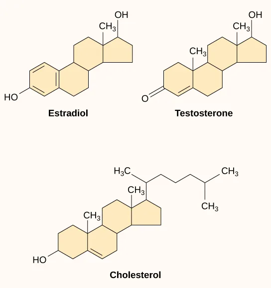
Small hydrophobic ligands can directly diffuse through the plasma membrane and interact with internal receptors. Important members of this class of ligands are the steroid hormones. Steroids are lipids that have a hydrocarbon skeleton with four fused rings; different steroids have different functional groups attached to the carbon skeleton. Steroid hormones include the female sex hormone, estradiol, which is a type of estrogen; the male sex hormone, testosterone; and cholesterol, which is an important structural component of biological membranes and a precursor of steroid hormones (below). Other hydrophobic hormones include thyroid hormones and vitamin D. In order to be soluble in blood, hydrophobic ligands must bind to carrier proteins while they are being transported through the bloodstream.
Water-Soluble Ligands
Water-soluble ligands are polar and, therefore, cannot pass through the plasma membrane unaided; sometimes, they are too large to pass through the membrane at all. Instead, most water-soluble ligands bind to the extracellular domain of cell-surface receptors. This group of ligands is quite diverse and includes small molecules, peptides, and proteins.
Other Ligands
Nitric oxide (NO) is a gas that also acts as a ligand. It is able to diffuse directly across the plasma membrane, and one of its roles is to interact with receptors in smooth muscle and induce relaxation of the tissue. NO has a very short half-life and, therefore, only functions over short distances. Nitroglycerin, a treatment for heart disease, acts by triggering the release of NO, which causes blood vessels to dilate (expand), thus restoring blood flow to the heart. NO has become better known recently because the pathway that it affects is targeted by prescription medications for erectile dysfunction, such as Viagra (erection involves dilated blood vessels).
Summary
Cells communicate by both inter- and intracellular signaling. Signaling cells secrete ligands that bind to target cells and initiate a chain of events within the target cell. The four categories of signaling in multicellular organisms are paracrine signaling, endocrine signaling, autocrine signaling, and direct signaling across gap junctions. Paracrine signaling takes place over short distances. Endocrine signals are carried long distances through the bloodstream by hormones, and autocrine signals are received by the same cell that sent the signal or other nearby cells of the same kind. Gap junctions allow small molecules, including signaling molecules, to flow between neighboring cells.
Internal receptors are found in the cell cytoplasm. Here, they bind ligand molecules that cross the plasma membrane; these receptor-ligand complexes move to the nucleus and interact directly with cellular DNA. Cell-surface receptors transmit a signal from outside the cell to the cytoplasm. Ion channel-linked receptors, when bound to their ligands, form a pore through the plasma membrane through which certain ions can pass. G-protein-linked receptors interact with a G-protein on the cytoplasmic side of the plasma membrane, promoting the exchange of bound GDP for GTP and interacting with other enzymes or ion channels to transmit a signal. Enzyme-linked receptors transmit a signal from outside the cell to an intracellular domain of a membrane-bound enzyme. Ligand binding causes activation of the enzyme. Small hydrophobic ligands (like steroids) are able to penetrate the plasma membrane and bind to internal receptors. Water-soluble hydrophilic ligands are unable to pass through the membrane; instead, they bind to cell-surface receptors, which transmit the signal to the inside of the cell.
Key terms
- autocrine signal — signal that is sent and received by the same or similar nearby cells
- cell-surface receptor — cell-surface protein that transmits a signal from the exterior of the cell to the interior, even though the ligand does not enter the cell
- chemical synapse — small space between axon terminals and dendrites of nerve cells where neurotransmitters function
- endocrine cell — cell that releases ligands involved in endocrine signaling (hormones)
- endocrine signal — long-distance signal that is delivered by ligands (hormones) traveling through an organism’s circulatory system from the signaling cell to the target cell
- enzyme-linked receptor — cell-surface receptor with intracellular domains that are associated with membrane-bound enzymes
- extracellular domain — region of a cell-surface receptor that is located on the cell surface
- G-protein-linked receptor — cell-surface receptor that activates membrane-bound G-proteins to transmit a signal from the receptor to nearby membrane components
- intercellular signaling — communication between cells
- internal receptor — (also, intracellular receptor) receptor protein that is located in the cytosol of a cell and binds to ligands that pass through the plasma membrane
- intracellular mediator — (also, second messenger) small molecule that transmits signals within a cell
- intracellular signaling — communication within cells
- ion channel-linked receptor — cell-surface receptor that forms a plasma membrane channel, which opens when a ligand binds to the extracellular domain (ligand-gated channels)
- ligand — molecule produced by a signaling cell that binds with a specific receptor, delivering a signal in the process
- neurotransmitter — chemical ligand that carries a signal from one nerve cell to the next
- paracrine signal — signal between nearby cells that is delivered by ligands traveling in the liquid medium in the space between the cells
- receptor — protein in or on a target cell that binds to ligands
- signaling cell — cell that releases signal molecules that allow communication with another cell
- synaptic signal — chemical signal (neurotransmitter) that travels between nerve cells
- target cell — cell that has a receptor for a signal or ligand from a signaling cell
Practice
Describe four types of signaling mechanisms found in multicellular organisms
A molecule produced by a signaling cell that binds a specific receptor, delivering a signal in the process, is called a ________.
Its own name says what it does: it binds.The secretion of hormones by the pituitary gland is an example of ________.
The pituitary’s hormones travel through the bloodstream to reach distant target cells.Endocrine signals are transmitted more slowly than paracrine signals because ________.
Compare how far each signal has to travel and by what route.What is the difference between intracellular signaling and intercellular signaling?
Show model answer
Did your answer mention:
How are the effects of paracrine signaling limited to an area near the signaling cells?
Show model answer
Did your answer mention:
Compare internal receptors with cell-surface receptors
What property prevents the ligands of cell-surface receptors from entering the cell?
Ask what kind of molecule can cross a lipid membrane unaided, and what kind cannot.Why are ion channels necessary to transport ions into or out of a cell?
Ions carry a charge; think about what kind of environment a charged particle can and cannot cross unaided.A scientist notices that when she adds a small, water-soluble molecule to a dish of cells, the cells turn off transcription of a gene. She hypothesizes that the ligand she added binds to a(n) ________ receptor.
A water-soluble molecule cannot cross the membrane, so it must act through a cell-surface receptor that starts a signaling chain reaching the nucleus.What are the differences between internal receptors and cell-surface receptors?
Show model answer
Did your answer mention:
Cells grown in the laboratory are mixed with a dye molecule that is unable to pass through the plasma membrane. If a ligand is added to the cells, observations show that the dye enters the cells. What type of receptor did the ligand bind to on the cell surface?
Show model answer
Did your answer mention:
Recognize the relationship between a ligand’s structure and its mechanism of action
A ligand that is small and hydrophobic, such as a steroid hormone, typically reaches its target by ________.
Ask whether the ligand’s chemical nature lets it cross the lipid membrane on its own.Small hydrophobic ligands (like steroids) are able to penetrate the plasma membrane and bind to ________.
This section’s summary distinguishes these from the receptors that water-soluble ligands must use instead.Insulin is a hormone that regulates blood sugar by binding to its receptor, insulin receptor tyrosine kinase. How does insulin’s behavior differ from steroid hormone signaling, and what can you infer about its structure?
Show model answer
Insulin’s receptor is an enzyme-linked transmembrane receptor, as can be determined from the “tyrosine kinase” in its name. This receptor is embedded in the plasma membrane, and insulin binds to its extracellular (outer) surface to initiate intracellular signaling cascades.
Normally, steroid hormones cross the plasma membrane to bind with intracellular receptors. These intracellular hormone-receptor complexes then interact directly with DNA to regulate transcription. This limits steroid hormones to be small, non-polar molecules so they can cross the plasma membrane. However, since insulin does not have to cross into the cell it could be large or polar (it is a small, polar molecule).
Did your answer mention:
This section is adapted from Biology 2e, Section 9.1: Signaling Molecules and Cellular Receptors by Mary Ann Clark, Jung Choi, Matthew Douglas, and OpenStax, © OpenStax, licensed under CC BY-NC-SA 4.0. Access the original for free at openstax.org. Changes: figures re-encoded as WebP, with five of the eight re-kinded from the manifest’s file-extension guess of “photo” to “diagram” after inspection (every one is a drawn illustration, not a photograph — only the 1866 cholera-poster reproduction is a true photo); a longdesc added to the five figures that are labeled diagrams or flow charts whose full reading is not carried by their captions (the synapse diagram, the intracellular-receptor diagram, the gated-ion-channel panels, the G-protein cycle, and the tyrosine-kinase-receptor panels); the synapse figure’s alt rewritten to clean up a source typo (“neutrotransmitter”/“neurotrasmitter”) and the intracellular-receptor figure’s alt rewritten to spell out “DNA” in place of the source’s letter-by-letter “D N A”; ion charges and calcium set as Unicode (Ca²⁺); inline references to figures changed from the source’s numbered pointers to descriptive phrases (“below,” “above”) since figures are not numbered here; Cao et al. 2011) omitted as bibliographic apparatus rather than section content; the Visual Connection question kept in the body immediately after its figure and rendered as multiple choice, since the source keys it to a lettered option; the Key Terms entry for “intercellular signaling,” whose glossary definition read “communication between a cell,” corrected to “communication between cells” to match the section’s own definition of the term (reported as a source defect); the end-of-section Review Questions and Critical Thinking Questions adapted into the closing interactive Practice block (multiple choice and self-check respectively); one Critical Thinking Question (the insulin/steroid comparison) placed under the third objective rather than the second, since it argues the relationship between ligand structure and receptor type; a multiple choice item written from the section’s own text on small hydrophobic ligands added to give the third objective an auto-graded item, since no source exercise or glossary term tests it directly; and one key-term recall item (ligand) added from the glossary to round out the first objective group; the Evolution Connection’s two reference citations are kept as a parenthetical after the sentence they support; four small source typos are corrected on the page — the glossary’s “bind” (for “binds”), a caption’s “synapatic”, an option’s “downsteam”, and the letter-spaced “G D P”/“G T P” in two alts are corrected on the page; rubric checkpoints added to each self-check, decomposing its model answer (the source solution) into check-off clauses with no new claims; and one summary-derived recall item (internal receptors) added under the third objective from the section summary’s own sentence. One claim is corrected with a visible Source note: GTP is expanded as guanosine triphosphate rather than the source’s “guanosine 3-phosphate” (erratum 395).