Propagation of the Signal
By the end of this section, you will be able to:
- Explain how the binding of a ligand initiates signal transduction throughout a cell
- Recognize the role of phosphorylation in the transmission of intracellular signals
- Evaluate the role of second messengers in signal transmission
Once a ligand binds to a receptor, the signal is transmitted through the membrane and into the cytoplasm. Continuation of a signal in this manner is called signal transduction. Signal transduction only occurs with cell-surface receptors, which cannot interact with most components of the cell such as DNA. Only internal receptors are able to interact directly with DNA in the nucleus to initiate protein synthesis.
When a ligand binds to its receptor, conformational changes occur that affect the receptor’s intracellular domain. Conformational changes of the extracellular domain upon ligand binding can propagate through the membrane region of the receptor and lead to activation of the intracellular domain or its associated proteins. In some cases, binding of the ligand causes dimerization of the receptor, which means that two receptors bind to each other to form a stable complex called a dimer. A dimer is a chemical compound formed when two molecules (often identical) join together. The binding of the receptors in this manner enables their intracellular domains to come into close contact and activate each other.
Binding Initiates a Signaling Pathway
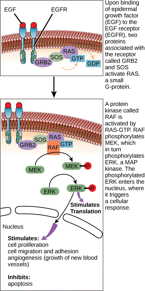
After the ligand binds to the cell-surface receptor, the activation of the receptor’s intracellular components sets off a chain of events that is called a signaling pathway, sometimes called a signaling cascade. In a signaling pathway, second messengers–enzymes–and activated proteins interact with specific proteins, which are in turn activated in a chain reaction that eventually leads to a change in the cell’s environment (below), such as an increase in metabolism or specific gene expression. The events in the cascade occur in a series, much like a current flows in a river. Interactions that occur before a certain point are defined as upstream events, and events after that point are called downstream events.
Extended description
Two stacked panels share the same membrane cross-section, drawn as cross-hatched red and tan bands, with two EGFR receptors (blue, Y-shaped) embedded side by side. Top panel: EGF (a red circle) is bound to the extracellular end of the right-hand receptor; on the cytoplasmic side, GRB2 and SOS are bound to the receptor tails, and a curved arrow shows SOS converting RAS from its GDP-bound to its GTP-bound state. Bottom panel: the same bound receptor pair with GRB2, SOS, and RAS-GTP is joined by RAF (an orange box). An arrow runs from RAF down to MEK, which becomes MEK-P (marked with a red P); a second arrow runs from MEK-P to ERK, which becomes ERK-P. From ERK-P, one arrow points right to “Stimulates Translation” in the cytoplasm, and a second, purple arrow curves down through two unlabeled oval outlines marking the nucleus to a bulleted list: “Stimulates: cell proliferation, cell migration and adhesion, angiogenesis (growth of new blood vessels)” and, below it, “Inhibits: apoptosis.”
In certain cancers, the GTPase activity of the RAS G-protein is inhibited. This means that the RAS protein can no longer hydrolyze GTP into GDP. What effect would this have on downstream cellular events?
Show model answer
Did your answer mention:
You can see that signaling pathways can get very complicated very quickly because most cellular proteins can affect different downstream events, depending on the conditions within the cell. A single pathway can branch off toward different endpoints based on the interplay between two or more signaling pathways, and the same ligands are often used to initiate different signals in different cell types. This variation in response is due to differences in protein expression in different cell types. Another complicating element is signal integration of the pathways, in which signals from two or more different cell-surface receptors merge to activate the same response in the cell. This process can ensure that multiple external requirements are met before a cell commits to a specific response.
The effects of extracellular signals can also be amplified by enzymatic cascades. At the initiation of the signal, a single ligand binds to a single receptor. However, activation of a receptor-linked enzyme can activate many copies of a component of the signaling cascade, which amplifies the signal.
Link to Learning
Observe an animation of cell signaling.
Methods of Intracellular Signaling
The induction of a signaling pathway depends on the modification of a cellular component by an enzyme. There are numerous enzymatic modifications that can occur, and they are recognized in turn by the next component downstream. The following are some of the more common events in intracellular signaling.
Phosphorylation
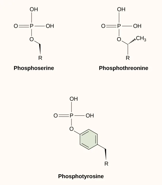
One of the most common chemical modifications that occurs in signaling pathways is the addition of a phosphate group (PO₄³⁻) to a molecule such as a protein in a process called phosphorylation. The phosphate can be added to a nucleotide such as GMP to form GDP or GTP. Phosphates are also often added to serine, threonine, and tyrosine residues of proteins, where they replace the hydroxyl group of the amino acid (below). The transfer of the phosphate is catalyzed by an enzyme called a kinase. Various kinases are named for the substrate they phosphorylate. Phosphorylation of serine and threonine residues often activates enzymes. Phosphorylation of tyrosine residues can either affect the activity of an enzyme or create a binding site that interacts with downstream components in the signaling cascade. Phosphorylation may activate or inactivate enzymes, and the reversal of phosphorylation, dephosphorylation by a phosphatase, will reverse the effect.
Extended description
Three skeletal structures: phosphoserine and phosphothreonine in a top row, phosphotyrosine below. Each begins with a phosphate group — a central P atom double-bonded to one O and single-bonded to two OH groups — connected through a single O to the amino acid side chain, labeled R at its open end. In phosphoserine, that O connects directly down to R. In phosphothreonine, the O connects to a carbon that also carries a CH₃ group on a dashed wedge bond, then down to R. In phosphotyrosine, the O connects to a six-membered aromatic ring, drawn with alternating double bonds, which connects down to R.
Second Messengers
Second messengers are small molecules that propagate a signal after it has been initiated by the binding of the signaling molecule to the receptor. These molecules help to spread a signal through the cytoplasm by altering the behavior of certain cellular proteins.
Calcium ion is a widely used second messenger. The free concentration of calcium ions (Ca²⁺) within a cell is very low because ion pumps in the plasma membrane continuously remove it by using adenosine-5’-triphosphate (ATP). For signaling purposes, Ca²⁺ is stored in cytoplasmic vesicles, such as the endoplasmic reticulum, or accessed from outside the cell. When signaling occurs, ligand-gated calcium ion channels allow the higher levels of Ca²⁺ that are present outside the cell (or in intracellular storage compartments) to flow into the cytoplasm, which raises the concentration of cytoplasmic Ca²⁺. The response to the increase in Ca²⁺ varies and depends on the cell type involved. For example, in the β-cells of the pancreas, Ca²⁺ signaling leads to the release of insulin, and in muscle cells, an increase in Ca²⁺ leads to muscle contractions.
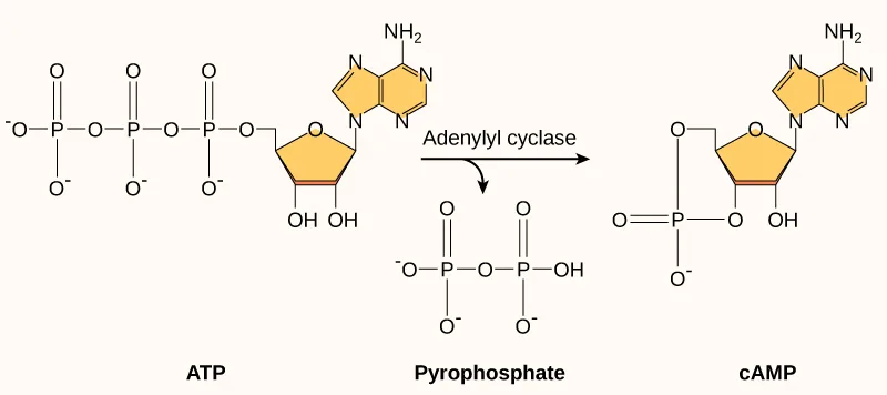
Another second messenger utilized in many different cell types is cyclic AMP (cAMP). Cyclic AMP is synthesized by the enzyme adenylyl cyclase from ATP (below). The main role of cAMP in cells is to bind to and activate an enzyme called cAMP-dependent kinase (A-kinase). A-kinase regulates many vital metabolic pathways: It phosphorylates serine and threonine residues of its target proteins, activating them in the process. A-kinase is found in many different types of cells, and the target proteins in each kind of cell are different. Differences give rise to the variation of the responses to cAMP in different cells.
Extended description
ATP (left) is drawn as three phosphate groups in a row — the terminal one a P with a double-bonded O and two negatively charged O atoms, the other two each with a double-bonded O, one negatively charged O, and a bridging O to the next group — attached through a ribose sugar ring to an adenine base (a fused five- and six-membered ring system with an NH₂ group) above it. An arrow labeled “Adenylyl cyclase” points right: the two outer phosphate groups leave together as pyrophosphate (below the arrow, still linked to each other), while the remaining phosphate closes into a second ring, bonded twice to the ribose, drawn on the right and labeled cAMP.
Present in small concentrations in the plasma membrane, inositol phospholipids are lipids that can also be converted into second messengers. Because these molecules are membrane components, they are located near membrane-bound receptors and can easily interact with them. Phosphatidylinositol (PI) is the main phospholipid that plays a role in cellular signaling. Enzymes known as kinases phosphorylate PI to form PI-phosphate (PIP) and PI-bisphosphate (PIP₂).
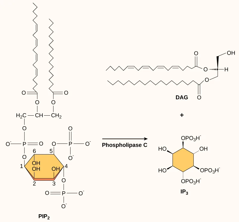
The enzyme phospholipase C cleaves PIP₂ to form diacylglycerol (DAG) and inositol triphosphate (IP₃) (below). These products of the cleavage of PIP₂ serve as second messengers. Diacylglycerol (DAG) remains in the plasma membrane and activates protein kinase C (PKC), which then phosphorylates serine and threonine residues in its target proteins. IP₃ diffuses into the cytoplasm and binds to ligand-gated calcium channels in the endoplasmic reticulum to release Ca²⁺ that continues the signal cascade.
Extended description
PIP₂ (bottom left) shows a glycerol backbone bonded to two long zigzag fatty-acid tails through ester linkages; the glycerol’s third carbon links through a phosphate to an inositol ring numbered 1 through 6, which carries additional phosphate groups at positions 4 and 5 and free hydroxyl groups at positions 2, 3, and 6. An arrow labeled “Phospholipase C” points from PIP₂ to its two cleavage products at the right: DAG (top), the same glycerol backbone and two fatty-acid tails, now ending in a free hydroxyl where the inositol ring detached; and IP₃ (bottom), the inositol ring alone, carrying three phosphate groups — at positions 1, 4, and 5 — and hydroxyl groups at 2, 3, and 6.
Summary
Ligand binding to the receptor allows for signal transduction through the cell. The chain of events that conveys the signal through the cell is called a signaling pathway or cascade. Signaling pathways are often very complex because of the interplay between different proteins. A major component of cell signaling cascades is the phosphorylation of molecules by enzymes known as kinases. Phosphorylation adds a phosphate group to serine, threonine, and tyrosine residues in a protein, changing their shapes, and activating or inactivating the protein. Small molecules like nucleotides can also be phosphorylated. Second messengers are small, non-protein molecules that are used to transmit a signal within a cell. Some examples of second messengers are calcium ions (Ca²⁺), cyclic AMP (cAMP), diacylglycerol (DAG), and inositol triphosphate (IP₃).
Key terms
- cyclic AMP (cAMP) — second messenger that is derived from ATP.
- cyclic AMP-dependent kinase — (also, protein kinase A, or PKA) kinase that is activated by binding to cAMP.
- diacylglycerol (DAG) — cleavage product of PIP₂ that is used for signaling within the plasma membrane.
- dimer — chemical compound formed when two molecules join together.
- dimerization — (of receptor proteins) interaction of two receptor proteins to form a functional complex called a dimer.
- inositol phospholipid — lipid present at small concentrations in the plasma membrane that is converted into a second messenger; it has inositol (a carbohydrate) as its hydrophilic head group.
- inositol triphosphate (IP₃) — cleavage product of PIP₂ that is used for signaling within the cell.
- kinase — enzyme that catalyzes the transfer of a phosphate group from ATP to another molecule.
- second messenger — small, non-protein molecule that propagates a signal within the cell after activation of a receptor causes its release.
- signal integration — interaction of signals from two or more different cell-surface receptors that merge to activate the same response in the cell.
- signal transduction — propagation of the signal through the cytoplasm (and sometimes also the nucleus) of the cell.
- signaling pathway — (also signaling cascade) chain of events that occurs in the cytoplasm of the cell to propagate the signal from the plasma membrane to produce a response.
Practice
Explain how the binding of a ligand initiates signal transduction throughout a cell
Histamine binds to the H1 G-protein-linked receptor to initiate the itchiness and airway constriction associated with an allergic response. If a mutation in the associated G-protein’s alpha subunit prevented the hydrolysis of GTP how would the allergic response change?
GTP hydrolysis is how the G-protein normally switches itself off; think about what happens to a signal if it can never be turned off.A scientist observes a mutation in the transmembrane region of EGFR that eliminates its ability to be stabilized by binding interactions during dimerization after ligand binding. Which hypothesis regarding the effect of this mutation on EGF signaling is most likely to be correct?
A less stable dimer breaks apart sooner, and the receptor’s intracellular domains only activate each other while the dimer holds together.What would happen if the intracellular domain of a cell-surface receptor was switched with the domain from another receptor?
Show model answer
Did your answer mention:
Recognize the role of phosphorylation in the transmission of intracellular signals
What property enables the residues of the amino acids serine, threonine, and tyrosine to be phosphorylated?
The phosphate group attaches in place of a specific functional group these three side chains all share.If a cell developed a mutation in its MAP2K1 gene (encodes the MEK protein) that prevented MEK from being recognized by phosphatases, how would the EGFR signaling cascade and the cell’s behavior change?
Show model answer
Did your answer mention:
An enzyme that catalyzes the transfer of a phosphate group from ATP to another molecule is called a ________.
Various ones are named for the substrate they phosphorylate.Evaluate the role of second messengers in signal transmission
Where do DAG and IP₃ originate?
Both come from breaking apart a single larger membrane lipid, not from a nucleotide or a diffusing hormone.The same second messengers are used in many different cells, but the response to second messengers is different in each cell. How is this possible?
Show model answer
Did your answer mention:
A small, non-protein molecule that propagates a signal within the cell after activation of a receptor causes its release is called a ________.
Calcium ions, cAMP, DAG, and IP₃ are all examples.This section is adapted from Biology 2e, Section 9.2: Propagation of the Signal by Mary Ann Clark, Jung Choi, Matthew Douglas, and OpenStax, © OpenStax, licensed under CC BY-NC-SA 4.0. Access the original for free at openstax.org. Changes: figures re-encoded as WebP, with three of the four re-kinded from the manifest’s file-extension guess of “photo” to “diagram” after inspection (all four are line-drawn molecular illustrations, not photographs); an extended description added to all four since none is fully carried by its caption; the EGFR-cascade figure’s alt shortened from over 600 characters to what the diagram shows, with its full walk-through moved into the longdesc, and its source alt’s letter-spaced “M E K,” “R A F,” and “E R K” set as ordinary acronyms; the cAMP-formation and PIP₂-cleavage figures’ alts similarly normalized from the source’s letter-spaced “A M P,” “A T P,” and subscript-spelled “P I P subscript 2 baseline” / “I P subscript 3 baseline” to ordinary text and Unicode subscripts; inline print references to the four figures changed to “below,” since figures are not numbered here; the phosphate-group ion charge (PO₄³⁻) and ion/second-messenger charges (Ca²⁺) set with Unicode superscripts; the Visual Connection question kept in the body immediately after its figure and rendered as a self-check, since the source keys it with a prose solution rather than a lettered option; the end-of-section Review Questions and Critical Thinking Questions adapted into the closing interactive Practice block (multiple choice and self-check respectively); and two key-term recall items (kinase, second messenger) added from the glossary so the phosphorylation and second-messenger objective groups each carry at least one auto-graded item beyond their single Review Question; rubric checkpoints added to each self-check, decomposing its model answer (the source solution) into check-off clauses with no new claims.