Cell Division
By the end of this section, you will be able to:
- Describe the structure of prokaryotic and eukaryotic genomes
- Distinguish between chromosomes, genes, and traits
- Describe the mechanisms of chromosome compaction
The continuity of life from one cell to another has its foundation in the reproduction of cells by way of the cell cycle. The cell cycle is an orderly sequence of events that describes the stages of a cell’s life from the division of a single parent cell to the production of two new genetically identical daughter cells.
Genomic DNA
Before discussing the steps a cell must undertake to replicate and divide its DNA, a deeper understanding of the structure and function of a cell’s genetic information is necessary. A cell’s DNA, packaged as a double-stranded DNA molecule, is called its genome. In prokaryotes, the genome is composed of a single, double-stranded DNA molecule in the form of a loop or circle. The region in the cell containing this genetic material is called a nucleoid. Some prokaryotes also have smaller loops of DNA called plasmids that are not essential for normal growth. Bacteria can exchange these plasmids with other bacteria, sometimes receiving beneficial new genes that the recipient can add to their chromosomal DNA. Antibiotic resistance is one trait that often spreads through a bacterial colony through plasmid exchange from resistant donors to recipient cells.
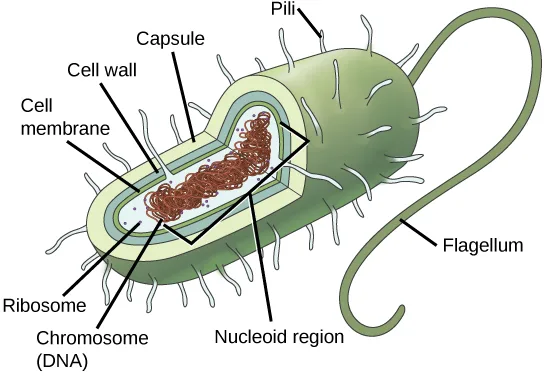
Extended description
A labeled illustration of a rod-shaped prokaryotic cell with a wedge cut away to show its interior. Short, hair-like pili cover the entire cell surface, and one long, looping flagellum extends from one end. The outer boundary is labeled, from outside in, as the capsule, the cell wall, and the cell membrane. Inside the cytoplasm, small dots are labeled ribosomes, and a tangled reddish-brown coil occupying the cell’s center is labeled chromosome (DNA), sitting within the region labeled the nucleoid region.
In eukaryotes, the genome consists of several double-stranded linear DNA molecules. Each species of eukaryotes has a characteristic number of chromosomes in the nuclei of its cells. Human body (somatic) cells have 46 chromosomes, while human gametes (sperm or eggs) have 23 chromosomes each. A typical body cell contains two matched or homologous sets of chromosomes (one set from each biological parent)—a configuration known as diploid. (Note: The letter n is used to represent a single set of chromosomes; therefore, a diploid organism is designated 2n.) Human cells that contain one set of chromosomes are called gametes, or sex cells; these are eggs and sperm, and are designated 1n, or haploid.
Upon fertilization, each gamete contributes one set of chromosomes, creating a diploid cell containing matched pairs of chromosomes called homologous (“same knowledge”) chromosomes. Homologous chromosomes are the same length and have specific nucleotide segments called genes in exactly the same location, or locus. Genes, the functional units of chromosomes, determine specific characteristics by coding for specific proteins. Traits are the variations of those characteristics. For example, hair color is a characteristic with traits that are blonde, brown, or black, and many colors in between.
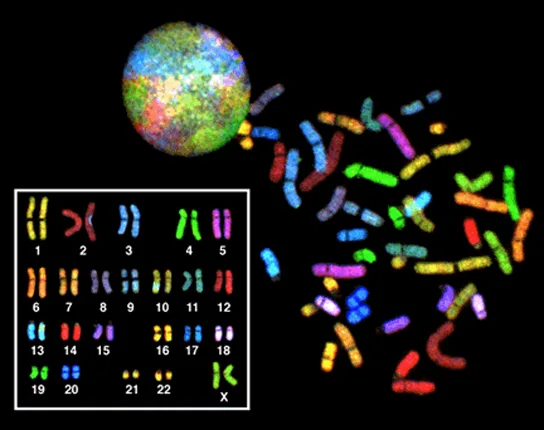
Extended description
A composite of three related images on a black background. Top left, a multicolored sphere is the interphase nucleus, its surface a patchwork of the many different colors that stain each chromosome’s territory. To its right, a loose scattering of thick, condensed chromosome bars in many distinct colors are the same chromosomes after the cell has entered mitosis and each one has coiled into a compact rod. Bottom left, a white-bordered inset box arranges the same chromosomes into 23 numbered and lettered groups—1 through 22, then X—each group holding a matched pair of similarly shaped, similarly colored bars, ordered from longest to shortest; this arranged grid is the karyotype.
Each copy of a homologous pair of chromosomes originates from a different parent; therefore, the different genes (alleles) themselves are not identical, although they code for the same traits such as “hair color.” The variation of individuals within a species is due to the specific combination of the genes inherited from both parents. Even a slightly altered sequence of nucleotides within a gene can result in an alternative trait. For example, there are three possible gene sequences on the human chromosome that code for blood type: sequence A, sequence B, and sequence O. Because all diploid human cells have two copies of the chromosome that determines blood type, the blood type (the trait) is determined by the two alleles of the marker gene that are inherited. It is possible to have two copies of the same gene sequence on both homologous chromosomes, with one on each (for example, AA, BB, or OO), or two different sequences, such as AB, AO, or BO.
Apparently minor variations of traits, such as blood type, eye color, and handedness, contribute to the natural variation found within a species, but even though they seem minor, these traits may be connected with the expression of other traits as of yet unknown. However, if the entire DNA sequence from any pair of human homologous chromosomes is compared, the difference is much less than one percent. The sex chromosomes, X and Y, are the single exception to the rule of homologous chromosome uniformity: Other than a small amount of homology that is necessary to accurately produce gametes, the genes found on the X and Y chromosomes are different.
Eukaryotic Chromosomal Structure and Compaction
If the DNA from all 46 chromosomes in a human cell nucleus were laid out end-to-end, it would measure approximately two meters; however, its diameter would be only 2 nm! Considering that the size of a typical human cell is about 10 µm (100,000 cells lined up to equal one meter), DNA must be tightly packaged to fit in the cell’s nucleus. At the same time, it must also be readily accessible for the genes to be expressed. For this reason, the long strands of DNA are condensed into compact chromosomes during certain stages of the cell cycle. There are a number of ways that chromosomes are compacted.
In the first level of compaction, short stretches of the DNA double helix wrap around a core of eight histone proteins at regular intervals along the entire length of the chromosome. The DNA-histone complex is called chromatin. The beadlike, histone DNA complex is called a nucleosome, and DNA connecting the nucleosomes is called linker DNA. A DNA molecule in this form is about seven times shorter than the double helix without the histones, and the beads are about 10 nm in diameter, in contrast with the 2-nm diameter of a DNA double helix.
The second level of compaction occurs as the nucleosomes and the linker DNA between them coil into a 30-nm chromatin fiber. This coiling further condenses the chromosome so that it is now about 50 times shorter than the extended form.
In the third level of compaction, a variety of fibrous proteins is used to “pack the chromatin.” These fibrous proteins also ensure that each chromosome in a non-dividing cell occupies a particular area of the nucleus that does not overlap with that of any other chromosome (see the top image above).
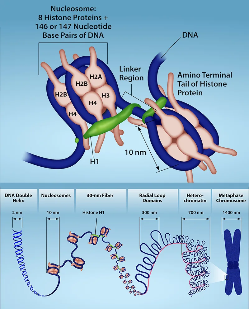
Extended description
Top panel: a nucleosome core of eight histone proteins, two each of H2A, H2B, H3, and H4, with a separate linker histone labeled H1 in the linker region, wrapped by a strand of DNA carrying 146 or 147 nucleotide base pairs; a green linker region connects the amino-terminal tail of one histone protein toward the next nucleosome, and the entering and exiting DNA strands are each labeled DNA, with the nucleosome’s 10-nm diameter marked by an arrow. Bottom panel: six labeled stages of DNA packing in a row, each drawn beneath its width in nanometers—the DNA double helix at 2 nm, nucleosomes strung like beads at 10 nm, a 30-nm fiber coiled with the help of histone H1, radial loop domains at 300 nm, heterochromatin at 700 nm, and the fully condensed metaphase chromosome at 1400 nm.
DNA replicates in the S phase of interphase, which technically is not a part of mitosis, but must always precede it. After replication, the chromosomes are composed of two linked sister chromatids. When fully compact, the pairs of identically packed chromosomes are bound to each other by cohesin proteins. The connection between the sister chromatids is closest in a region called the centromere. The conjoined sister chromatids, with a diameter of about 1 µm, are visible under a light microscope. The centromeric region is highly condensed and thus will appear as a constricted area.
Summary
Prokaryotes have a single circular chromosome composed of double-stranded DNA, whereas eukaryotes have multiple, linear chromosomes composed of chromatin wrapped around histones, all of which are surrounded by a nuclear membrane. The 46 chromosomes of human somatic cells are composed of 22 pairs of autosomes (matched pairs) and a pair of sex chromosomes, which may or may not be matched. This is the 2n or diploid state. Human gametes have 23 chromosomes, or one complete set of chromosomes; a set of chromosomes is complete with either one of the sex chromosomes, X or Y. This is the n or haploid state. Genes are segments of DNA that code for a specific functional molecule (a protein or RNA). An organism’s traits are determined by the genes inherited from each parent. Duplicated chromosomes are composed of two sister chromatids. Chromosomes are compacted using a variety of mechanisms during certain stages of the cell cycle. Several classes of protein are involved in the organization and packing of the chromosomal DNA into a highly condensed structure. The condensing complex compacts chromosomes, and the resulting condensed structure is necessary for chromosomal segregation during mitosis.
Key terms
- centromere — region at which sister chromatids are bound together; a constricted area in condensed chromosomes
- chromatid — single DNA molecule of two strands of duplicated DNA and associated proteins held together at the centromere
- diploid — cell, nucleus, or organism containing two sets of chromosomes (2n)
- gamete — haploid reproductive cell or sex cell (sperm, pollen grain, or egg)
- gene — physical and functional unit of heredity, a sequence of DNA that codes for a protein.
- genome — total genetic information of a cell or organism
- haploid — cell, nucleus, or organism containing one set of chromosomes (n)
- histone — one of several similar, highly conserved, low molecular weight, basic proteins found in the chromatin of all eukaryotic cells; associates with DNA to form nucleosomes
- homologous chromosomes — chromosomes of the same morphology with genes in the same location; diploid organisms have pairs of homologous chromosomes (homologs), with each homolog derived from a different parent
- locus — position of a gene on a chromosome
- nucleosome — subunit of chromatin composed of a short length of DNA wrapped around a core of histone proteins
Practice
Describe the structure of prokaryotic and eukaryotic genomes
A diploid cell has ________ the number of chromosomes as a haploid cell.
A diploid cell carries two matched sets of chromosomes, while a haploid cell carries only one — compare the section’s own numbers for human somatic cells (46) and gametes (23).The total genetic information of a cell or organism is called its ________.
In prokaryotes it is a single circular DNA molecule; in eukaryotes it is several linear ones.Compare and contrast a human somatic cell to a human gamete.
Show model answer
Did your answer mention:
Distinguish between chromosomes, genes, and traits
An organism’s traits are determined by the specific combination of inherited ________.
Traits like hair color trace back to specific variants of these hereditary units, not directly to the cells or proteins they help build.A physical and functional unit of heredity — a sequence of DNA that codes for a protein — is called a ________.
Homologous chromosomes carry these in exactly the same location, or locus.What is the relationship between a genome, chromosomes, and genes?
Show model answer
Did your answer mention:
Describe the mechanisms of chromosome compaction
The first level of DNA organization in a eukaryotic cell is maintained by which molecule?
Short stretches of DNA wrap around a core of eight of these proteins to form the beadlike nucleosome.Identical copies of chromatin held together by cohesin at the centromere are called ________.
These identical copies form after DNA replication in the S phase and stay joined until they separate during mitosis.The beadlike subunit of chromatin, formed by DNA wrapped around a core of histone proteins, is called a ________.
Eight histone proteins form the core that a short stretch of DNA wraps around to form this structure.Chromosome compaction happens during certain stages of the ________.
The orderly sequence of a cell’s life from one division to the next; compaction peaks just before mitosis.Eukaryotic chromosomes are thousands of times longer than a typical cell. Explain how chromosomes can fit inside a eukaryotic nucleus.
Show model answer
Did your answer mention:
This section is adapted from Biology 2e, Section 10.1: Cell Division by Mary Ann Clark, Jung Choi, Matthew Douglas, and OpenStax, © OpenStax, licensed under CC BY-NC-SA 4.0. Access the original for free at openstax.org. Changes: figures re-encoded as WebP, with two of the three re-kinded from the manifest’s file-extension guess of “photo” to “diagram” after inspection (the prokaryotic-cell illustration and the nucleosome/compaction diagram are both drawings; only the karyotype composite is a true photograph); a longdesc added for all three figures, which are labeled diagrams or a composite whose full reading is not carried by their captions alone; the karyotype figure’s alt shortened from the source’s process-focused walkthrough to what is visible, with that detail moved into the longdesc; the nucleosome/compaction figure’s alt rewritten to remove the source’s letter-spaced “D N A” artifact and shortened, with its full labeled walkthrough moved into the longdesc; the interactive note rendered as a Link to Learning callout with descriptive link text, keeping the source’s URL; Review Question 1’s keyed answer corrected from “half” to “twice” and reported as a source defect (module m66477, exercise fs-id1414909): a diploid cell has twice, not half, the chromosome number of a haploid cell, per the section’s own worked numbers (46 vs. 23 chromosomes); the end-of-section Review Questions and Critical Thinking Questions adapted into the closing interactive Practice block (multiple choice and self-check respectively); and three key-term recall items (genome, gene, nucleosome) added from the glossary to round out every objective group with a second auto-graded item; rubric checkpoints added to each self-check, decomposing its model answer (the source solution) into check-off clauses with no new claims; and one summary-derived recall item added under the compaction objective from the section summary’s own sentence.