Control of the Cell Cycle
By the end of this section, you will be able to:
- Understand how the cell cycle is controlled by mechanisms that are both internal and external to the cell
- Explain how the three internal “control checkpoints” occur at the end of G₁, at the G₂/M transition, and during metaphase
- Describe the molecules that control the cell cycle through positive and negative regulation
The length of the cell cycle is highly variable, even within the cells of a single organism. In humans, the frequency of cell turnover ranges from a few hours in early embryonic development, to an average of two to five days for epithelial cells, and to an entire human lifetime spent in G₀ by specialized cells, such as cortical neurons or cardiac muscle cells.
There is also variation in the time that a cell spends in each phase of the cell cycle. When rapidly dividing mammalian cells are grown in a culture (outside the body under optimal growing conditions), the length of the cell cycle is about 24 hours. In rapidly dividing human cells with a 24-hour cell cycle, the G₁ phase lasts approximately nine hours, the S phase lasts 10 hours, the G₂ phase lasts about four and one-half hours, and the M phase lasts approximately one-half hour. By comparison, in fertilized eggs (and early embryos) of fruit flies, the cell cycle is completed in about eight minutes. This is because the nucleus of the fertilized egg divides many times by mitosis but does not go through cytokinesis until a multinucleate “zygote” has been produced, with many nuclei located along the periphery of the cell membrane, thereby shortening the time of the cell division cycle. The timing of events in the cell cycle of both “invertebrates” and “vertebrates” is controlled by mechanisms that are both internal and external to the cell.
Regulation of the Cell Cycle by External Events
Both the initiation and inhibition of cell division are triggered by events external to the cell when it is about to begin the replication process. An event may be as simple as the death of nearby cells or as sweeping as the release of growth-promoting hormones, such as human growth hormone (HGH or hGH). A lack of HGH can inhibit cell division, resulting in dwarfism, whereas too much HGH can result in gigantism. Crowding of cells can also inhibit cell division. In contrast, a factor that can initiate cell division is the size of the cell: As a cell grows, it becomes physiologically inefficient due to its decreasing surface-to-volume ratio. The solution to this problem is to divide.
Whatever the source of the message, the cell receives the signal, and a series of events within the cell allows it to proceed into interphase. Moving forward from this initiation point, every parameter required during each cell cycle phase must be met or the cycle cannot progress.
Regulation at Internal Checkpoints
It is essential that the daughter cells produced be exact duplicates of the parent cell. Mistakes in the duplication or distribution of the chromosomes lead to mutations that may be passed forward to every new cell produced from an abnormal cell. To prevent a compromised cell from continuing to divide, there are internal control mechanisms that operate at three main cell-cycle checkpoints: A checkpoint is one of several points in the eukaryotic cell cycle at which the progression of a cell to the next stage in the cycle can be halted until conditions are favorable. These checkpoints occur near the end of G₁, at the G₂/M transition, and during metaphase (below).
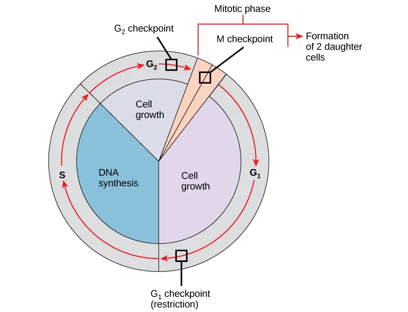
Extended description
Reading clockwise from the bottom: a checkpoint box labeled ‘G₁ checkpoint (restriction)’ sits between the G₁ and S segments; the cell then moves through S (DNA synthesis) to a checkpoint box labeled ‘G₂ checkpoint’ between G₂ and the mitotic phase; a checkpoint box labeled ‘M checkpoint’ sits at the start of the wedge-shaped mitotic phase, which is bracketed at the top with the label ‘Mitotic phase’; an arrow leaving the wedge is labeled ‘Formation of 2 daughter cells.’
The G₁ Checkpoint
The G₁ checkpoint determines whether all conditions are favorable for cell division to proceed. The G₁ checkpoint, also called the restriction point (in yeast), is a point at which the cell irreversibly commits to the cell division process. External influences, such as growth factors, play a large role in carrying the cell past the G₁ checkpoint. In addition to adequate reserves and cell size, there is a check for genomic DNA damage at the G₁ checkpoint. A cell that does not meet all the requirements will not be allowed to progress into the S phase. The cell can halt the cycle and attempt to remedy the problematic condition, or the cell can advance into G₀ and await further signals when conditions improve.
The G₂ Checkpoint
The G₂ checkpoint bars entry into the mitotic phase if certain conditions are not met. As at the G₁ checkpoint, cell size and protein reserves are assessed. However, the most important role of the G₂ checkpoint is to ensure that all of the chromosomes have been replicated and that the replicated DNA is not damaged. If the checkpoint mechanisms detect problems with the DNA, the cell cycle is halted, and the cell attempts to either complete DNA replication or repair the damaged DNA.
The M Checkpoint
The M checkpoint occurs near the end of the metaphase stage of karyokinesis. The M checkpoint is also known as the spindle checkpoint, because it determines whether all the sister chromatids are correctly attached to the spindle microtubules. Because the separation of the sister chromatids during anaphase is an irreversible step, the cycle will not proceed until the kinetochores of each pair of sister chromatids are firmly anchored to at least two spindle fibers arising from opposite poles of the cell.
Link to Learning
Watch what occurs at the G₁, G₂, and M checkpoints by visiting this animation of the cell cycle.
Regulator Molecules of the Cell Cycle
In addition to the internally controlled checkpoints, there are two groups of intracellular molecules that regulate the cell cycle. These regulatory molecules either promote progress of the cell to the next phase (positive regulation) or halt the cycle (negative regulation). Regulator molecules may act individually, or they can influence the activity or production of other regulatory proteins. Therefore, the failure of a single regulator may have almost no effect on the cell cycle, especially if more than one mechanism controls the same event. However, the effect of a deficient or non-functioning regulator can be wide-ranging and possibly fatal to the cell if multiple processes are affected.
Positive Regulation of the Cell Cycle
Two groups of proteins, called cyclins and cyclin-dependent kinases (Cdks), are termed positive regulators. They are responsible for the progress of the cell through the various checkpoints. The levels of the four cyclin proteins fluctuate throughout the cell cycle in a predictable pattern (below). Increases in the concentration of cyclin proteins are triggered by both external and internal signals. After the cell moves to the next stage of the cell cycle, the cyclins that were active in the previous stage are degraded by cytoplasmic enzymes, as shown below.
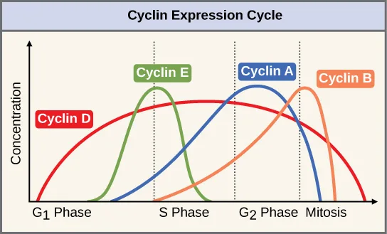
Extended description
Four overlapping curves, left to right across the phase axis. Cyclin D rises from the start of G₁ to a broad peak spanning G₁ and S phase, then declines gradually through G₂ and mitosis. Cyclin E rises during G₁ and peaks near the G₁/S boundary, falling away during S phase. Cyclin A rises during S phase and peaks in G₂, falling during mitosis. Cyclin B rises during G₂, peaks at the start of mitosis, and falls sharply before mitosis ends.
Cyclins regulate the cell cycle only when they are tightly bound to Cdks. To be fully active, the Cdk/cyclin complex must also be phosphorylated in specific locations to activate the complex. Like all kinases, Cdks are enzymes (kinases) that in turn phosphorylate other proteins. Phosphorylation activates the protein by changing its shape. The proteins phosphorylated by Cdks are involved in advancing the cell to the next phase (below). The levels of Cdk proteins are relatively stable throughout the cell cycle; however, the concentrations of cyclin fluctuate and determine when Cdk/cyclin complexes form. The different cyclins and Cdks bind at specific points in the cell cycle and thus regulate different checkpoints.
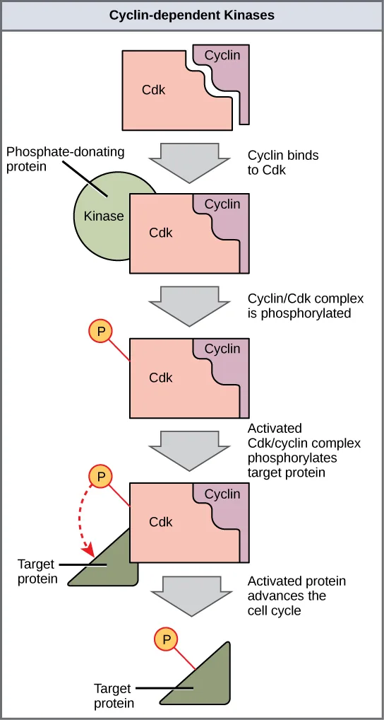
Extended description
Top to bottom: a rectangular Cdk shape and a smaller cyclin shape sit apart, with an arrow labeled ‘Cyclin binds to Cdk’ leading to them joined into one Cdk/cyclin complex. A green circle labeled ‘Kinase’ (the phosphate-donating protein) touches the complex, with an arrow labeled ‘Cyclin/Cdk complex is phosphorylated’ leading to the complex now carrying a yellow phosphate (P) marker. A further arrow labeled ‘Activated Cdk/cyclin complex phosphorylates target protein’ shows the phosphate moving by a dashed line from the complex to a dark triangular target protein. A final arrow labeled ‘Activated protein advances the cell cycle’ leads to the target protein, shown alone, now carrying its own phosphate marker.
Because the cyclic fluctuations of cyclin levels are largely based on the timing of the cell cycle and not on specific events, regulation of the cell cycle usually occurs by either the Cdk molecules alone or the Cdk/cyclin complexes. Without a specific concentration of fully activated cyclin/Cdk complexes, the cell cycle cannot proceed through the checkpoints.
Although the cyclins are the main regulatory molecules that determine the forward momentum of the cell cycle, there are several other mechanisms that fine-tune the progress of the cycle with negative, rather than positive, effects. These mechanisms essentially block the progression of the cell cycle until problematic conditions are resolved. Molecules that prevent the full activation of Cdks are called Cdk inhibitors. Many of these inhibitor molecules directly or indirectly monitor a particular cell-cycle event. The block placed on Cdks by inhibitor molecules will not be removed until the specific event that the inhibitor monitors is completed.
Negative Regulation of the Cell Cycle
The second group of cell-cycle regulatory molecules are negative regulators, which stop the cell cycle. Remember that in positive regulation, active molecules cause the cycle to progress.
The best understood negative regulatory molecules are retinoblastoma protein (Rb), p53, and p21. Retinoblastoma proteins are a group of tumor-suppressor proteins common in many cells. We should note here that the 53 and 21 designations refer to the functional molecular masses of the proteins (p) in kilodaltons (a dalton is equal to an atomic mass unit, which is equal to one proton or one neutron or 1 g/mol). Much of what is known about cell-cycle regulation comes from research conducted with cells that have lost regulatory control. All three of these regulatory proteins were discovered to be damaged or non-functional in cells that had begun to replicate uncontrollably (i.e., became cancerous). In each case, the main cause of the unchecked progress through the cell cycle was a faulty copy of the regulatory protein.
Rb, p53, and p21 act primarily at the G₁ checkpoint. p53 is a multi-functional protein that has a major impact on the commitment of a cell to division because it acts when there is damaged DNA in cells that are undergoing the preparatory processes during G₁. If damaged DNA is detected, p53 halts the cell cycle and then recruits specific enzymes to repair the DNA. If the DNA cannot be repaired, p53 can trigger apoptosis, or cell suicide, to prevent the duplication of damaged chromosomes. As p53 levels rise, the production of p21 is triggered. p21 enforces the halt in the cycle dictated by p53 by binding to and inhibiting the activity of the Cdk/cyclin complexes. As a cell is exposed to more stress, higher levels of p53 and p21 accumulate, making it less likely that the cell will move into the S phase.
Rb, which largely monitors cell size, exerts its regulatory influence on other positive regulator proteins. In the active, dephosphorylated state, Rb binds to proteins called transcription factors, most commonly, E2F (below). Transcription factors “turn on” specific genes, allowing the production of proteins encoded by that gene. When Rb is bound to E2F, production of proteins necessary for the G₁/S transition is blocked. As the cell increases in size, Rb is slowly phosphorylated until it becomes inactivated. Rb releases E2F, which can now turn on the gene that produces the transition protein, and this particular block is removed. For the cell to move past each of the checkpoints, all positive regulators must be “turned on,” and all negative regulators must be “turned off.”
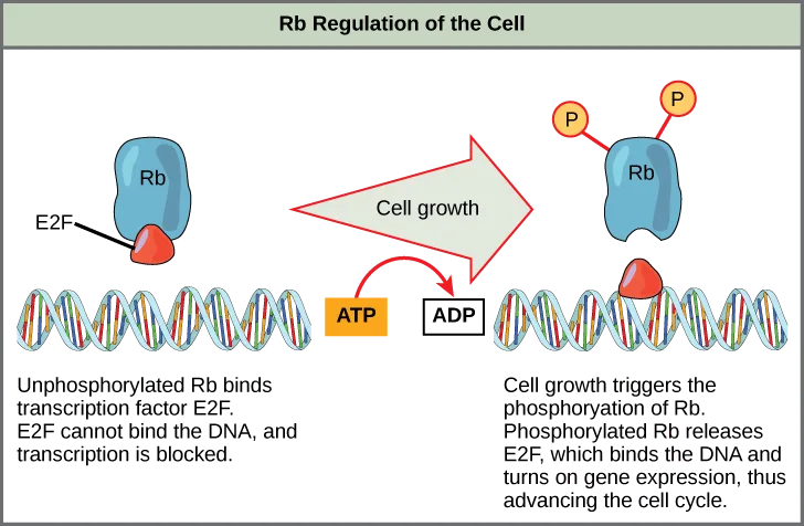
Extended description
Left panel: a blue Rb protein shape binds a red E2F shape above a DNA double helix; captioned text reads ‘Unphosphorylated Rb binds transcription factor E2F. E2F cannot bind the DNA, and transcription is blocked.’ A wide arrow labeled ‘Cell growth,’ drawn beside an ATP box turning into an ADP box, points from the left panel to the right panel. Right panel: the same Rb shape now carries two yellow phosphate (P) markers and no longer touches E2F, which has moved down to bind the DNA strand; captioned text reads ‘Cell growth triggers the phosphorylation of Rb. Phosphorylated Rb releases E2F, which binds the DNA and turns on gene expression, thus advancing the cell cycle.’
Rb and other proteins that negatively regulate the cell cycle are sometimes called tumor suppressors. Why do you think the name tumor suppressor might be appropriate for these proteins?
Show model answer
Did your answer mention:
Summary
Each step of the cell cycle is monitored by internal controls called checkpoints. There are three major checkpoints in the cell cycle: one near the end of G₁, a second at the G₂/M transition, and the third during metaphase. Positive regulator molecules allow the cell cycle to advance to the next stage of cell division. Negative regulator molecules monitor cellular conditions and can halt the cycle until specific requirements are met.
Key terms
- cell-cycle checkpoint — mechanism that monitors the preparedness of a eukaryotic cell to advance through the various cell-cycle stages
- cyclin — one of a group of proteins that act in conjunction with cyclin-dependent kinases to help regulate the cell cycle by phosphorylating key proteins; the concentrations of cyclins fluctuate throughout the cell cycle
- cyclin-dependent kinase (Cdk) — one of a group of protein kinases that helps to regulate the cell cycle when bound to cyclin; it functions to phosphorylate other proteins that are either activated or inactivated by phosphorylation
- p21 — cell-cycle regulatory protein that inhibits the cell cycle; its levels are controlled by p53
- p53 — cell-cycle regulatory protein that regulates cell growth and monitors DNA damage; it halts the progression of the cell cycle in cases of DNA damage and may induce apoptosis
- retinoblastoma protein (Rb) — regulatory molecule that exhibits negative effects on the cell cycle by interacting with a transcription factor (E2F)
Practice
Understand how the cell cycle is controlled by mechanisms that are both internal and external to the cell
At which of the cell-cycle checkpoints do external forces have the greatest influence?
Think about which checkpoint is most associated with signals like growth factors arriving from outside the cell.A mechanism that monitors a eukaryotic cell’s preparedness to advance through the various cell-cycle stages is called a cell-cycle ________.
Three of these occur in the cell cycle: near the end of G1, at the G2/M transition, and during metaphase.According to the section, a lack of human growth hormone (HGH) can inhibit cell division, resulting in ________.
Too little of the growth-promoting hormone stunts growth; too much produces the opposite condition.Explain how the three internal “control checkpoints” occur at the end of G₁, at the G₂/M transition, and during metaphase
What is the main prerequisite for clearance at the G2 checkpoint?
The G2 checkpoint’s most important role is confirming that every chromosome was correctly copied.If the M checkpoint is not cleared, what stage of mitosis will be blocked?
The M checkpoint confirms that every kinetochore is anchored to spindle fibers before the sister chromatids separate.Describe the general conditions that must be met at each of the three main cell-cycle checkpoints.
Show model answer
Did your answer mention:
Describe the molecules that control the cell cycle through positive and negative regulation
Which protein is a positive regulator that phosphorylates other proteins when activated?
Positive regulators advance the cell cycle; only one of these options is itself an enzyme that adds phosphate groups to other proteins.Many of the negative regulator proteins of the cell cycle were discovered in what type of cells?
These regulators were found to be damaged or non-functional in cells that had begun to replicate uncontrollably.Which negative regulatory molecule can trigger cell suicide (apoptosis) if vital cell cycle events do not occur?
This protein halts the cycle when it detects damaged DNA and can trigger apoptosis if the damage cannot be repaired.Compare and contrast the roles of the positive cell-cycle regulators and negative regulators.
Show model answer
Did your answer mention:
What steps are necessary for Cdk to become fully active?
Show model answer
Did your answer mention:
Rb is a negative regulator that blocks the cell cycle at the G1 checkpoint until the cell achieves a requisite size. What molecular mechanism does Rb employ to halt the cell cycle?
Show model answer
Did your answer mention:
This section is adapted from Biology 2e, Section 10.3: Control of the Cell Cycle by Mary Ann Clark, Jung Choi, Matthew Douglas, and OpenStax, © OpenStax, licensed under CC BY-NC-SA 4.0. Access the original for free at openstax.org. Changes: figures re-encoded as WebP and three of the four re-kinded from the manifest’s file-extension guess of “photo” to “diagram” (each is a labeled illustration or graph, not a photograph; the fourth was already guessed “diagram”), with a longdesc added to each since none of the four captions carries the full reading of its labels, arrows, or curves; the interactive note rendered as a Link to Learning callout with descriptive link text in place of “this website”; the Visual Connection question kept in the body immediately after its figure and rendered as a self-check (with a hint added), since the source keys it with a prose solution rather than a lettered option, and not repeated in the closing Practice block; inline references to the source’s print figure numbers ("()", “below”) changed to “(below)” or “below” since figures are not numbered here; the end-of-section Review Questions and Critical Thinking Questions adapted into the closing interactive Practice block (multiple choice and self-check respectively); and one key-term recall item (cell-cycle checkpoint) added from the glossary to complete the first objective’s group with an auto-graded item; rubric checkpoints added to each self-check (the body Visual Connection included), decomposing its model answer (the source solution) into check-off clauses with no new claims; and one locally written multiple choice (HGH and dwarfism) added under the first objective, built strictly from the section’s own sentence and disclosed in the ledger.