Cancer and the Cell Cycle
By the end of this section, you will be able to:
- Describe how cancer is caused by uncontrolled cell growth
- Understand how proto-oncogenes are normal cell genes that, when mutated, become oncogenes
- Describe how tumor suppressors function
- Explain how mutant tumor suppressors cause cancer
Cancer comprises many different diseases caused by a common mechanism: uncontrolled cell growth. Despite the redundancy and overlapping levels of cell-cycle control, errors do occur. One of the critical processes monitored by the cell-cycle checkpoint surveillance mechanism is the proper replication of DNA during the S phase. Even when all of the cell-cycle controls are fully functional, a small percentage of replication errors (mutations) will be passed on to the daughter cells. If changes to the DNA nucleotide sequence occur within a coding portion of a gene and are not corrected, a gene mutation results. All cancers start when a gene mutation gives rise to a faulty protein that plays a key role in cell reproduction.
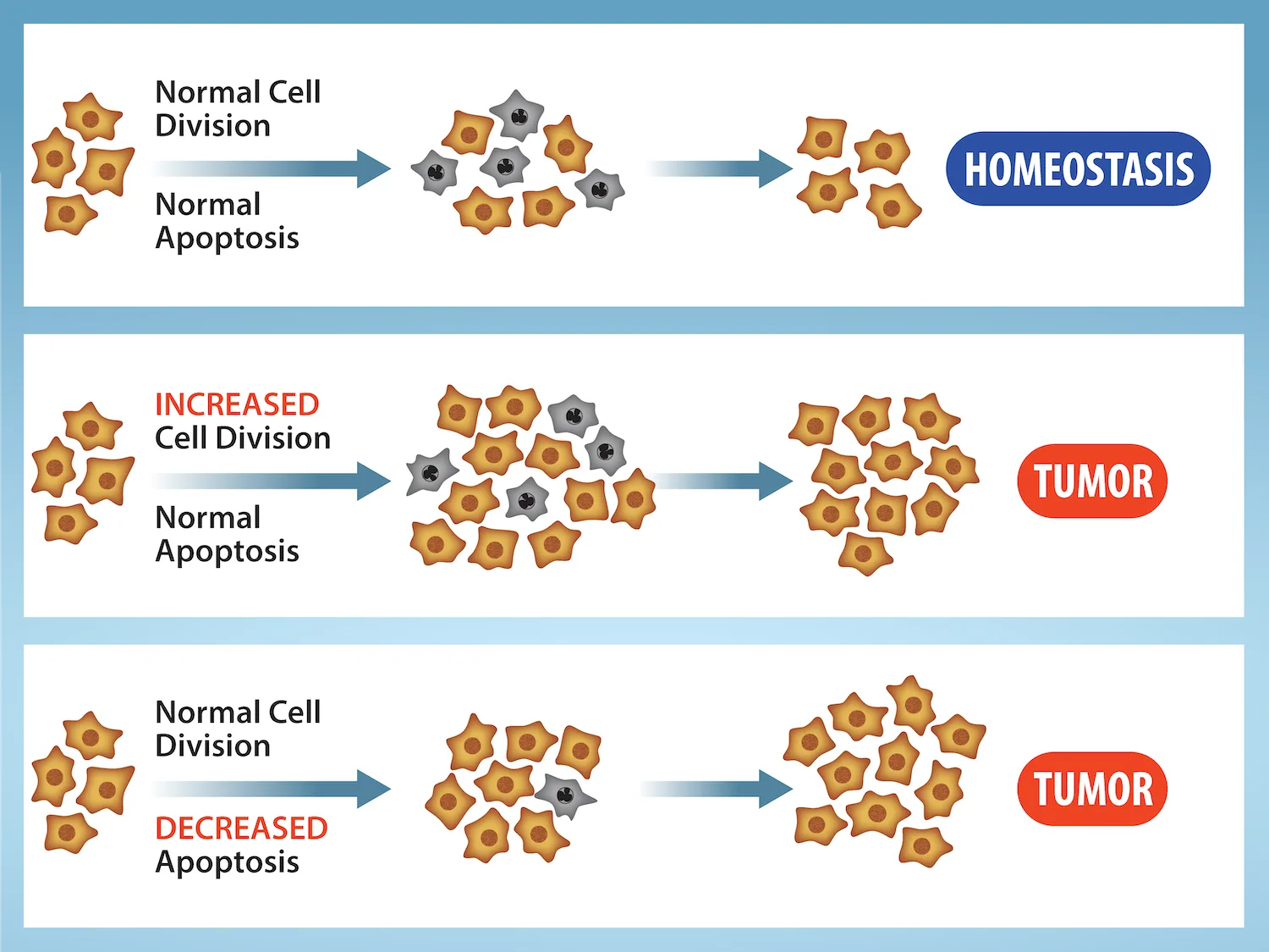
The change in the cell that results from the malformed protein may be minor: perhaps a slight delay in the binding of Cdk to cyclin or an Rb protein that detaches from its target DNA while still phosphorylated. Even minor mistakes, however, may allow subsequent mistakes to occur more readily. Over and over, small uncorrected errors are passed from the parent cell to the daughter cells and amplified as each generation produces more non-functional proteins from uncorrected DNA damage. Eventually, the pace of the cell cycle speeds up as the effectiveness of the control and repair mechanisms decreases. Uncontrolled growth of the mutated cells outpaces the growth of normal cells in the area, and a tumor ("-oma") can result.
Extended description
Three horizontal panels, each showing an arrow from a small starting cluster of orange, cell-shaped icons to a larger middle cluster (with a few grey, star-shaped icons mixed in), then a second arrow to a final cluster beside a rounded label. Top panel: labels ‘Normal Cell Division’ and ‘Normal Apoptosis’ lead to a small, even final cluster and a blue oval reading ‘HOMEOSTASIS.’ Middle panel: ‘INCREASED Cell Division’ (in red) and ‘Normal Apoptosis’ lead to a much larger, denser final cluster and a red oval reading ‘TUMOR.’ Bottom panel: ‘Normal Cell Division’ and ‘DECREASED Apoptosis’ (in red) lead to a large final cluster and a red oval reading ‘TUMOR.’
Proto-oncogenes
The genes that code for the positive cell-cycle regulators are called proto-oncogenes. Proto-oncogenes are normal genes that, when mutated in certain ways, become oncogenes—genes that cause a cell to become cancerous. Consider what might happen to the cell cycle in a cell with a recently acquired oncogene. In most instances, the alteration of the DNA sequence will result in a less functional (or non-functional) protein. The result is detrimental to the cell and will likely prevent the cell from completing the cell cycle; however, the organism is not harmed because the mutation will not be carried forward. If a cell cannot reproduce, the mutation is not propagated and the damage is minimal. Occasionally, however, a gene mutation causes a change that increases the activity of a positive regulator. For example, a mutation that allows Cdk to be activated without being partnered with cyclin could push the cell cycle past a checkpoint before all of the required conditions are met. If the resulting daughter cells are too damaged to undergo further cell divisions, the mutation would not be propagated and no harm would come to the organism. However, if the atypical daughter cells are able to undergo further cell divisions, subsequent generations of cells may accumulate even more mutations, some possibly in additional genes that regulate the cell cycle.
The Cdk gene in the above example is only one of many genes that are considered proto-oncogenes. In addition to the cell-cycle regulatory proteins, any protein that influences the cycle can be altered in such a way as to override cell-cycle checkpoints. An oncogene is any gene that, when altered, leads to an increase in the rate of cell-cycle progression.
Tumor Suppressor Genes
Like proto-oncogenes, many of the negative cell-cycle regulatory proteins were discovered in cells that had become cancerous. Tumor suppressor genes are segments of DNA that code for negative regulator proteins, the type of regulators that, when activated, can prevent the cell from undergoing uncontrolled division. The collective function of the best-understood tumor suppressor gene proteins, Rb, p53, and p21, is to put up a roadblock to cell-cycle progression until certain events are completed. A cell that carries a mutated form of a negative regulator might not be able to halt the cell cycle if there is a problem. Tumor suppressors are similar to brakes in a vehicle: Malfunctioning brakes can contribute to a car crash!
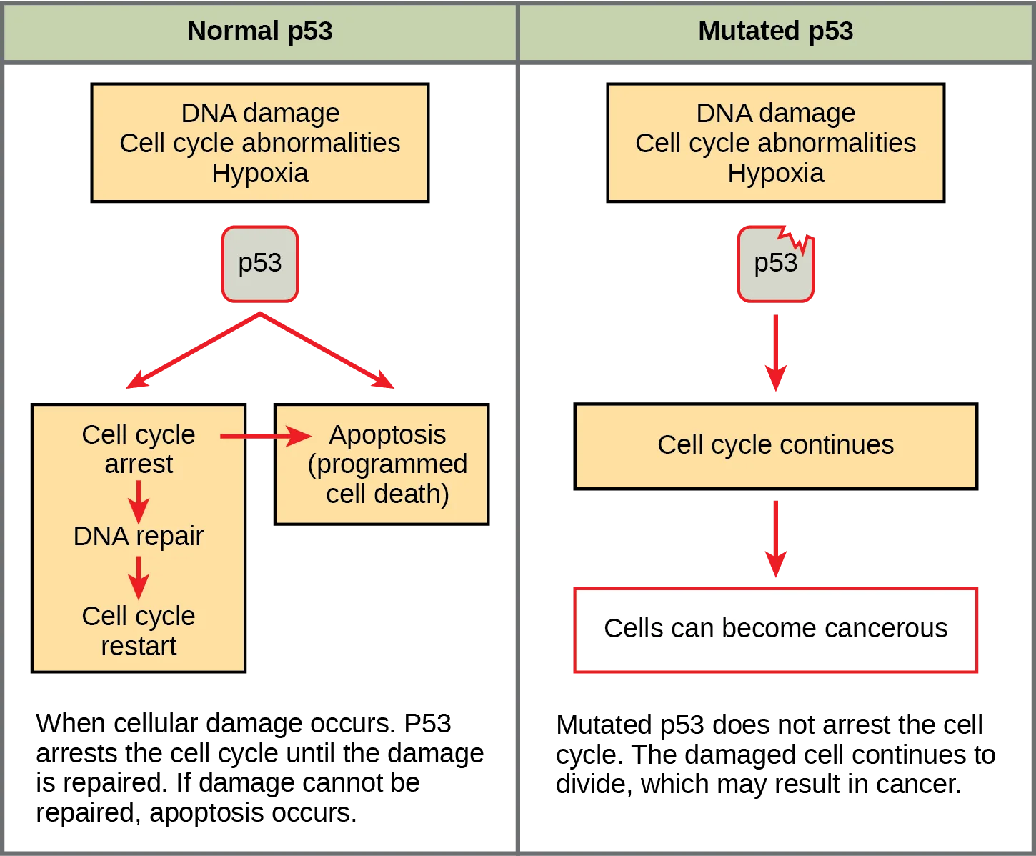
Mutated p53 genes have been identified in more than 50 percent of all human tumor cells. This discovery is not surprising in light of the multiple roles that the p53 protein plays at the G₁ checkpoint. A cell with a faulty p53 may fail to detect errors present in the genomic DNA (see the diagram below). Even if a partially functional p53 does identify the mutations, it may no longer be able to signal the necessary DNA repair enzymes. Either way, damaged DNA will remain uncorrected. At this point, a functional p53 will deem the cell unsalvageable and trigger programmed cell death (apoptosis). The damaged version of p53 found in cancer cells, however, cannot trigger apoptosis.
Extended description
Two side-by-side panels under a green header row reading ‘Normal p53’ and ‘Mutated p53.’ In both panels, an orange box reading ‘DNA damage, Cell cycle abnormalities, Hypoxia’ points down to a grey box labeled ‘p53.’ In the left panel, red arrows branch from ‘p53’ to two orange boxes, ‘Cell cycle arrest’ and ‘Apoptosis (programmed cell death),’ which are joined by a red arrow; ‘Cell cycle arrest’ also points down through ‘DNA repair’ to ‘Cell cycle restart.’ Text below the left panel reads: ‘When cellular damage occurs. P53 arrests the cell cycle until the damage is repaired. If damage cannot be repaired, apoptosis occurs.’ In the right panel, a jagged red stress mark sits on the ‘p53’ box, and a single red arrow leads from it to an orange box, ‘Cell cycle continues,’ which arrows down to a red-outlined box, ‘Cells can become cancerous.’ Text below the right panel reads: ‘Mutated p53 does not arrest the cell cycle. The damaged cell continues to divide, which may result in cancer.’
Human papillomavirus can cause cervical cancer. The virus encodes E6, a protein that binds p53. Based on this fact and what you know about p53, what effect do you think E6 binding has on p53 activity?
A virus that wants to escape p53’s apoptosis signal needs p53 gone entirely, not merely blocked in place.The loss of p53 function has other repercussions for the cell cycle. Mutated p53 might lose its ability to trigger p21 production. Without adequate levels of p21, there is no effective block on Cdk activation. Essentially, without a fully functional p53, the G₁ checkpoint is severely compromised and the cell proceeds directly from G₁ to S regardless of internal and external conditions. At the completion of this shortened cell cycle, two daughter cells are produced that have inherited the mutated p53 gene. Given the non-optimal conditions under which the parent cell reproduced, it is likely that the daughter cells will have acquired other mutations in addition to the faulty tumor-suppressor gene. Cells such as these daughter cells quickly accumulate both oncogenes and non-functional tumor-suppressor genes. Again, the result is tumor growth.
Summary
Cancer is the result of unchecked cell division caused by a breakdown of the mechanisms that regulate the cell cycle. The loss of control begins with a change in the DNA sequence of a gene that codes for one of the regulatory molecules. Faulty instructions lead to a protein that does not function as it should. Any disruption of the monitoring system can allow other mistakes to be passed on to the daughter cells. Each successive cell division will give rise to daughter cells with even more accumulated damage. Eventually, all checkpoints become nonfunctional, and rapidly reproducing cells crowd out normal cells, resulting in a tumor or leukemia (blood cancer).
Key terms
- oncogene — mutated version of a normal gene involved in the positive regulation of the cell cycle
- proto-oncogene — normal gene that when mutated becomes an oncogene
- tumor suppressor gene — segment of DNA that codes for regulator proteins that prevent the cell from undergoing uncontrolled division
Practice
Describe how cancer is caused by uncontrolled cell growth
___________ are changes to the order of nucleotides in a segment of DNA that codes for a protein.
Re-read the section’s opening paragraph on what a change to the DNA nucleotide sequence is called.Outline the steps that lead to a cell becoming cancerous.
Show model answer
Did your answer mention:
Rapidly reproducing cancer cells that crowd out normal blood cells cause the blood cancer called ________.
A cancer of the blood rather than a solid tumor — the summary names it in parentheses.Understand how proto-oncogenes are normal cell genes that, when mutated, become oncogenes
A gene that codes for a positive cell-cycle regulator is called a(n) _____.
This is the normal, unmutated version of the gene.A mutated gene that codes for an altered version of Cdk that is active in the absence of cyclin is a(n) _____.
This is the mutated form that increases the rate of cell-cycle progression.A mutated version of a normal gene involved in the positive regulation of the cell cycle is called a(n) ________.
It is the mutated, cancer-causing form of a normal gene that promotes cell division; its prefix means ’tumor.'A normal gene that becomes an oncogene when it is mutated is called a(n) ________.
It codes for a positive cell-cycle regulator before any mutation occurs.Describe how tumor suppressors function
A segment of DNA that codes for regulator proteins that prevent a cell from undergoing uncontrolled division is called a(n) ________.
Rb, p53, and p21 are the best-understood proteins in this category.Which molecule is a Cdk inhibitor that is controlled by p53?
Mutated p53 loses the ability to trigger production of this molecule.Explain the difference between a proto-oncogene and a tumor-suppressor gene.
Show model answer
Did your answer mention:
Explain how mutant tumor suppressors cause cancer
According to this section, when a cell’s p53 is no longer fully functional and the G₁ checkpoint is severely compromised, which transition does the cell proceed directly through, regardless of internal and external conditions?
Re-read the paragraph describing what happens once p53 can no longer trigger enough p21 to block Cdk.List the regulatory mechanisms that might be lost in a cell producing faulty p53.
Show model answer
Did your answer mention:
p53 can trigger apoptosis if certain cell-cycle events fail. How does this regulatory outcome benefit a multicellular organism?
Show model answer
Did your answer mention:
This section is adapted from Biology 2e, Section 10.4: Cancer and the Cell Cycle by Mary Ann Clark, Jung Choi, Matthew Douglas, and OpenStax, © OpenStax, licensed under CC BY-NC-SA 4.0. Access the original for free at openstax.org. Changes: figures re-encoded as WebP (both kinds matched the manifest’s “diagram” guess after inspection); an extended description added to both figures, since neither flow chart’s arrows, colors, and box labels are fully carried by its caption; the source alt’s letter-by-letter spelling (“lower case p 5 3”) rewritten as “p53”; the G₁-checkpoint cross-reference to a numbered figure changed to a descriptive “see the diagram below” since figures are not numbered here; the two feature boxes (the note wrapping the Visual Connection question, and the interactive animation link) rendered as, respectively, the figure plus a multiple choice kept in the body, and a Link to Learning callout with descriptive link text; the end-of-section Review Questions and Critical Thinking Questions adapted into the closing interactive Practice block (multiple choice and self-check respectively); three key-term recall items (oncogene, proto-oncogene, tumor suppressor gene) added from the glossary; and one additional multiple choice written locally, strictly from the section’s own paragraph on the G₁-to-S transition, since the module’s four keyed Review Questions all map to the first three objectives and left the fourth objective (“Explain how mutant tumor suppressors cause cancer”) without a second auto-graded item; rubric checkpoints added to each self-check, decomposing its model answer (the source solution) into check-off clauses with no new claims; and one summary-derived recall item (leukemia) added under the first objective from the section summary’s own sentence.