Modern Understandings of Inheritance
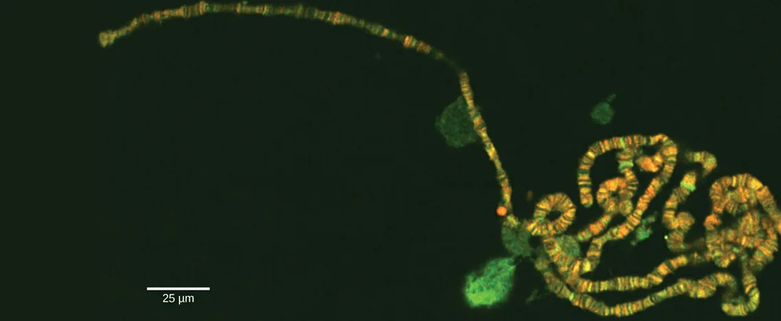
The gene is the physical unit of inheritance, and genes are arranged in a linear order on chromosomes. Chromosome behavior and interaction during meiosis explain, at a cellular level, inheritance patterns that we observe in populations. Genetic disorders involving alterations in chromosome number or structure may have dramatic effects and can prevent a fertilized egg from developing.
Sections
- Chromosomal Theory and Genetic Linkage — Sutton’s Chromosomal Theory of Inheritance, genetic linkage, how homologous recombination (crossing over) works, chromosome mapping, and calculating gene distances with a three-point test cross.
- Chromosomal Basis of Inherited Disorders — how a karyogram is created, how nondisjunction leads to disorders in chromosome number, the disorders aneuploidy causes, and how inversions and translocations produce errors in chromosome structure.
This chapter is adapted from Biology 2e, Chapter 13: Modern Understandings of Inheritance by Mary Ann Clark, Jung Choi, Matthew Douglas, and OpenStax, © OpenStax, licensed under CC BY-NC-SA 4.0. Access the original for free at openstax.org. Each section page records its own changes from the source. Changes: the chapter-opening micrograph is the source’s, re-encoded for the web with its credit kept in the caption; its alt text is rewritten from the image, because the source alt calls the picture an electron micrograph while the image is a color, dye-stained light micrograph and the caption itself says the chromosomes were stained with dye and visualized under a microscope — reported as a source defect.