Historical Basis of Modern Understanding
By the end of this section, you will be able to:
- Explain transformation of DNA
- Describe the key experiments that helped identify that DNA is the genetic material
- State and explain Chargaff’s rules
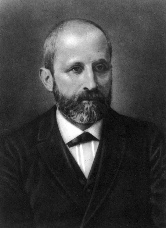
Our current understanding of DNA began with the discovery of nucleic acids followed by the development of the double-helix model. In the 1860s, Friedrich Miescher, a physician by profession, isolated phosphate-rich chemicals from white blood cells (leukocytes). He named these chemicals (which would eventually be known as DNA) nuclein because they were isolated from the nuclei of the cells.
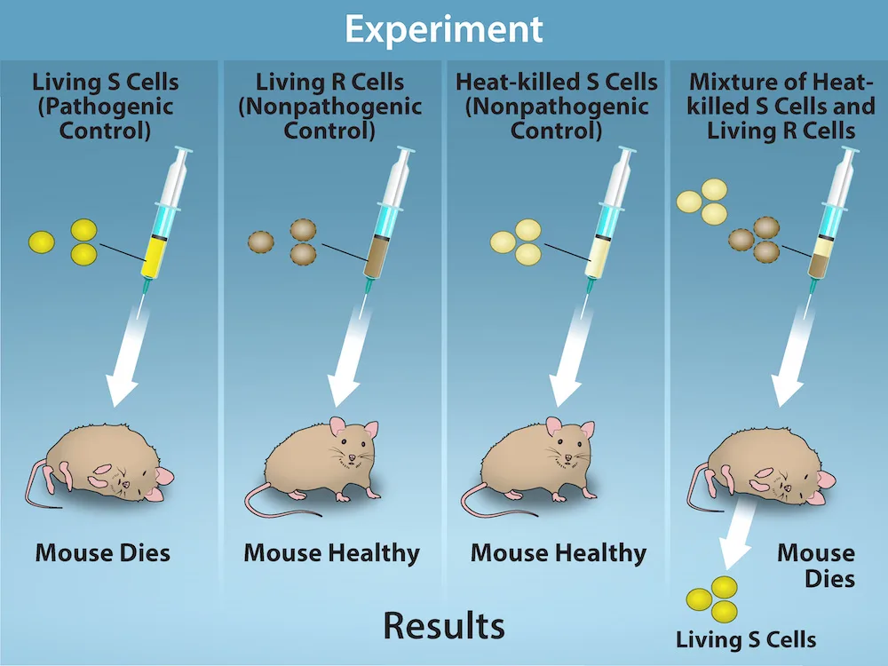
A half century later, in 1928, British bacteriologist Frederick Griffith reported the first demonstration of bacterial transformation—a process in which external DNA is taken up by a cell, thereby changing its morphology and physiology. Griffith conducted his experiments with Streptococcus pneumoniae, a bacterium that causes pneumonia. Griffith worked with two strains of this bacterium called rough (R) and smooth (S). (The two cell types were called “rough” and “smooth” after the appearance of their colonies grown on a nutrient agar plate.)
The R strain is non-pathogenic (does not cause disease). The S strain is pathogenic (disease-causing), and has a capsule outside its cell wall. The capsule allows the cell to escape the immune responses of the host mouse.
When Griffith injected the living S strain into mice, they died from pneumonia. In contrast, when Griffith injected the live R strain into mice, they survived. In another experiment, when he injected mice with the heat-killed S strain, they also survived. This experiment showed that the capsule alone was not the cause of death. In a third set of experiments, a mixture of live R strain and heat-killed S strain were injected into mice, and—to his surprise—the mice died. Upon isolating the live bacteria from the dead mouse, only the S strain of bacteria was recovered. When this isolated S strain was injected into fresh mice, the mice died. Griffith concluded that something had passed from the heat-killed S strain into the live R strain and transformed it into the pathogenic S strain. He called this the transforming principle. These experiments are now known as Griffith’s transformation experiments.
Extended description
A four-column diagram titled “Experiment.” Column 1, “Living S Cells (Pathogenic Control)”: three yellow dots drawn into a syringe and injected into a mouse; the mouse dies. Column 2, “Living R Cells (Nonpathogenic Control)”: three brown dots injected into a mouse; the mouse stays healthy. Column 3, “Heat-killed S Cells (Nonpathogenic Control)”: three pale yellow dots injected into a mouse; the mouse stays healthy. Column 4, “Mixture of Heat-killed S Cells and Living R Cells”: yellow and brown dots injected together into a mouse; the mouse dies, and an arrow points down to three yellow dots labeled “Living S Cells,” recovered from it.
Scientists Oswald Avery, Colin MacLeod, and Maclyn McCarty (1944) were interested in exploring this transforming principle further. They isolated the S strain from the dead mice and isolated the proteins and nucleic acids (RNA and DNA) as these were possible candidates for the molecule of heredity. They used enzymes that specifically degraded each component and then used each mixture separately to transform the R strain. They found that when DNA was degraded, the resulting mixture was no longer able to transform the bacteria, whereas all of the other combinations were able to transform the bacteria. This led them to conclude that DNA was the transforming principle.
Career Connection. Forensic Scientist.
Forensic Scientists used DNA analysis evidence for the first time to solve an immigration case. The story started with a teenage boy returning to London from Ghana to be with his mother. Immigration authorities at the airport were suspicious of him, thinking that he was traveling on a forged passport. After much persuasion, he was allowed to go live with his mother, but the immigration authorities did not drop the case against him. All types of evidence, including photographs, were provided to the authorities, but deportation proceedings were started nevertheless. Around the same time, Dr. Alec Jeffreys of Leicester University in the United Kingdom had invented a technique known as DNA fingerprinting. The immigration authorities approached Dr. Jeffreys for help. He took DNA samples from the mother and three of her children, as well as an unrelated mother, and compared the samples with the boy’s DNA. Because the biological father was not in the picture, DNA from the three children was compared with the boy’s DNA. He found a match in the boy’s DNA for both the mother and his three siblings. He concluded that the boy was indeed the mother’s son.
Forensic scientists analyze many items, including documents, handwriting, firearms, and biological samples. They analyze the DNA content of hair, semen, saliva, and blood, and compare it with a database of DNA profiles of known criminals. Analysis includes DNA isolation, sequencing, and sequence analysis. Forensic scientists are expected to appear at court hearings to present their findings. They are usually employed in crime labs of city and state government agencies. Geneticists experimenting with DNA techniques also work for scientific and research organizations, pharmaceutical industries, and college and university labs. Students wishing to pursue a career as a forensic scientist should have at least a bachelor’s degree in chemistry, biology, or physics, and preferably some experience working in a laboratory.
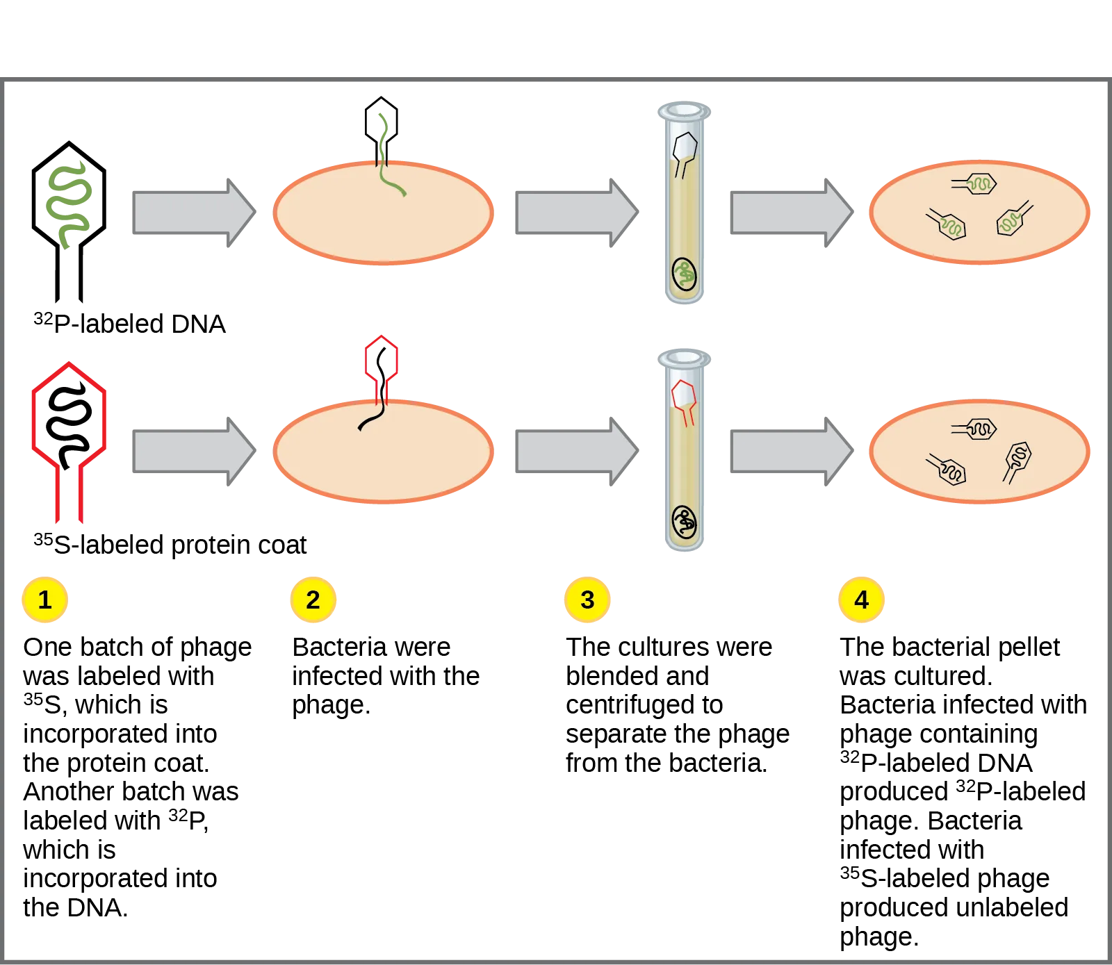
Although the experiments of Avery, McCarty and McLeod had demonstrated that DNA was the informational component transferred during transformation, DNA was still considered to be too simple a molecule to carry biological information. Proteins, with their 20 different amino acids, were regarded as more likely candidates. The decisive experiment, conducted by Martha Chase and Alfred Hershey in 1952, provided confirmatory evidence that DNA was indeed the genetic material and not proteins. Chase and Hershey were studying a bacteriophage—a virus that infects bacteria. Viruses typically have a simple structure: a protein coat, called the capsid, and a nucleic acid core that contains the genetic material (either DNA or RNA). The bacteriophage infects the host bacterial cell by attaching to its surface, and then it injects its nucleic acids inside the cell. The phage DNA makes multiple copies of itself using the host machinery, and eventually the host cell bursts, releasing a large number of bacteriophages. Hershey and Chase selected radioactive elements that would specifically distinguish the protein from the DNA in infected cells. They labeled one batch of phage with radioactive sulfur, ³⁵S, to label the protein coat. Another batch of phage were labeled with radioactive phosphorus, ³²P. Because phosphorous is found in DNA, but not protein, the DNA and not the protein would be tagged with radioactive phosphorus. Likewise, sulfur is absent from DNA, but present in several amino acids such as methionine and cysteine.
Each batch of phage was allowed to infect the cells separately. After infection, the phage bacterial suspension was put in a blender, which caused the phage coat to detach from the host cell. Cells exposed long enough for infection to occur were then examined to see which of the two radioactive molecules had entered the cell. The phage and bacterial suspension was spun down in a centrifuge. The heavier bacterial cells settled down and formed a pellet, whereas the lighter phage particles stayed in the supernatant. In the tube that contained phage labeled with ³⁵S, the supernatant contained the radioactively labeled phage, whereas no radioactivity was detected in the pellet. In the tube that contained the phage labeled with ³²P, the radioactivity was detected in the pellet that contained the heavier bacterial cells, and no radioactivity was detected in the supernatant. Hershey and Chase concluded that it was the phage DNA that was injected into the cell and carried information to produce more phage particles, thus providing evidence that DNA was the genetic material and not proteins.
Extended description
Two rows of icons run left to right through four numbered steps. Step 1: an outlined phage icon with a green coiled line inside is labeled “³²P-labeled DNA” above a second phage icon outlined in red with a black coiled line inside, labeled “³⁵S-labeled protein coat.” Step 2, “Bacteria were infected with the phage”: each phage’s tail contacts an oval bacterial cell, and only the coiled line — green in the top row, black in the bottom row — passes inside it. Step 3, “The cultures were blended and centrifuged to separate the phage from the bacteria”: a test tube for each row shows an empty phage shell floating above a pellet at the bottom holding the infected bacterial cell. Step 4, “The bacterial pellet was cultured”: the top row’s bacterial cell yields three new phage particles each again drawn with the green coil, labeled as ³²P-labeled progeny; the bottom row’s bacterial cell yields three new phage particles drawn without color, labeled as unlabeled phage.
Around this same time, Austrian biochemist Erwin Chargaff examined the content of DNA in different species and found that the amounts of adenine, thymine, guanine, and cytosine were not found in equal quantities, and that relative concentrations of the four nucleotide bases varied from species to species, but not within tissues of the same individual or between individuals of the same species. He also discovered something unexpected: That the amount of adenine equaled the amount of thymine, and the amount of cytosine equaled the amount of guanine (that is, A = T and G = C). Different species had equal amounts of purines (A+G) and pyrimidines (T + C), but different ratios of A+T to G+C. These observations became known as Chargaff’s rules. Chargaff’s findings proved immensely useful when Watson and Crick were getting ready to propose their DNA double helix model! You can see after reading the past few pages how science builds upon previous discoveries, sometimes in a slow and laborious process.
Summary
DNA was first isolated from white blood cells by Friedrich Miescher, who called it nuclein because it was isolated from nuclei. Frederick Griffith’s experiments with strains of Streptococcus pneumoniae provided the first hint that DNA may be the transforming principle. Avery, MacLeod, and McCarty showed that DNA is required for the transformation of bacteria. Later experiments by Hershey and Chase using bacteriophage T2 proved that DNA is the genetic material. Chargaff found that the ratio of A = T and C = G, and that the percentage content of A, T, G, and C is different for different species.
Key terms
- transformation — process in which external DNA is taken up by a cell
Practice
Explain transformation of DNA
Bacterial transformation is a major concern in many medical settings. Why might health care providers be concerned?
Think about the different pathways above — pathogenicity, drug resistance, and toxin genes — that DNA transfer between bacteria opens up.Explain Griffith’s transformation experiments. What did he conclude from them?
Show model answer
Did your answer mention:
A process in which external DNA is taken up by a cell is called ________.
Griffith named this process after observing it turn non-pathogenic R strain bacteria into the pathogenic S strain.Describe the key experiments that helped identify that DNA is the genetic material
The experiments by Hershey and Chase helped confirm that DNA was the hereditary material on the basis of the finding that:
Phosphorus, not sulfur, is a component of DNA — check which labeled molecule the centrifuge separated into the bacterial pellet.Why were radioactive sulfur and phosphorous used to label bacteriophage in Hershey and Chase’s experiments?
Show model answer
Did your answer mention:
Later experiments by Hershey and Chase using ________ proved that DNA is the genetic material.
This is the specific virus strain the section summary names, not just any bacteriophage.State and explain Chargaff’s rules
If DNA of a particular species was analyzed and it was found that it contains 27 percent A, what would be the percentage of C?
Since A = T, T is also 27%; the remaining 46% is split evenly between G and C because G = C.When Chargaff was performing his experiments, the tetranucleotide hypothesis, which stated that DNA was composed of GACT nucleotide repeats, was the most widely accepted view of DNA’s composition. How did Chargaff disprove this hypothesis?
Show model answer
Did your answer mention:
Chargaff found that the ratio of A = T and C = G, and that the percentage content of A, T, G, and C is ________.
This is the second half of the summary’s sentence about Chargaff’s findings — species vary in overall base composition even though A always equals T and G always equals C.This section is adapted from Biology 2e, Section 14.1: Historical Basis of Modern Understanding by Mary Ann Clark, Jung Choi, Matthew Douglas, and OpenStax, © OpenStax, licensed under CC BY-NC-SA 4.0. Access the original for free at openstax.org. Changes: figures re-encoded as WebP; Figure_14_01_01’s bare manifest alt (“Photo of Friedrich Miescher.”) rewritten to describe the portrait as drawn; Figure_14_01_02 re-kinded from the manifest’s file-extension guess of “photo” to “diagram” after inspection (it is a colored illustration, not a photograph), and given a longdesc walking its four columns since the panel layout, colors, and counts are not carried by the caption’s prose narrative; Figure_14_01_03’s source alt (a letter-spaced, screen-reader-style description narrating “superscript 35 baseline upper case S” and “D N A”) rewritten as a plain description from the image, with the full four-step walkthrough moved into a longdesc, and the isotope labels set as Unicode superscripts (³⁵S, ³²P) in the alt, longdesc, and caption in place of the source’s <sup> markup; the interactive note rendered as a Link to Learning callout with descriptive link text in place of the source’s bare “this review”; the Career Connection box’s title set in italics per house style; the end-of-section Review Questions and Critical Thinking Questions adapted into the closing interactive Practice block; one key-term recall item (transformation) added from the glossary; and, because the “describe the key experiments” and “state and explain Chargaff’s rules” objective groups had only two source items each, two summary-derived cloze textins were added — one naming bacteriophage T2 from the summary’s Hershey-Chase sentence, one naming “different for different species” from the summary’s Chargaff sentence — built strictly from the section’s own Section Summary sentences with no new claim; rubric checkpoints added to each self-check, decomposing its model answer (the source solution) into check-off clauses with no new claims.