DNA Structure and Sequencing
By the end of this section, you will be able to:
- Describe the structure of DNA
- Explain the Sanger method of DNA sequencing
- Discuss the similarities and differences between eukaryotic and prokaryotic DNA
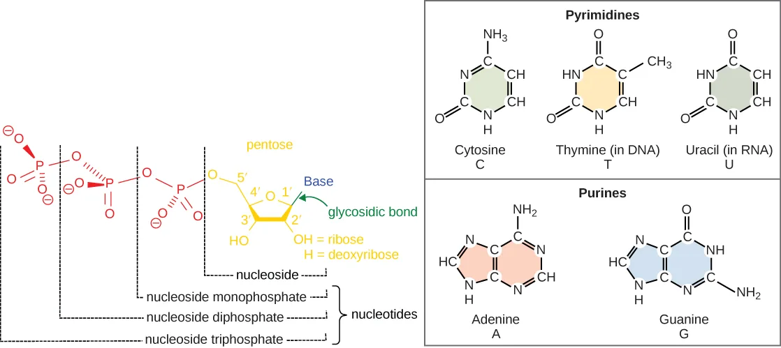
The building blocks of DNA are nucleotides. The important components of the nucleotide are a nitrogenous (nitrogen-bearing) base, a 5-carbon sugar (pentose), and a phosphate group. The nucleotide is named depending on the nitrogenous base. The nitrogenous base can be a purine such as adenine (A) and guanine (G), or a pyrimidine such as cytosine (C) and thymine (T).
Extended description
Left panel, reading right to left along the chain: a pentose sugar ring with carbons numbered 5′ (bearing the phosphate chain), 4′, 1′ (bearing a green arrow labeled glycosidic bond pointing to a blue Base label), 3′ (bearing an OH group), and 2′, with a note that OH at 2′ means ribose and H at 2′ means deoxyribose; one, two, or three phosphate groups (each shown with negative charges) extend from the 5′ carbon. Brackets beneath the sugar mark, from shortest to longest phosphate chain: nucleoside (sugar plus base only), then the three nucleotides — nucleoside monophosphate, diphosphate, and triphosphate. Right panel, two boxed groups of ring diagrams with nitrogen and carbon positions labeled: the Pyrimidines box shows cytosine, thymine, and uracil, each a single six-membered ring with its own attached side groups; the Purines box shows adenine and guanine, each a six-membered ring fused to a five-membered ring, likewise with attached side groups.
The images above illustrate the five bases of DNA and RNA. Examine the images and explain why these are called “nitrogenous bases.” How are the purines different from the pyrimidines? How is one purine or pyrimidine different from another, e.g., adenine from guanine? How is a nucleoside different from a nucleotide?
Show model answer
Did your answer mention:
The purines have a double ring structure with a six-membered ring fused to a five-membered ring. Pyrimidines are smaller in size; they have a single six-membered ring structure.
The sugar is deoxyribose in DNA and ribose in RNA. The carbon atoms of the five-carbon sugar are numbered 1′, 2′, 3′, 4′, and 5′ (1′ is read as “one prime”). The phosphate, which makes DNA and RNA acidic, is connected to the 5′ carbon of the sugar by the formation of an ester linkage between phosphoric acid and the 5′-OH group (an ester is an acid + an alcohol). In DNA nucleotides, the 3′ carbon of the sugar deoxyribose is attached to a hydroxyl (OH) group. In RNA nucleotides, the 2′ carbon of the sugar ribose also contains a hydroxyl group. The base is attached to the 1′ carbon of the sugar.
The nucleotides combine with each other to produce phosphodiester bonds. The phosphate residue attached to the 5′ carbon of the sugar of one nucleotide forms a second ester linkage with the hydroxyl group of the 3′ carbon of the sugar of the next nucleotide, thereby forming a 5′–3′ phosphodiester bond. In a polynucleotide, one end of the chain has a free 5′ phosphate, and the other end has a free 3′-OH. These are called the 5′ and 3′ ends of the chain. A base attached to a five-carbon sugar represents a nucleoside. A nucleotide is composed of one, two, or three phosphate groups attached to a nucleoside.
In the 1950s, Francis Crick and James Watson worked together to determine the structure of DNA at the University of Cambridge, England. Other scientists like Linus Pauling and Maurice Wilkins were also actively exploring this field. Pauling previously had discovered the secondary structure of proteins using X-ray crystallography. In Wilkins’ lab, researcher Rosalind Franklin was using X-ray diffraction methods to understand the structure of DNA. Watson and Crick were able to piece together the puzzle of the DNA molecule on the basis of Franklin’s data because Crick had also studied X-ray diffraction. In 1962, James Watson, Francis Crick, and Maurice Wilkins were awarded the Nobel Prize in Medicine. Unfortunately, by then Franklin had died, and Nobel prizes are not awarded posthumously.
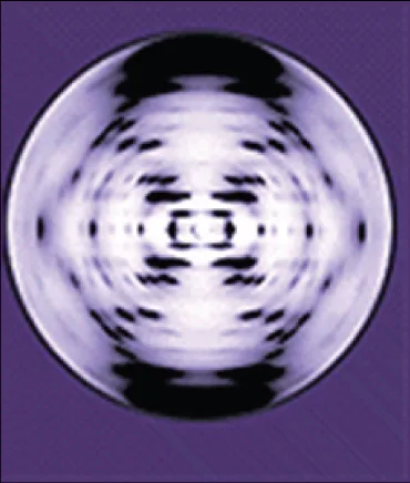
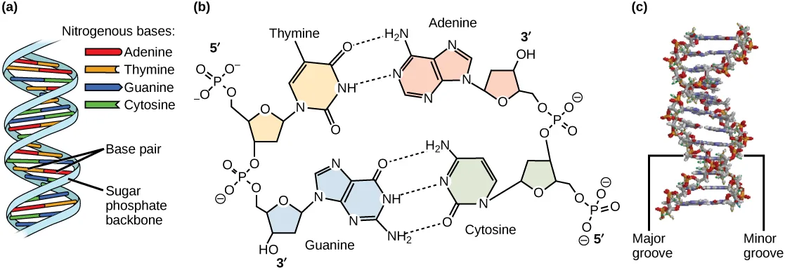
Watson and Crick proposed that DNA is made up of two strands that are twisted around each other to form a right-handed helix. Base pairing takes place between a purine and pyrimidine on opposite strands, so that A pairs with T, and G pairs with C (suggested by Chargaff’s Rules). Thus, adenine and thymine are complementary base pairs, and cytosine and guanine are also complementary base pairs. The base pairs are stabilized by hydrogen bonds: adenine and thymine form two hydrogen bonds and cytosine and guanine form three hydrogen bonds. The two strands are anti-parallel in nature; that is, the 3′ end of one strand faces the 5′ end of the other strand. The sugar and phosphate of the nucleotides form the backbone of the structure, whereas the nitrogenous bases are stacked inside, like the rungs of a ladder. Each base pair is separated from the next base pair by a distance of 0.34 nm, and each turn of the helix measures 3.4 nm. Therefore, 10 base pairs are present per turn of the helix. The diameter of the DNA double-helix is 2 nm, and it is uniform throughout. Only the pairing between a purine and pyrimidine and the antiparallel orientation of the two DNA strands can explain the uniform diameter. The twisting of the two strands around each other results in the formation of uniformly spaced major and minor grooves.
Extended description
Panel (a): a light blue double helix with paired rungs colored red for adenine, orange for thymine, blue for guanine, and green for cytosine, arrows labeling one rung “base pair” and the outer strand “sugar phosphate backbone.” Panel (b): at top, thymine and adenine drawn as ring structures, each attached to a sugar-phosphate unit, joined by two dashed hydrogen bonds; the thymine strand’s phosphate end is marked 5′ and its sugar end 3′, while the adenine strand runs the opposite way, sugar end 3′ and phosphate end 5′. At bottom, guanine and cytosine are drawn the same way, joined by three dashed hydrogen bonds, with matching 5′/3′ end labels running antiparallel to the pair above. Panel (c): an upright space-filling model of the helix in white, gray, and red atoms, with a bracket on one side spanning a wider surface gap labeled major groove and a bracket on the other spanning a narrower gap labeled minor groove.
DNA Sequencing Techniques
Until the 1990s, the sequencing of DNA (reading the sequence of DNA) was a relatively expensive and long process. Using radiolabeled nucleotides also compounded the problem through safety concerns. With currently available technology and automated machines, the process is cheaper, safer, and can be completed in a matter of hours. Fred Sanger developed the sequencing method used for the human genome sequencing project, which is widely used today.
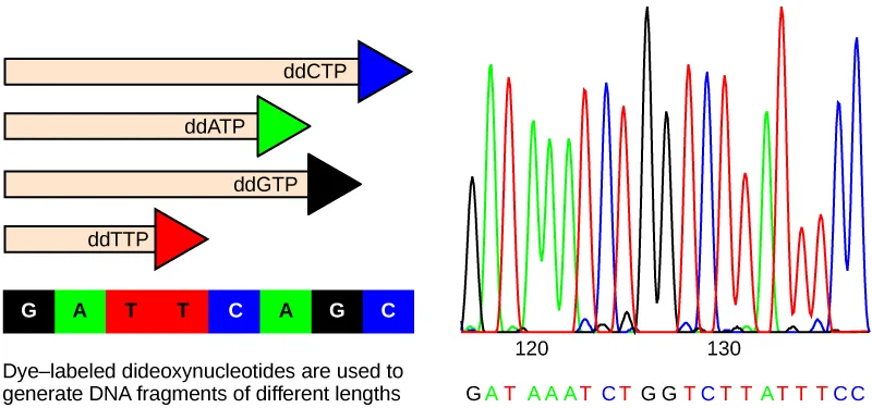
The sequencing method is known as the dideoxy chain termination method. The method is based on the use of chain terminators, the dideoxynucleotides (ddNTPs). The ddNTPs differ from the deoxynucleotides by the lack of a free 3′ OH group on the five-carbon sugar. If a ddNTP is added to a growing DNA strand, the chain cannot be extended any further because the free 3′ OH group needed to add another nucleotide is not available. By using a predetermined ratio of deoxynucleotides to dideoxynucleotides, it is possible to generate DNA fragments of different sizes.
Extended description
Panel (a): four arrow-shaped strands of increasing length, each capped by a triangular arrowhead colored to match its terminating dideoxynucleotide — black for ddGTP, green for ddATP, red for ddTTP, blue for ddCTP — stacked above a single row of eight colored boxes reading, left to right, G (black), A (green), T (red), T (red), C (blue), A (green), G (black), C (blue), captioned “Dye-labeled dideoxynucleotides are used to generate DNA fragments of different lengths.” Panel (b): a trace of overlapping black, green, red, and blue peaks running left to right above the axis positions 120 and 130, with the base-call sequence “GATAAATCTGGTCTTATTTCC” printed in matching colors beneath the peaks.
The DNA sample to be sequenced is denatured (separated into two strands by heating it to high temperatures). The DNA is divided into four tubes in which a primer, DNA polymerase, and all four nucleoside triphosphates (A, T, G, and C) are added. In addition, limited quantities of one of the four dideoxynucleoside triphosphates (ddCTP, ddATP, ddGTP, and ddTTP) are added to each tube respectively. The tubes are labeled as A, T, G, and C according to the ddNTP added. For detection purposes, each of the four dideoxynucleotides carries a different fluorescent label. Chain elongation continues until a fluorescent dideoxy nucleotide is incorporated, after which no further elongation takes place. After the reaction is over, electrophoresis is performed. Even a difference in length of a single base can be detected. The sequence is read from a laser scanner that detects the fluorescent marker of each fragment. For his work on DNA sequencing, Sanger received a Nobel Prize in Chemistry in 1980.
Gel electrophoresis is a technique used to separate DNA fragments of different sizes. Usually the gel is made of a chemical called agarose (a polysaccharide polymer extracted from seaweed that is high in galactose residues). Agarose powder is added to a buffer and heated. After cooling, the gel solution is poured into a casting tray. Once the gel has solidified, the DNA is loaded on the gel and electric current is applied. The DNA has a net negative charge and moves from the negative electrode toward the positive electrode. The electric current is applied for sufficient time to let the DNA separate according to size; the smallest fragments will be farthest from the well (where the DNA was loaded), and the heavier molecular weight fragments will be closest to the well. Once the DNA is separated, the gel is stained with a DNA-specific dye for viewing it.
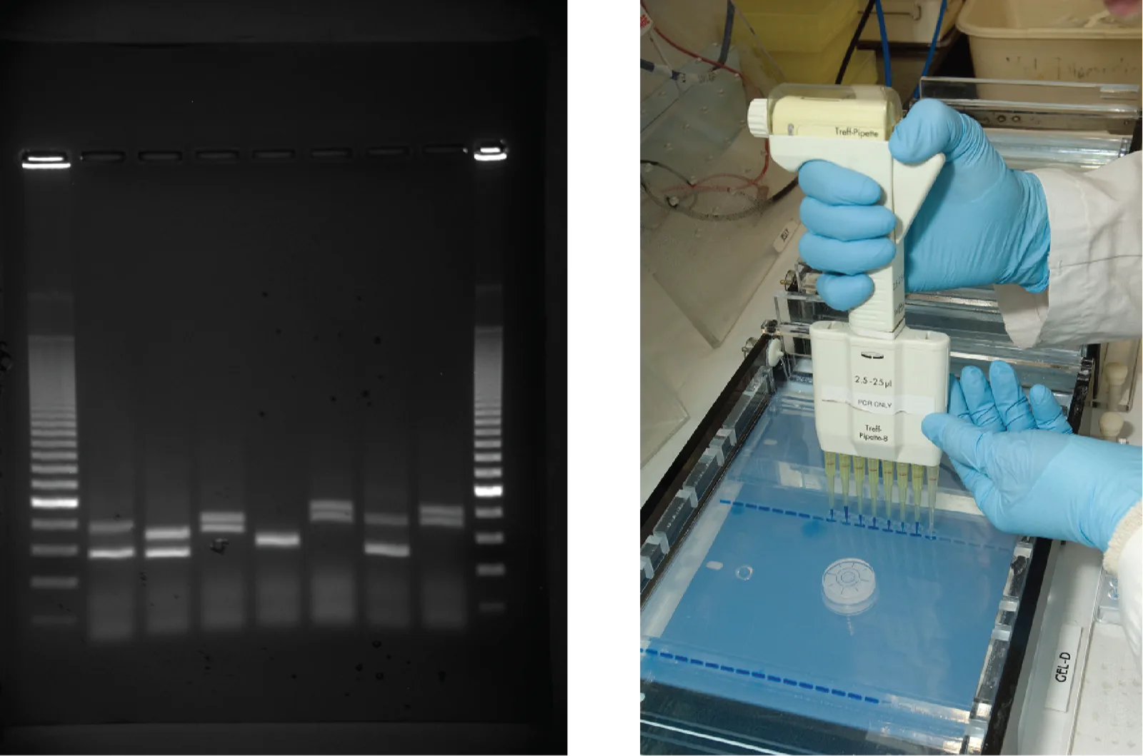
Extended description
First photo: an agarose gel, nine lanes across, imaged under UV light so the DNA fluoresces as thin white bands on a black background. Lanes one and nine (the outer lanes) each show many bands from a DNA size standard, closely spaced near the top of the gel and progressively farther apart toward the bottom. Lanes two through eight each show one or two bands; some sample lanes carry bands that run the same distance into the gel as a neighboring lane’s (matching sizes), while others run a slightly different distance (a small size difference). Second photo: a gloved hand grips a handheld multichannel pipette fitted with eight tips and lowers it toward a row of sample wells at the top of a gel tray that already holds several previously loaded lanes.
Evolution Connection. Neanderthal Genome: How Are We Related?
The first draft sequence of the Neanderthal genome was recently published by Richard E. Green et al. in 2010 (Richard E. Green et al., “A Draft Sequence of the Neandertal Genome,” Science 328 (2010): 710-22.). Neanderthals are the closest ancestors of present-day humans. They were known to have lived in Europe and Western Asia (and now, perhaps, in Northern Africa) before they disappeared from fossil records approximately 30,000 years ago. Green’s team studied almost 40,000-year-old fossil remains that were selected from sites across the world. Extremely sophisticated means of sample preparation and DNA sequencing were employed because of the fragile nature of the bones and heavy microbial contamination. In their study, the scientists were able to sequence some four billion base pairs. The Neanderthal sequence was compared with that of present-day humans from across the world. After comparing the sequences, the researchers found that the Neanderthal genome had 2 to 3 percent greater similarity to people living outside Africa than to people in Africa. While current theories have suggested that all present-day humans can be traced to a small ancestral population in Africa, the data from the Neanderthal genome suggest some interbreeding between Neanderthals and early modern humans.
Green and his colleagues also discovered DNA segments among people in Europe and Asia that are more similar to Neanderthal sequences than to other contemporary human sequences. Another interesting observation was that Neanderthals are as closely related to people from Papua New Guinea as to those from China or France. This is surprising because Neanderthal fossil remains have been located only in Europe and West Asia. Most likely, genetic exchange took place between Neanderthals and modern humans as modern humans emerged out of Africa, before the divergence of Europeans, East Asians, and Papua New Guineans.
Several genes seem to have undergone changes from Neanderthals during the evolution of present-day humans. These genes are involved in cranial structure, metabolism, skin morphology, and cognitive development. One of the genes that is of particular interest is RUNX2, which is different in modern day humans and Neanderthals. This gene is responsible for the prominent frontal bone, bell-shaped rib cage, and dental differences seen in Neanderthals. It is speculated that an evolutionary change in RUNX2 was important in the origin of modern-day humans, and this affected the cranium and the upper body.
DNA Packaging in Cells
Prokaryotes are much simpler than eukaryotes in many of their features. Most prokaryotes contain a single, circular chromosome that is found in an area of the cytoplasm called the nucleoid region.
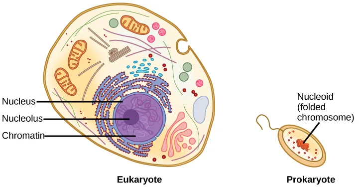
Extended description
Eukaryotic cell (left): a large yellow oval cell with a rounded nucleus, ringed by a blue dotted nuclear envelope, containing purple coiled chromatin strands and a dark purple nucleolus; arrows labeled Nucleus, Nucleolus, and Chromatin point to those structures. The surrounding cytoplasm holds several reddish oval mitochondria, a blue coiled network (endoplasmic reticulum) studded with small dots (ribosomes), a pink stack of flattened sacs (Golgi apparatus), and scattered green and pink circles (vesicles). Prokaryotic cell (right), drawn much smaller than the eukaryotic cell at the same scale: a plain yellow oval with no internal membrane-bound structures except a compact, string-like red region near its center, labeled by an arrow reading Nucleoid (folded chromosome).
In eukaryotic cells, DNA and RNA synthesis occur in a separate compartment from protein synthesis. In prokaryotic cells, both processes occur together. What advantages might there be to separating the processes? What advantages might there be to having them occur together?
Show model answer
Did your answer mention:
The size of the genome in one of the most well-studied prokaryotes, E.coli, is 4.6 million base pairs (approximately 1.6 mm, if cut and stretched out). (Source note: the source says 1.1 mm; at the 0.34 nm per base pair this section gives above, 4.6 million base pairs measure about 1.6 mm.) So how does this fit inside a small bacterial cell? The DNA is twisted by what is known as supercoiling. Supercoiling suggests that DNA is either “under-wound” (less than one turn of the helix per 10 base pairs) or “over-wound” (more than 1 turn per 10 base pairs) from its normal relaxed state. Some proteins are known to be involved in the supercoiling; other proteins and enzymes such as DNA gyrase help in maintaining the supercoiled structure.
Eukaryotes, whose chromosomes each consist of a linear DNA molecule, employ a different type of packing strategy to fit their DNA inside the nucleus. At the most basic level, DNA is wrapped around proteins known as histones to form structures called nucleosomes. The histones are evolutionarily conserved proteins that are rich in basic amino acids and form an octamer composed of two molecules of each of four different histones. Their composition and properties are important to understanding gene expression, and were partially uncovered based on research by Marie M. Daly and Alfred E. Mirsky in the early 1950s. The DNA (remember, it is negatively charged because of the phosphate groups) is wrapped tightly around the histone core. This nucleosome is linked to the next one with the help of a linker DNA. This is also known as the “beads on a string” structure. With the help of a fifth histone, a string of nucleosomes is further compacted into a 30-nm fiber, which is the diameter of the structure. Metaphase chromosomes are even further condensed by association with scaffolding proteins. At the metaphase stage, the chromosomes are at their most compact, approximately 700 nm in width.
In interphase, eukaryotic chromosomes have two distinct regions that can be distinguished by staining. The tightly packaged region is known as heterochromatin, and the less dense region is known as euchromatin. Heterochromatin usually contains genes that are not expressed, and is found in the regions of the centromere and telomeres. The euchromatin usually contains genes that are transcribed, with DNA packaged around nucleosomes but not further compacted.
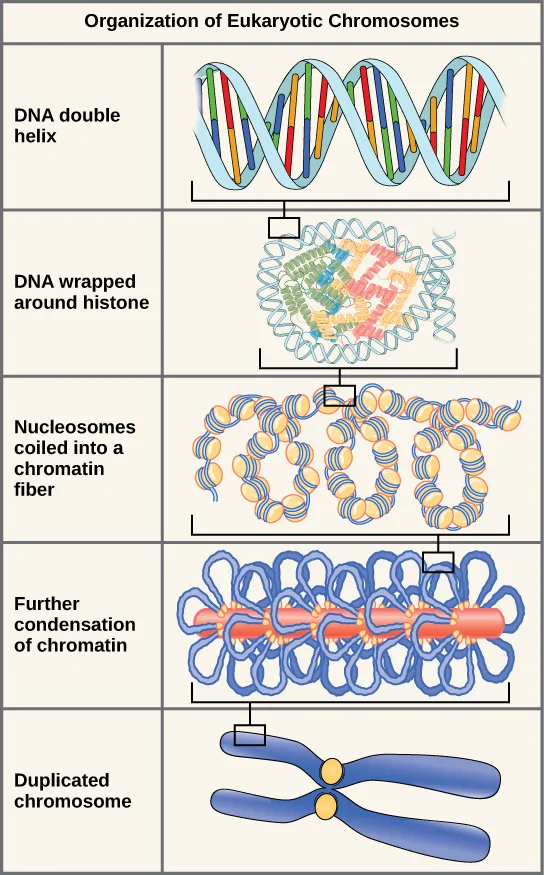
Extended description
Row 1, “DNA double helix”: a light blue double helix with colored rungs, brackets under one section pointing down to row 2. Row 2, “DNA wrapped around histone”: the double helix coiled around a cluster of colored globular histone proteins, brackets pointing down to row 3. Row 3, “Nucleosomes coiled into a chromatin fiber”: a row of the histone-DNA beads strung together and coiled into a thicker fiber, brackets pointing down to row 4. Row 4, “Further condensation of chromatin”: the fiber shown looped repeatedly around a central red rod-like scaffold, brackets pointing down to row 5. Row 5, “Duplicated chromosome”: two identical blue X-shaped chromosome arms joined at a yellow centromere, the most condensed and final structure in the sequence.
Summary
The currently accepted model of the double-helix structure of DNA was proposed by Watson and Crick. Some of the salient features are that the two strands that make up the double helix have complementary base sequences and anti-parallel orientations. Alternating deoxyribose sugars and phosphates form the backbone of the structure, and the nitrogenous bases are stacked like rungs inside. The diameter of the double helix, 2 nm, is uniform throughout. A purine always pairs with a pyrimidine; A pairs with T, and G pairs with C. One turn of the helix has 10 base pairs. Prokaryotes are much simpler than eukaryotes in many of their features. Most prokaryotes contain a single, circular chromosome. In general, eukaryotic chromosomes contain a linear DNA molecule packaged into nucleosomes, and have two distinct regions that can be distinguished by staining, reflecting different states of packaging and compaction.
Key terms
- electrophoresis — technique used to separate DNA fragments according to size
Practice
Describe the structure of DNA
DNA double helix does not have which of the following?
Uracil replaces thymine only in RNA, not in DNA.According to the section summary, the currently accepted model of the double-helix structure of DNA was proposed by ________.
They worked together at the University of Cambridge in the 1950s, piecing the structure together from Franklin’s X-ray diffraction data.Describe the structure and complementary base pairing of DNA.
Show model answer
Did your answer mention:
Explain the Sanger method of DNA sequencing
Provide a brief summary of the Sanger sequencing method.
Show model answer
Did your answer mention:
The technique used to separate DNA fragments according to size is called ________.
The section names the gel-based version of this technique with a two-word phrase starting with “gel.”In the dideoxy chain termination method, a ddNTP halts strand synthesis because it lacks a free ________ needed to add the next nucleotide.
Compare a dideoxynucleotide to a normal deoxynucleotide: which group is missing from its sugar?Discuss the similarities and differences between eukaryotic and prokaryotic DNA
In eukaryotes, what is the DNA wrapped around?
This protein forms an octamer, and the negatively charged DNA winds tightly around it to form a nucleosome.Prokaryotes have a single circular chromosome while eukaryotes have linear chromosomes. Describe one advantage and one disadvantage to the eukaryotic genome packaging compared to the prokaryotes.
Show model answer
Did your answer mention:
According to the section summary, most prokaryotes contain a single, ________.
This is the shape of the one chromosome that carries nearly all of a prokaryote’s DNA.This section is adapted from Biology 2e, Section 14.2: DNA Structure and Sequencing by Mary Ann Clark, Jung Choi, Matthew Douglas, and OpenStax, © OpenStax, licensed under CC BY-NC-SA 4.0. Access the original for free at openstax.org. Changes: figures re-encoded as WebP; Figure_14_02_01, Figure_14_02_03abc-c0c6, Figure_14_02_04ab-b7be, Figure_14_02_06_new, and Figure_14_02_07-de13 re-kinded from the manifest’s file-extension guess of “photo” to “diagram” (each is a colored illustration or rendered model, not a photograph), and Figure_14_02_09 re-kinded from “diagram” to “photo” (it is two real photographs — an illuminated gel and a person pipetting); every figure’s alt rewritten from the image — several source alts were letter-spaced screen-reader spellings (“D N A”), Figure_14_02_01’s was corrupted with its leading letter missing (“llustration depicts…” — reported as a source defect), and the rest were plain prose that did not say what each figure teaches; longdescs added for the labeled diagrams and flow charts (Figures 14.2.1, 14.2.3, 14.2.4, 14.2.6, 14.2.7) and for the gel-electrophoresis photo pair (Figure 14.2.9), whose original alt also ran well past the 600-character accessibility limit; both source Visual Connection notes kept in the body immediately after their figures — the first (Figure 14.2.1, on nitrogenous bases and nucleosides/nucleotides) has no matching exercise or solution anywhere in the CNXML, so its self-check model answer is composed strictly from the section’s own definitions of nitrogenous base, purine/pyrimidine ring count, and nucleoside/nucleotide, plus what the figure itself shows for how one purine or pyrimidine differs from another; the second (Figure 14.2.6, on compartmentalization) rendered as a self-check from its keyed prose solution; the Neanderthal Genome evolution connection kept with its title and its bibliographic footnote folded into a parenthetical citation after the sentence it supports; all three interactive notes rendered as Link to Learning callouts with descriptive link text in place of the source’s bare “this site”/“here”; the end-of-section Review Questions and Critical Thinking Questions adapted into the closing interactive Practice block; one key-term recall item (electrophoresis) added from the glossary; because the section summary never mentions DNA sequencing at all, one Practice item for “Explain the Sanger method of DNA sequencing” is author-written strictly from the section’s own sentences about why a dideoxynucleotide halts strand synthesis; two further summary-derived items were added to raise their objective groups to three — a select-the-name multiple choice on who proposed the double-helix model, and a cloze text-in on the shape of the prokaryotic chromosome, each built strictly from the section’s own Section Summary sentences; and rubric checkpoints added to every self-check (both in the body and in Practice), decomposing each model answer into check-off clauses with no new claims. One number is corrected with a visible Source note: the stretched-out E. coli chromosome is about 1.6 mm by this section’s own 0.34 nm per base pair, not 1.1 mm (erratum 400).