DNA Replication in Prokaryotes
By the end of this section, you will be able to:
- Explain the process of DNA replication in prokaryotes
- Discuss the role of different enzymes and proteins in supporting this process
DNA replication has been well studied in prokaryotes primarily because of the small size of the genome and because of the large variety of mutants that are available. E. coli has 4.6 million base pairs in a single circular chromosome and all of it gets replicated in approximately 42 minutes, starting from a single site along the chromosome and proceeding around the circle in both directions. This means that approximately 1000 nucleotides are added per second. Thus, the process is quite rapid and occurs without many mistakes.
DNA replication employs a large number of structural proteins and enzymes, each of which plays a critical role during the process. One of the key players is the enzyme DNA polymerase, also known as DNA pol, which adds nucleotides one-by-one to the growing DNA chain that is complementary to the template strand. The addition of nucleotides requires energy; this energy is obtained from the nucleoside triphosphates dATP, dGTP, dTTP and dCTP. Like ATP, the other NTPs (nucleoside triphosphates) are high-energy molecules that can serve both as the source of DNA nucleotides and the source of energy to drive the polymerization. When the bond between the phosphates is “broken,” the energy released is used to form the phosphodiester bond between the incoming nucleotide and the growing chain. In prokaryotes, three main types of polymerases are known: DNA pol I, DNA pol II, and DNA pol III. It is now known that DNA pol III is the enzyme required for DNA synthesis; DNA pol I is an important accessory enzyme in DNA replication, and along with DNA pol II, is primarily required for repair.
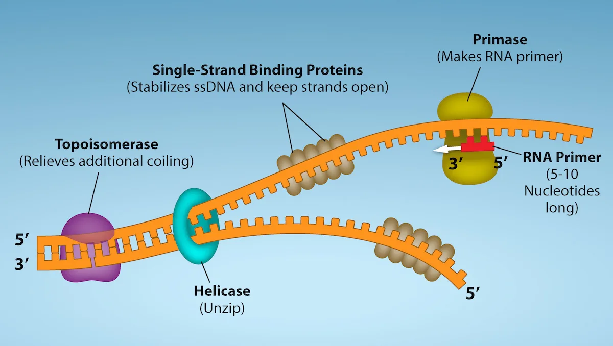
How does the replication machinery know where to begin? It turns out that there are specific nucleotide sequences called origins of replication where replication begins. In E. coli, which has a single origin of replication on its one chromosome (as do most prokaryotes), this origin of replication is approximately 245 base pairs long and is rich in AT sequences. The origin of replication is recognized by certain proteins that bind to this site. An enzyme called helicase unwinds the DNA by breaking the hydrogen bonds between the nitrogenous base pairs. ATP hydrolysis is required for this process. As the DNA opens up, Y-shaped structures called replication forks are formed. Two replication forks are formed at the origin of replication and these get extended bi-directionally as replication proceeds. Single-strand binding proteins coat the single strands of DNA near the replication fork to prevent the single-stranded DNA from winding back into a double helix.
DNA polymerase has two important restrictions: it is able to add nucleotides only in the 5′ to 3′ direction (a new DNA strand can be only extended in this direction). It also requires a free 3′-OH group to which it can add nucleotides by forming a phosphodiester bond between the 3′-OH end and the 5′ phosphate of the next nucleotide. This essentially means that it cannot add nucleotides if a free 3′-OH group is not available. Then how does it add the first nucleotide? The problem is solved with the help of a primer that provides the free 3′-OH end. Another enzyme, RNA primase, synthesizes an RNA segment that is about five to ten nucleotides long and complementary to the template DNA. Because this sequence primes the DNA synthesis, it is appropriately called the primer. DNA polymerase can now extend this RNA primer, adding nucleotides one-by-one that are complementary to the template strand (the figure below).
Extended description
The diagram reads left to right along one DNA molecule drawn as an orange ladder-like ribbon. At the far left the ribbon is still a closed double helix, its two strands labeled 5′ (top) and 3′ (bottom); a purple blob labeled Topoisomerase sits on top of the helix there. Just to its right a teal ring labeled Helicase marks the point where the double helix opens into two single strands. From the helicase, the upper single strand curves up and to the right; partway along it, a diagonal cluster of tan oval beads labeled Single-Strand Binding Proteins coats the strand. Farther right on this same strand, an olive-green blob labeled Primase sits on the strand beside a short red segment labeled RNA Primer, with 3′ marked at the segment’s left edge and 5′ at its right edge. The lower single strand curves down and then back to the right below the upper strand; a second, smaller cluster of tan Single-Strand Binding Protein beads coats it partway along, and the strand ends at the right labeled 5′ with no further protein bound.
You isolate a cell strain in which the joining of Okazaki fragments is impaired and suspect that a mutation has occurred in an enzyme found at the replication fork. Which enzyme is most likely to be mutated?
Show model answer
Did your answer mention:
The replication fork moves at the rate of 1000 nucleotides per second. Topoisomerase prevents the over-winding of the DNA double helix ahead of the replication fork as the DNA is opening up; it does so by causing temporary nicks in the DNA helix and then resealing it. Because DNA polymerase can only extend in the 5′ to 3′ direction, and because the DNA double helix is antiparallel, there is a slight problem at the replication fork. The two template DNA strands have opposing orientations: one strand is in the 5′ to 3′ direction and the other is oriented in the 3′ to 5′ direction. Only one new DNA strand, the one that is complementary to the 3′ to 5′ parental DNA strand, can be synthesized continuously towards the replication fork. This continuously synthesized strand is known as the leading strand. The other strand, complementary to the 5′ to 3′ parental DNA, is extended away from the replication fork, in small fragments known as Okazaki fragments, each requiring a primer to start the synthesis. New primer segments are laid down in the direction of the replication fork, but each pointing away from it. (Okazaki fragments are named after the Japanese scientist who first discovered them. The strand with the Okazaki fragments is known as the lagging strand.)
The leading strand can be extended from a single primer, whereas the lagging strand needs a new primer for each of the short Okazaki fragments. The overall direction of the lagging strand will be 3′ to 5′, and that of the leading strand 5′ to 3′. A protein called the sliding clamp holds the DNA polymerase in place as it continues to add nucleotides. The sliding clamp is a ring-shaped protein that binds to the DNA and holds the polymerase in place. As synthesis proceeds, the RNA primers are replaced by DNA. The primers are removed by the exonuclease activity of DNA pol I, which uses DNA behind the RNA as its own primer and fills in the gaps left by removal of the RNA nucleotides by the addition of DNA nucleotides. The nicks that remain between the newly synthesized DNA (that replaced the RNA primer) and the previously synthesized DNA are sealed by the enzyme DNA ligase, which catalyzes the formation of phosphodiester linkages between the 3′-OH end of one nucleotide and the 5′ phosphate end of the other fragment.
Once the chromosome has been completely replicated, the two DNA copies move into two different cells during cell division.
The process of DNA replication can be summarized as follows:
- DNA unwinds at the origin of replication.
- Helicase opens up the DNA-forming replication forks; these are extended bidirectionally.
- Single-strand binding proteins coat the DNA around the replication fork to prevent rewinding of the DNA.
- Topoisomerase binds at the region ahead of the replication fork to prevent supercoiling.
- Primase synthesizes RNA primers complementary to the DNA strand.
- DNA polymerase III starts adding nucleotides to the 3′-OH end of the primer.
- Elongation of both the lagging and the leading strand continues.
- RNA primers are removed by exonuclease activity.
- Gaps are filled by DNA pol I by adding dNTPs.
- The gap between the two DNA fragments is sealed by DNA ligase, which helps in the formation of phosphodiester bonds.
The following table summarizes the enzymes involved in prokaryotic DNA replication and the functions of each.
Prokaryotic DNA Replication: Enzymes and Their Function
| Enzyme/protein | Specific Function |
|---|---|
| DNA pol I | Removes RNA primer and replaces it with newly synthesized DNA |
| DNA pol III | Main enzyme that adds nucleotides in the 5′-3′ direction |
| Helicase | Opens the DNA helix by breaking hydrogen bonds between the nitrogenous bases |
| Ligase | Seals the gaps between the Okazaki fragments to create one continuous DNA strand |
| Primase | Synthesizes RNA primers needed to start replication |
| Sliding Clamp | Helps to hold the DNA polymerase in place when nucleotides are being added |
| Topoisomerase | Helps relieve the strain on DNA when unwinding by causing breaks, and then resealing the DNA |
| Single-strand binding proteins (SSB) | Binds to single-stranded DNA to prevent DNA from rewinding back. |
Summary
Replication in prokaryotes starts from a sequence found on the chromosome called the origin of replication—the point at which the DNA opens up. Helicase opens up the DNA double helix, resulting in the formation of the replication fork. Single-strand binding proteins bind to the single-stranded DNA near the replication fork to keep the fork open. Primase synthesizes an RNA primer to initiate synthesis by DNA polymerase, which can add nucleotides only to the 3′ end of a previously synthesized primer strand. Both new DNA strands grow according to their respective 5′-3′ directions. One strand is synthesized continuously in the direction of the replication fork; this is called the leading strand. The other strand is synthesized in a direction away from the replication fork, in short stretches of DNA known as Okazaki fragments. This strand is known as the lagging strand. Once replication is completed, the RNA primers are replaced by DNA nucleotides and the DNA is sealed with DNA ligase, which creates phosphodiester bonds between the 3′-OH of one end and the 5′ phosphate of the other strand.
Key terms
- exonuclease — enzymes that cleave nucleotides one at a time from the end of a polynucleotide chain
- helicase — during replication, this enzyme helps to open up the DNA helix by breaking the hydrogen bonds
- lagging strand — during replication, the strand that is replicated in short fragments and away from the replication fork
- leading strand — strand that is synthesized continuously in the 5′-3′ direction, which is synthesized in the direction of the replication fork
- ligase — enzyme that catalyzes the formation of a phosphodiester linkage between the 3′ OH and 5′ phosphate ends of the DNA
- Okazaki fragment — DNA fragment that is synthesized in short stretches on the lagging strand
- primase — enzyme that synthesizes the RNA primer; the primer is needed for DNA pol to start synthesis of a new DNA strand
- primer — short stretch of nucleotides that is required to initiate replication; in the case of replication, the primer has RNA nucleotides
- replication fork — Y-shaped structure formed during initiation of replication
- single-strand binding protein — during replication, protein that binds to the single-stranded DNA; this helps in keeping the two strands of DNA apart so that they may serve as templates
- sliding clamp — ring-shaped protein that holds the DNA pol on the DNA strand
- topoisomerase — enzyme that prevents overwinding of DNA when DNA replication is taking place
Practice
Explain the process of DNA replication in prokaryotes
In which direction does DNA replication take place?
DNA polymerase can only add nucleotides to a free 3′-OH end of a growing strand.DNA replication is bidirectional and discontinuous; explain your understanding of those concepts.
Show model answer
Did your answer mention:
What are Okazaki fragments and how are they formed?
Show model answer
Did your answer mention:
If the rate of replication in a particular prokaryote is 900 nucleotides per second, how long would it take 1.2 million base pair genomes to make two copies?
Show model answer
Did your answer mention:
Explain the events taking place at the replication fork. If the gene for helicase is mutated, what part of replication will be affected?
Show model answer
Did your answer mention:
Discuss the role of different enzymes and proteins in supporting this process
Which of the following components is not involved during the formation of the replication fork?
Ligase seals gaps late in replication, after new strands are made — think about what forming the fork itself requires.Which of the following does the enzyme primase synthesize?
Primase is itself an RNA polymerase — what kind of nucleic acid segment does it lay down?A scientist randomly mutates the DNA of a bacterium. She then sequences the bacterium’s daughter cells, and finds that the daughters have many errors in their replicated DNA. The parent bacterium likely acquired a mutation in which enzyme?
The section names which polymerase, alongside DNA pol I, is primarily required for repair rather than for the main synthesis reaction.What is the role of a primer in DNA replication? What would happen if you forgot to add a primer in a tube containing the reaction mix for a DNA sequencing reaction?
Show model answer
Did your answer mention:
Quinolone antibiotics treat bacterial infections by blocking the activity of topoisomerase. Why does this treatment work? Explain what occurs at the molecular level.
Show model answer
Did your answer mention:
This section is adapted from Biology 2e, Section 14.4: DNA Replication in Prokaryotes by Mary Ann Clark, Jung Choi, Matthew Douglas, and OpenStax, © OpenStax, licensed under CC BY-NC-SA 4.0. Access the original for free at openstax.org. Changes: the figure re-encoded as WebP and re-kinded from the manifest’s file-extension guess of “photo” to “diagram” after inspection (it is a colored illustrated schematic of the replication fork, not a photograph); its source alt (a letter-spaced, screen-reader spelling — “D N A”, “R N A”, “s s D N A” — with a typo, “stablize”) rewritten from the image, and a longdesc added describing the drawing’s enzymes, strand curves, and 5′/3′ labels as positioned, since that spatial layout is not carried by the caption; the Visual Connection note kept in the body immediately after its figure, rendered as a self-check because its solution is prose rather than a lettered option; the interactive note rendered as a Link to Learning callout with descriptive anchor text in place of the source’s bare “here”; every prime mark set as the Unicode ′ in place of a straight apostrophe or the source’s right single quotation mark throughout the prose, figure caption, table, key terms, and exercise text; the end-of-section Review Questions (4) and Critical Thinking Questions (6) adapted into the closing interactive Practice block, split across the section’s two objectives; rubric checkpoints added to every self-check (the body item and the six Practice self-checks), decomposing each model answer — the source’s own solution — into check-off clauses with no new claims. No summary-derived or locally written items were needed: the ten source exercises alone meet this book’s floor for both objective groups. The comparison table of enzymes and their functions is kept as a Markdown table only; its columns name an enzyme and its function rather than two categories to sort between, so it is not rendered as a sort-into-bins exercise.