DNA Repair
By the end of this section, you will be able to:
- Discuss the different types of mutations in DNA
- Explain DNA repair mechanisms
DNA replication is a highly accurate process, but mistakes can occasionally occur, such as a DNA polymerase inserting a wrong base. Uncorrected mistakes may sometimes lead to serious consequences, such as cancer. Repair mechanisms correct the mistakes. In rare cases, mistakes are not corrected, leading to mutations; in other cases, repair enzymes are themselves mutated or defective.
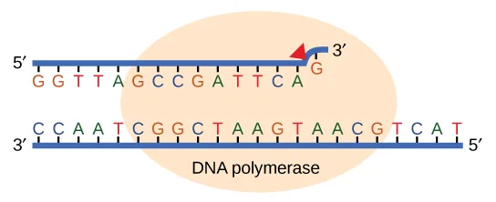
Most of the mistakes during DNA replication are promptly corrected by the proofreading ability of DNA polymerase itself, shown below. In proofreading, the DNA pol reads the newly added base before adding the next one, so a correction can be made. The polymerase checks whether the newly added base has paired correctly with the base in the template strand. If it is the right base, the next nucleotide is added. If an incorrect base has been added, the enzyme makes a cut at the phosphodiester bond and releases the wrong nucleotide. This is performed by the 3′ exonuclease action of DNA pol. Once the incorrect nucleotide has been removed, it can be replaced by the correct one.
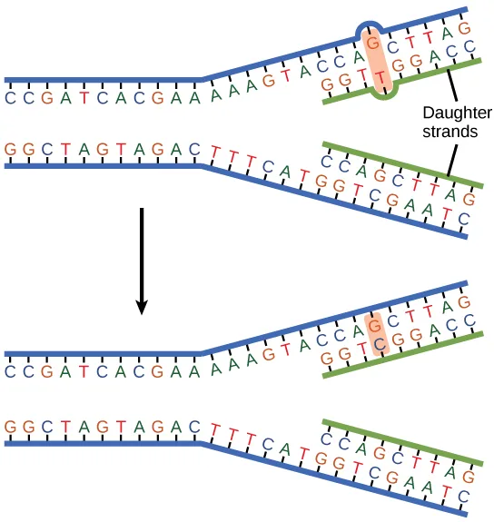
Some errors are not corrected during replication, but are instead corrected after replication is completed; this type of repair is known as mismatch repair, shown below. Specific repair enzymes recognize the mispaired nucleotide and excise part of the strand that contains it; the excised region is then resynthesized. If the mismatch remains uncorrected, it may lead to more permanent damage when the mismatched DNA is replicated. How do mismatch repair enzymes recognize which of the two bases is the incorrect one? In E. coli, after replication, the nitrogenous base adenine acquires a methyl group; the parental DNA strand will have methyl groups, whereas the newly synthesized strand lacks them. Thus, DNA polymerase is able to remove the wrongly incorporated bases from the newly synthesized, non-methylated strand. In eukaryotes, the mechanism is not very well understood, but it is believed to involve recognition of unsealed nicks in the new strand, as well as a short-term continuing association of some of the replication proteins with the new daughter strand after replication has completed.
Extended description
Each of the two stacked diagrams shows a replication fork: two parental template strands, both drawn in blue, running into a Y-shaped junction and continuing on the right, where the newly synthesized daughter-strand segments, drawn in green, pair with them. In the top diagram, one base pair just past the fork is highlighted with an orange oval — the daughter strand carries a mismatched base opposite its partner on the parental strand. A leader line from the label ‘Daughter strands’ points to the two newly synthesized strands. A downward arrow separates the top diagram from an identical one below it, where the same highlighted position now shows the mismatched base replaced with the correctly paired base, so the double helix is fully paired at that position.
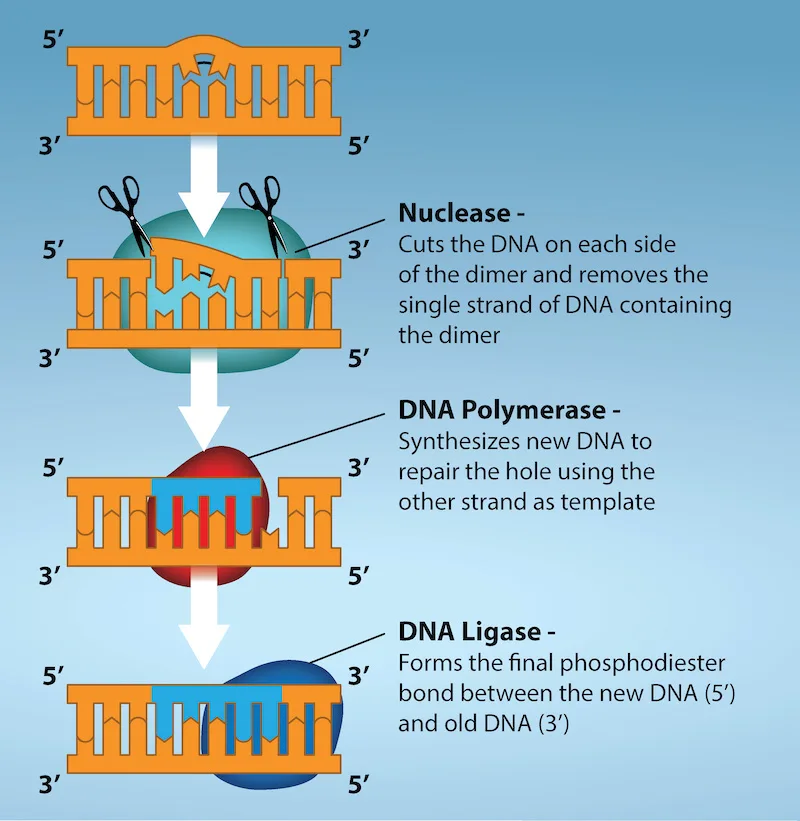
Another type of repair mechanism, nucleotide excision repair, is similar to mismatch repair, except that it is used to remove damaged bases rather than mismatched ones. The repair enzymes replace abnormal bases by making a cut on both the 3′ and 5′ ends of the damaged base, as shown below. The segment of DNA is removed and replaced with the correctly paired nucleotides by the action of DNA pol. Once the bases are filled in, the remaining gap is sealed with a phosphodiester linkage catalyzed by DNA ligase. This repair mechanism is often employed when UV exposure causes the formation of pyrimidine dimers.
Extended description
Four orange DNA-ladder diagrams are stacked top to bottom, each labeled 5′ and 3′ at both ends of both strands, connected by white downward arrows. Step 1 (top): the ladder has one distorted, bent rung representing the thymine dimer. Step 2, labeled ‘Nuclease – Cuts the DNA on each side of the dimer and removes the single strand of DNA containing the dimer’: a teal shape covers the damaged region, scissors icons mark cuts on either side of the dimer, and the ladder now shows a gap with missing rungs. Step 3, labeled ‘DNA Polymerase – Synthesizes new DNA to repair the hole using the other strand as template’: a red shape covers the same region, and blue rungs now fill part of the gap. Step 4, labeled ‘DNA Ligase – Forms the final phosphodiester bond between the new DNA (5′) and old DNA (3′)’: a dark blue shape covers the region, and the ladder is shown fully repaired with no remaining gap.
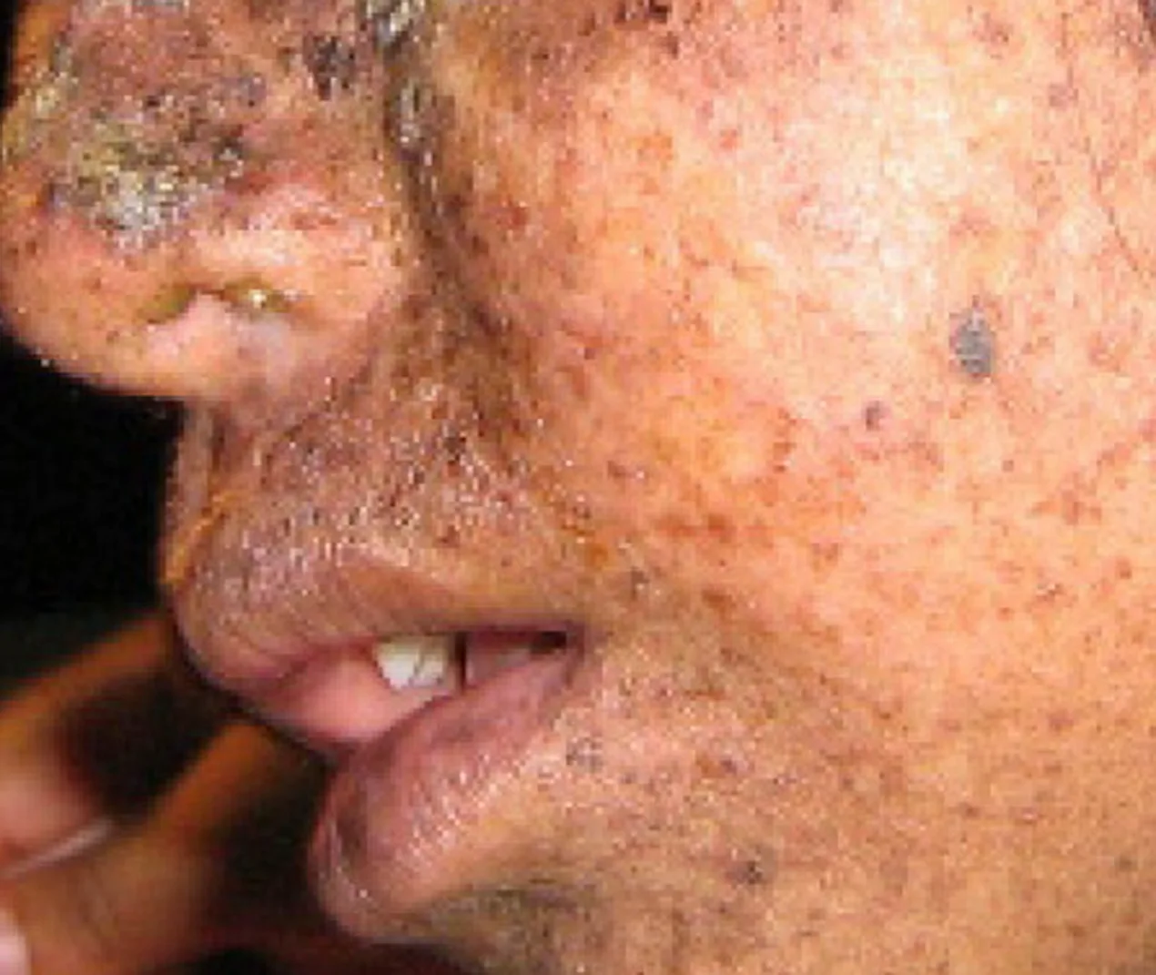
A well-studied example of mistakes not being corrected is seen in people suffering from xeroderma pigmentosa, shown below. Affected individuals have skin that is highly sensitive to UV rays from the sun. When individuals are exposed to UV light, pyrimidine dimers, especially those of thymine, are formed; people with xeroderma pigmentosa are not able to repair the damage. These are not repaired because of a defect in the nucleotide excision repair enzymes, whereas in normal individuals, the thymine dimers are excised and the defect is corrected. The thymine dimers distort the structure of the DNA double helix, and this may cause problems during DNA replication. People with xeroderma pigmentosa may have a higher risk of contracting skin cancer than those who don’t have the condition.
Errors during DNA replication are not the only reason why mutations arise in DNA. Mutations, variations in the nucleotide sequence of a genome, can also occur because of damage to DNA. Such mutations may be of two types: induced or spontaneous. Induced mutations are those that result from an exposure to chemicals, UV rays, x-rays, or some other environmental agent. For example, Charlotte Auerbach and J.M Robson discovered the mutation-inducing effects of mustard gas. Spontaneous mutations occur without any exposure to any environmental agent; they are a result of natural reactions taking place within the body.
Mutations may have a wide range of effects. Point mutations are those mutations that affect a single base pair. The most common nucleotide mutations are substitutions, in which one base is replaced by another. These substitutions can be of two types, either transitions or transversions. Transition substitution refers to a purine or pyrimidine being replaced by a base of the same kind; for example, a purine such as adenine may be replaced by the purine guanine. Transversion substitution refers to a purine being replaced by a pyrimidine, or vice versa; for example, cytosine, a pyrimidine, is replaced by adenine, a purine. Some point mutations are not detectable in the final product; these are known as silent mutations. Silent mutations are usually due to a substitution in the third base of a codon, which often represents the same amino acid as the original codon. Other point mutations can result in the replacement of one amino acid by another, which may alter the function of the protein. Point mutations that generate a stop codon can terminate a protein early.
Some mutations can result in an increased number of copies of the same codon. These are called trinucleotide repeat expansions and result in repeated regions of the same amino acid. Mutations can also be the result of the addition of a base, known as an insertion, or the removal of a base, also known as deletion. If an insertion or deletion results in the alteration of the translational reading frame (a frameshift mutation), the resultant protein is usually nonfunctional. Sometimes a piece of DNA from one chromosome may get translocated to another chromosome or to another region of the same chromosome; this is also known as translocation. These mutation types are shown below.
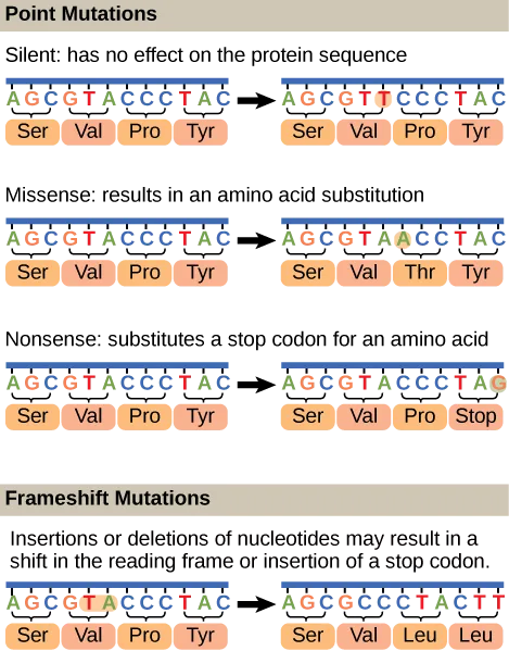
Extended description
Point Mutations box: all three rows start from the same original sequence AGC-GTA-CCC-TAC, translating to Ser-Val-Pro-Tyr, with an arrow to a changed sequence. Row 1, ‘Silent: has no effect on the protein sequence’: the sixth base changes (A to T, inside the second codon), giving AGC-GTT-CCC-TAC, still Ser-Val-Pro-Tyr. Row 2, ‘Missense: results in an amino acid substitution’: the seventh base changes (C to A, inside the third codon), giving AGC-GTA-ACC-TAC, translated Ser-Val-Thr-Tyr. Row 3, ‘Nonsense: substitutes a stop codon for an amino acid’: the twelfth base changes (C to G, inside the fourth codon), giving AGC-GTA-CCC-TAG, translated Ser-Val-Pro-Stop. Frameshift Mutations box, below the heading ‘Insertions or deletions of nucleotides may result in a shift in the reading frame or insertion of a stop codon’: the same original sequence AGC-GTA-CCC-TAC (with the fifth and sixth bases, T and A, circled) changes to AGC-GCC-CTA-CTT, which the figure’s printed labels translate Ser-Val-Leu-Leu (the Val label on the GCC codon is a defect in the source artwork — GCC encodes Ala) — the circled bases are removed and every codon boundary after them shifts, changing the last two amino acids from the original Pro-Tyr.
A frameshift mutation that results in the insertion of three nucleotides is often less deleterious than a mutation that results in the insertion of one nucleotide. Why?
Show model answer
Did your answer mention:
Mutations in repair genes have been known to cause cancer. Many mutated repair genes have been implicated in certain forms of pancreatic cancer, colon cancer, and colorectal cancer. Mutations can affect either somatic cells or germ cells. If many mutations accumulate in a somatic cell, they may lead to problems such as the uncontrolled cell division observed in cancer. If a mutation takes place in germ cells, the mutation will be passed on to the next generation, as in the case of hemophilia and xeroderma pigmentosa.
Summary
DNA polymerase can make mistakes while adding nucleotides. It edits the DNA by proofreading every newly added base. Incorrect bases are removed and replaced by the correct base before proceeding with elongation. Most mistakes are corrected during replication, although when this does not happen, the mismatch repair mechanism is employed. Mismatch repair enzymes recognize the wrongly incorporated base and excise it from the DNA, replacing it with the correct base. In yet another type of repair, nucleotide excision repair, a damaged base is removed along with a few bases on the 5′ and 3′ end, and these are replaced by copying the template with the help of DNA polymerase. The ends of the newly synthesized fragment are attached to the rest of the DNA using DNA ligase, which creates a phosphodiester bond.
Most mistakes are corrected, and if they are not, they may result in a mutation, defined as a permanent change in the DNA sequence. Mutations can be of many types, such as substitution, deletion, insertion, and trinucleotide repeat expansions. Mutations in repair genes may lead to serious consequences such as cancer. Mutations can be induced or may occur spontaneously.
Key terms
- induced mutation — mutation that results from exposure to chemicals or environmental agents
- mutation — variation in the nucleotide sequence of a genome
- mismatch repair — type of repair mechanism in which mismatched bases are removed after replication
- nucleotide excision repair — type of DNA repair mechanism in which the wrong base, along with a few nucleotides upstream or downstream, are removed
- proofreading — function of DNA pol in which it reads the newly added base before adding the next one
- point mutation — mutation that affects a single base
- silent mutation — mutation that is not expressed
- spontaneous mutation — mutation that takes place in the cells as a result of chemical reactions taking place naturally without exposure to any external agent
- transition substitution — when a purine is replaced with a purine or a pyrimidine is replaced with another pyrimidine
- transversion substitution — when a purine is replaced by a pyrimidine or a pyrimidine is replaced by a purine
Practice
Discuss the different types of mutations in DNA
An adult with a history of tanning has his genome sequenced. The beginning of a protein-coding region of his DNA reads ATGGGGATATGGCAT. If the protein-coding region of a healthy adult reads ATGGGGATATGAGCAT, identify the site and type of mutation.
Show model answer
This is a frameshift mutation with a deletion of an “A” in the 12th position of the coding region.
- Patient: ATGGGGATATGGCAT
- Normal: ATGGGGATATGAGCAT
Did your answer mention:
A mutation that results from exposure to chemicals, UV rays, x-rays, or some other environmental agent is called a(n) ________.
Charlotte Auerbach and J.M Robson discovered that mustard gas can cause this kind of mutation.A mutation that occurs without any exposure to an environmental agent, as a result of natural reactions taking place within the body, is called a(n) ________.
It is the opposite category from a mutation caused by chemicals, UV rays, or x-rays.Replacing a purine with a different purine, or a pyrimidine with a different pyrimidine, is a ________.
The text’s example is a purine such as adenine being replaced by the purine guanine.Replacing a purine with a pyrimidine, or a pyrimidine with a purine, is a ________.
The text’s example is cytosine, a pyrimidine, being replaced by adenine, a purine.Explain DNA repair mechanisms
During proofreading, which of the following enzymes reads the DNA?
This is the same enzyme that adds nucleotides during replication and can back up to remove a mismatched one.The initial mechanism for repairing nucleotide errors in DNA is ________.
Think about which repair mechanism acts first, during replication itself, before any other repair pathway is needed.A scientist creates fruit fly larvae with a mutation that eliminates the exonuclease function of DNA pol. Which prediction about the mutational load in the adult fruit flies is most likely to be correct?
The exonuclease function lets DNA polymerase proofread and remove a wrongly added base, but it is only one of several repair mechanisms working in the cell.What is the consequence of mutation of a mismatch repair enzyme? How will this affect the function of a gene?
Show model answer
Did your answer mention:
The type of repair mechanism in which mismatched bases are removed after replication is called ________.
In E. coli, this repair machinery can tell the two strands apart because only the parental strand carries methyl groups.The type of DNA repair mechanism in which the wrong base, along with a few nucleotides upstream or downstream, is removed is called ________.
This mechanism is often used to remove thymine dimers formed after UV exposure.The function of DNA polymerase in which it reads the newly added base before adding the next one is called ________.
A wrongly added base is removed by the 3′ exonuclease action of this same enzyme.This section is adapted from Biology 2e, Section 14.6: DNA Repair by Mary Ann Clark, Jung Choi, Matthew Douglas, and OpenStax, © OpenStax, licensed under CC BY-NC-SA 4.0. Access the original for free at openstax.org. Changes: figures re-encoded as WebP; Figure_14_06_01, Figure_14_06_02, and Figure_14_06_03-3edf re-kinded from the manifest’s file-extension guess of “photo” to “diagram” after inspection (all three are colored line illustrations, not photographs); all five source alts rewritten from the images (three were letter-spaced screen-reader spellings — “Illustration shows D N A polymerase…” — and the xeroderma pigmentosa photo’s alt was a bare “Photo shows…” that did not describe what is visible), with a full panel-by-panel or step-by-step walkthrough moved into a longdesc for the mismatch-repair diagram, the nucleotide-excision-repair flow chart, and the point-mutation/frameshift-mutation diagram, since none of their one-line captions carries that detail; the Visual Connection note kept in the body immediately after its figure and rendered as a self-check, since the source keys it to a prose solution rather than a lettered option; the end-of-section Review Questions and Critical Thinking Questions adapted into the closing interactive Practice block; seven key-term recall items added from the glossary; rubric checkpoints added to each self-check, decomposing its model answer (the source solution) into check-off clauses with no new claims; and the Critical Thinking solution’s typographical “wont” corrected to “won’t” (reported as a source defect).