RNA Processing in Eukaryotes
By the end of this section, you will be able to:
- Describe the different steps in RNA processing
- Understand the significance of exons, introns, and splicing for mRNAs
- Explain how tRNAs and rRNAs are processed
After transcription, eukaryotic pre-mRNAs must undergo several processing steps before they can be translated. Eukaryotic (and prokaryotic) tRNAs and rRNAs also undergo processing before they can function as components in the protein-synthesis machinery.
mRNA Processing
The eukaryotic pre-mRNA undergoes extensive processing before it is ready to be translated. Eukaryotic protein-coding sequences are not continuous, as they are in prokaryotes. The coding sequences (exons) are interrupted by noncoding introns, which must be removed to make a translatable mRNA. The additional steps involved in eukaryotic mRNA maturation also create a molecule with a much longer half-life than a prokaryotic mRNA. Eukaryotic mRNAs last for several hours, whereas the typical E. coli mRNA lasts only a few minutes. (Source note: the source says “no more than five seconds”; bacterial mRNA half-lives average a few minutes, from under two to over twenty in E. coli.)
Pre-mRNAs are first coated in RNA-stabilizing proteins; these protect the pre-mRNA from degradation while it is processed and exported out of the nucleus. The three most important steps of pre-mRNA processing are the addition of stabilizing and signaling factors at the 5′ and 3′ ends of the molecule, and the removal of the introns (see the diagram below). In rare cases, the mRNA transcript can be “edited” after it is transcribed.
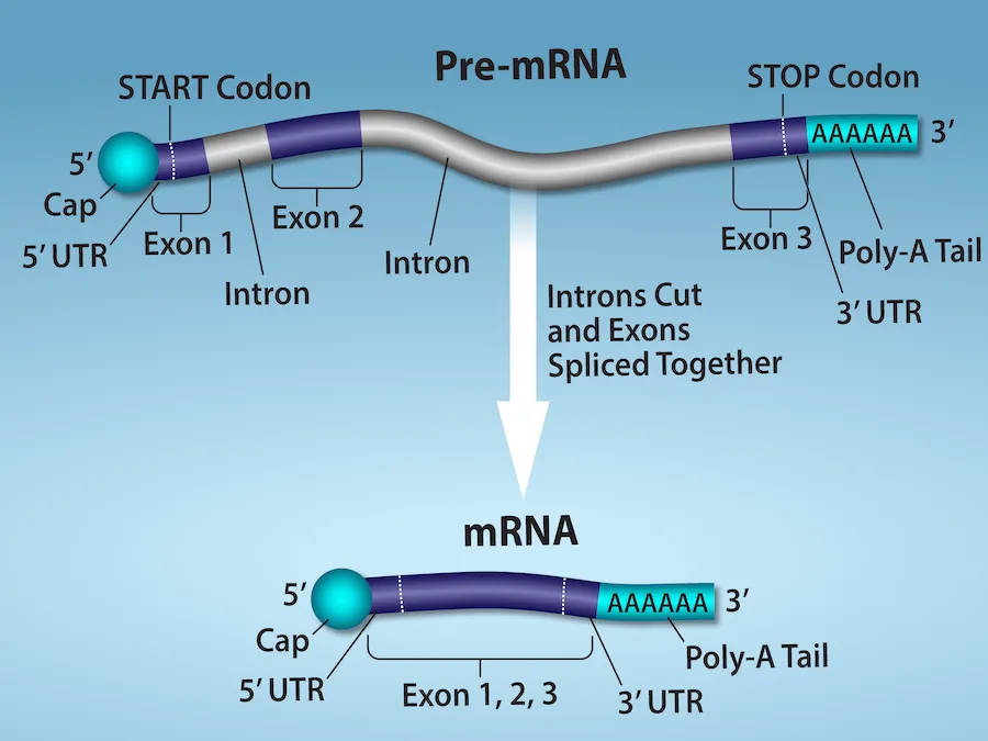
Extended description
A gray tube-shaped strand labeled ‘Pre-mRNA’ runs left to right. At its far left end sits a teal ball labeled ‘5′ Cap,’ with ‘5′ UTR’ bracketing the short stretch just after it. A vertical dashed line labeled ‘START Codon’ marks the start of a purple segment labeled ‘Exon 1’; the strand narrows and lightens for a segment labeled ‘Intron,’ widens again into purple for ‘Exon 2,’ narrows for a second ‘Intron,’ and widens into purple for ‘Exon 3.’ A second dashed line labeled ‘STOP Codon’ marks the end of Exon 3, followed by a teal box of A’s labeled ‘Poly-A Tail’ and a ‘3′’ label; ‘3′ UTR’ brackets the stretch from the STOP codon line to the end of the strand. A white arrow labeled ‘Introns Cut and Exons Spliced Together’ points down to a second, shorter strand labeled ‘mRNA.’ This strand keeps the same teal ‘5′ Cap’ and ‘5′ UTR’ at its left end, a single purple segment now labeled ‘Exon 1, 2, 3’ with no intervening introns, and the same ‘3′ UTR,’ teal ‘Poly-A Tail,’ and ‘3′’ at its right end.
Evolution Connection. RNA Editing in Trypanosomes
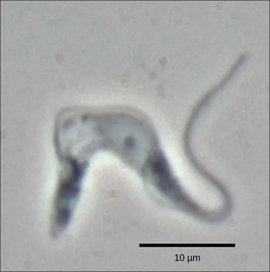
The trypanosomes are a group of protozoa that include the pathogen Trypanosoma brucei, which causes nagana in cattle and sleeping sickness in humans throughout great areas of Africa (see the micrograph below). The trypanosome is carried by biting flies in the genus Glossina (commonly called tsetse flies). Trypanosomes, and virtually all other eukaryotes, have organelles called mitochondria that supply the cell with chemical energy. Mitochondria are organelles that express their own DNA and are believed to be the remnants of a symbiotic relationship between a eukaryote and an engulfed prokaryote. The mitochondrial DNA of trypanosomes exhibit an interesting exception to the central dogma: their pre-mRNAs do not have the correct information to specify a functional protein. Usually, this is because the mRNA is missing several U nucleotides. The cell performs an additional RNA processing step called RNA editing to remedy this.
Other genes in the mitochondrial genome encode 40- to 80-nucleotide guide RNAs. One or more of these molecules interacts by complementary base pairing with some of the nucleotides in the pre-mRNA transcript. However, the guide RNA has more A nucleotides than the pre-mRNA has U nucleotides with which to bind. In these regions, the guide RNA loops out. The 3′ ends of guide RNAs have a long poly-U tail, and these U bases are inserted in regions of the pre-mRNA transcript at which the guide RNAs are looped. This process is entirely mediated by RNA molecules. That is, guide RNAs—rather than proteins—serve as the catalysts in RNA editing.
RNA editing is not just a phenomenon of trypanosomes. In the mitochondria of some plants, almost all pre-mRNAs are edited. RNA editing has also been identified in mammals such as rats, rabbits, and even humans. What could be the evolutionary reason for this additional step in pre-mRNA processing? One possibility is that the mitochondria, being remnants of ancient prokaryotes, have an equally ancient RNA-based method for regulating gene expression. In support of this hypothesis, edits made to pre-mRNAs differ depending on cellular conditions. Although speculative, the process of RNA editing may be a holdover from a primordial time when RNA molecules, instead of proteins, were responsible for catalyzing reactions.
5′ Capping
While the pre-mRNA is still being synthesized, a 7-methylguanosine cap is added to the 5′ end of the growing transcript by a phosphate linkage. This functional group protects the nascent mRNA from degradation. In addition, factors involved in protein synthesis recognize the cap to help initiate translation by ribosomes.
3′ Poly-A Tail
Once elongation is complete, the pre-mRNA is cleaved by an endonuclease between an AAUAAA consensus sequence and a GU-rich sequence, leaving the AAUAAA sequence on the pre-mRNA. An enzyme called poly-A polymerase then adds a string of approximately 200 A residues, called the poly-A tail. This modification further protects the pre-mRNA from degradation and is also the binding site for a protein necessary for exporting the processed mRNA to the cytoplasm.
Pre-mRNA Splicing
Eukaryotic genes are composed of exons, which correspond to protein-coding sequences (ex-on signifies that they are expressed), and intervening sequences called introns (int-ron denotes their intervening role), which may be involved in gene regulation but are removed from the pre-mRNA during processing. Intron sequences in mRNA do not encode functional proteins.
The discovery of introns came as a surprise to researchers in the 1970s who expected that pre-mRNAs would specify protein sequences without further processing, as they had observed in prokaryotes. The genes of higher eukaryotes very often contain one or more introns. These regions may correspond to regulatory sequences; however, the biological significance of having many introns or having very long introns in a gene is unclear. It is possible that introns slow down gene expression because it takes longer to transcribe pre-mRNAs with lots of introns. Alternatively, introns may be nonfunctional sequence remnants left over from the fusion of ancient genes throughout the course of evolution. This is supported by the fact that separate exons often encode separate protein subunits or domains. For the most part, the sequences of introns can be mutated without ultimately affecting the protein product.
All of a pre-mRNA’s introns must be completely and precisely removed before protein synthesis. If the process errs by even a single nucleotide, the reading frame of the rejoined exons would shift, and the resulting protein would be dysfunctional. The process of removing introns and reconnecting exons is called splicing (see the diagram below). Introns are removed and degraded while the pre-mRNA is still in the nucleus. Splicing occurs by a sequence-specific mechanism that ensures introns will be removed and exons rejoined with the accuracy and precision of a single nucleotide. Although the intron itself is noncoding, the beginning and end of each intron is marked with specific nucleotides: GU at the 5′ end and AG at the 3′ end of the intron. The splicing of pre-mRNAs is conducted by complexes of proteins and RNA molecules called spliceosomes.
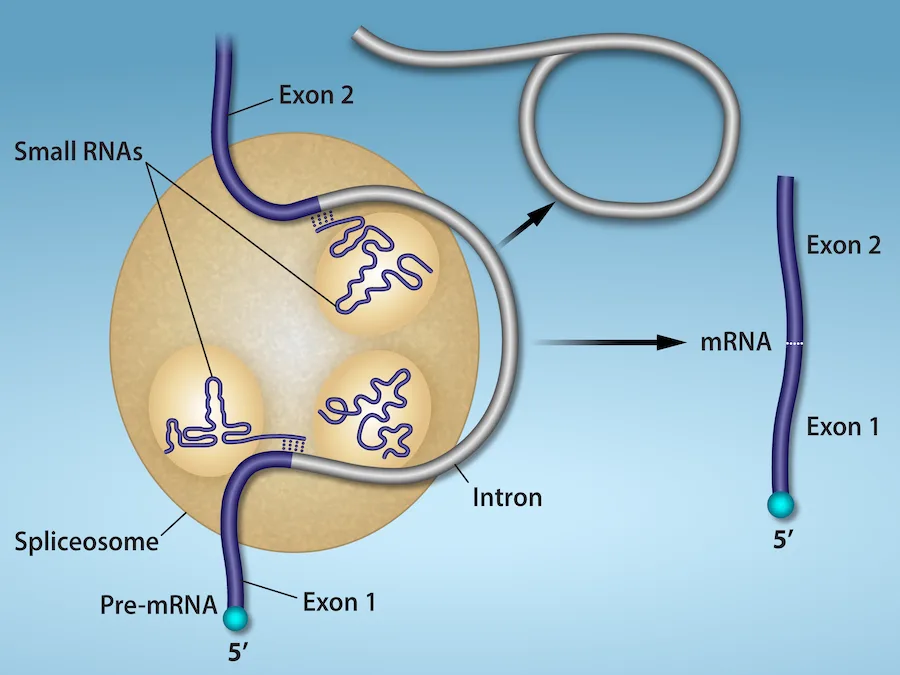
Extended description
A tan circle representing the spliceosome sits left of center. A purple strand labeled ‘Pre-mRNA’ enters from the bottom, labeled ‘5′’ at its tip, and is labeled ‘Exon 1’ where it meets the circle; a second purple strand labeled ‘Exon 2’ enters the circle from the top. Between them, inside the circle, a thin gray strand labeled ‘Intron’ loops past three small tan circles labeled ‘Small RNAs,’ each containing a squiggled purple loop. The gray intron strand exits the top of the spliceosome and loops into a large open circle at upper right. A black arrow points right from the spliceosome to a separate purple strand at the far right labeled ‘mRNA,’ with ‘Exon 2’ above and ‘Exon 1’ below, joined at a white dashed line, and ‘5′’ labeled at its lower end.
Errors in splicing are implicated in cancers and other human diseases. What kinds of mutations might lead to splicing errors? Think of different possible outcomes if splicing errors occur.
Show model answer
Did your answer mention:
Note that more than 70 individual introns can be present, and each has to undergo the process of splicing—in addition to 5′ capping and the addition of a poly-A tail—just to generate a single, translatable mRNA molecule.
Processing of tRNAs and rRNAs
The tRNAs and rRNAs are structural molecules that have roles in protein synthesis; however, these RNAs are not themselves translated. Pre-rRNAs are transcribed, processed, and assembled into ribosomes in the nucleolus. Pre-tRNAs are transcribed and processed in the nucleus and then released into the cytoplasm where they are linked to free amino acids for protein synthesis.
Most of the tRNAs and rRNAs in eukaryotes and prokaryotes are first transcribed as a long precursor molecule that spans multiple rRNAs or tRNAs. Enzymes then cleave the precursors into subunits corresponding to each structural RNA. Some of the bases of pre-rRNAs are methylated; that is, a –CH₃ methyl functional group is added for stability. Pre-tRNA molecules also undergo methylation. As with pre-mRNAs, subunit excision occurs in eukaryotic pre-RNAs destined to become tRNAs or rRNAs.
Mature rRNAs make up approximately 50 percent of each ribosome. Some of a ribosome’s RNA molecules are purely structural, whereas others have catalytic or binding activities. Mature tRNAs take on a three-dimensional structure through local regions of base pairing stabilized by intramolecular hydrogen bonding. The tRNA folds to position the amino acid binding site at one end and the anticodon at the other end (see the model below). The anticodon is a three-nucleotide sequence in a tRNA that interacts with an mRNA codon through complementary base pairing.
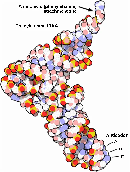
Extended description
A folded, L-shaped molecule built of overlapping spheres colored white, red, yellow, pink, and light blue represents the phenylalanine tRNA. At the upper end, a small cluster of spheres is labeled ‘Amino acid (phenylalanine) attachment site’ with an arrow pointing to it; the label ‘Phenylalanine tRNA’ sits beside this upper arm. The molecule bends near its middle and extends down and to the left before curving back down and to the right into a second arm. At the lower end of this second arm, three consecutive spheres are labeled, top to bottom, ‘A,’ ‘A,’ and ‘G’ under the heading ‘Anticodon.’
Summary
Eukaryotic pre-mRNAs are modified with a 5′ methylguanosine cap and a poly-A tail. These structures protect the mature mRNA from degradation and help export it from the nucleus. Pre-mRNAs also undergo splicing, in which introns are removed and exons are reconnected with single-nucleotide accuracy. Only finished mRNAs that have undergone 5′ capping, 3′ polyadenylation, and intron splicing are exported from the nucleus to the cytoplasm. Pre-rRNAs and pre-tRNAs may be processed by intramolecular cleavage, splicing, methylation, and chemical conversion of nucleotides. Rarely, RNA editing is also performed to insert missing bases after an mRNA has been synthesized.
Key terms
- 7-methylguanosine cap — modification added to the 5′ end of pre-mRNAs to protect mRNA from degradation and assist translation
- anticodon — three-nucleotide sequence in a tRNA molecule that corresponds to an mRNA codon
- exon — sequence present in protein-coding mRNA after completion of pre-mRNA splicing
- intron — non–protein-coding intervening sequences that are spliced from mRNA during processing
- poly-A tail — modification added to the 3′ end of pre-mRNAs to protect mRNA from degradation and assist mRNA export from the nucleus
- RNA editing — direct alteration of one or more nucleotides in an mRNA that has already been synthesized
- splicing — process of removing introns and reconnecting exons in a pre-mRNA
Practice
Describe the different steps in RNA processing
Which pre-mRNA processing step is important for initiating translation?
Re-read which of the four modifications is recognized by factors involved in protein synthesis so that ribosomes can begin translation.Chronic lymphocytic leukemia patients often harbor nonsense mutations in their spliceosome machinery. Describe how this mutation of the spliceosome would change the final location and sequence of a pre-mRNA.
Show model answer
Did your answer mention:
The modification added to the 5′ end of pre-mRNAs to protect the transcript from degradation and assist translation is called the ________.
It is added by a phosphate linkage while the transcript is still being synthesized.The direct alteration of one or more nucleotides in an mRNA that has already been synthesized is called ________.
Trypanosome mitochondria use this rare processing step to insert nucleotides a pre-mRNA is otherwise missing.The approximately 200-adenine-residue modification added to the 3′ end of a pre-mRNA to protect it from degradation and assist its export from the nucleus is called the ________.
An enzyme called poly-A polymerase adds this string of A residues after cleavage at the AAUAAA site.Understand the significance of exons, introns, and splicing for mRNAs
The sequence present in protein-coding mRNA after completion of pre-mRNA splicing is called a(n) ________.
Its name signals that it is the part of the sequence that gets expressed.A non–protein-coding intervening sequence that is spliced out of mRNA during processing is called a(n) ________.
Its name signals that it intervenes between the coding sequences.The process of removing introns and reconnecting exons in a pre-mRNA is called ________.
Complexes of proteins and RNA molecules called spliceosomes carry out this process with single-nucleotide accuracy.
A scientist identifies a pre-mRNA with the structure shown in the diagram above. What is the predicted size of the corresponding mature mRNA in base pairs (bp), excluding the 5′ cap and 3′ poly-A tail?
Add together the lengths of the three exons; the introns are removed during splicing and are not part of the mature mRNA.Explain how tRNAs and rRNAs are processed
What processing step enhances the stability of pre-tRNAs and pre-rRNAs?
Which chemical modification does the section describe as being added to some bases of pre-rRNAs, and separately to pre-tRNAs, for stability?The three-nucleotide sequence in a tRNA molecule that corresponds to an mRNA codon is called the ________.
It sits at the opposite end of the folded tRNA from the amino-acid attachment site.Pre-rRNAs and pre-tRNAs may be processed by ________, splicing, methylation, and chemical conversion of nucleotides.
Enzymes cut a single long precursor molecule into the separate rRNA or tRNA subunits it spans.This section is adapted from Biology 2e, Section 15.4: RNA Processing in Eukaryotes by Mary Ann Clark, Jung Choi, Matthew Douglas, and OpenStax, © OpenStax, licensed under CC BY-NC-SA 4.0. Access the original for free at openstax.org. Changes: figures re-encoded as WebP; Figure_15_04_03 re-kinded from the manifest’s file-extension guess of “photo” to “diagram” after inspection (it is a computer-rendered space-filling molecular model, not a photograph); a longdesc added for Figure_15_03_02, Figure_15_04_02, and Figure_15_04_03 (labeled diagrams whose full arrow/label layout is not carried by the caption alone); each source alt’s letter-by-letter TTS spelling (“m R N A,” “R N A,” “t R N A”) rewritten as “mRNA,” “RNA,” and “tRNA”; figure cross-references to numbered figures changed to descriptive references (“see the diagram below,” “see the model below”) since figures are not numbered here; the Evolution Connection and Link to Learning feature boxes rendered as callouts, the latter with descriptive link text in place of the source’s bare “at this website”; the Visual Connection note kept in the body immediately after its figure, rendered as a self-check since the source keys it to a prose solution rather than a lettered option; the end-of-section Review Questions and Critical Thinking Question adapted into the closing interactive Practice block; the third Review Question’s diagram, embedded directly in the source problem statement rather than shown in the body, added as its own mediafigure (with a locally written caption, since the source item carries none) immediately before the multiple choice that depends on it; all seven Key Terms recall items added from the glossary; and one summary-derived cloze text-recall item (“intramolecular cleavage”) added under the third objective, since the module’s three Review Questions and one Critical Thinking Question left that objective’s group short of the book’s three-item floor; rubric checkpoints added to each self-check, decomposing its model answer (the source solution) into check-off clauses with no new claims. One number is corrected with a visible Source note: a typical E. coli mRNA lasts a few minutes, not five seconds (erratum 403).