Ribosomes and Protein Synthesis
By the end of this section, you will be able to:
- Describe the different steps in protein synthesis
- Discuss the role of ribosomes in protein synthesis
The synthesis of proteins consumes more of a cell’s energy than any other metabolic process. In turn, proteins account for more mass than any other component of living organisms (with the exception of water), and proteins perform virtually every function of a cell. The process of translation, or protein synthesis, involves the decoding of an mRNA message into a polypeptide product. Amino acids are covalently strung together by interlinking peptide bonds in lengths ranging from approximately 50 to more than 1000 amino acid residues. Each individual amino acid has an amino group (NH₂) and a carboxyl (COOH) group. Polypeptides are formed when the amino group of one amino acid forms an amide (i.e., peptide) bond with the carboxyl group of another amino acid (see the figure below). This reaction is catalyzed by ribosomes and generates one water molecule.
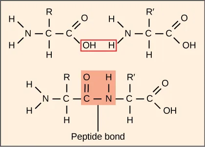
Extended description
Top panel: the left amino acid is drawn with H₂N– at upper left, a central carbon bearing R above and H below, connecting right to a carbonyl carbon double-bonded to O with an OH below it; the right amino acid mirrors this with R′ and its own carbonyl. A red-outlined box surrounds the left amino acid’s OH and the right amino acid’s amino H, sitting between the two molecules. Bottom panel repeats the same two structures, now bonded through a new C–N linkage sitting above an orange-outlined box labeled ‘Peptide bond’; the previously boxed OH and H are shown grouped together inside that orange box, and the outer amino and carboxyl groups (H₂N– on the left, –COOH on the right) remain unreacted.
The Protein Synthesis Machinery
In addition to the mRNA template, many molecules and macromolecules contribute to the process of translation. The composition of each component may vary across species; for example, ribosomes may consist of different numbers of rRNAs and polypeptides depending on the organism. However, the general structures and functions of the protein synthesis machinery are comparable from bacteria to human cells. Translation requires the input of an mRNA template, ribosomes, tRNAs, and various enzymatic factors. (Note: A ribosome can be thought of as an enzyme whose amino acid binding sites are specified by mRNA.)
Ribosomes
Even before an mRNA is translated, a cell must invest energy to build each of its ribosomes. In E. coli, there are between 10,000 and 70,000 ribosomes present in each cell at any given time. A ribosome is a complex macromolecule composed of structural and catalytic rRNAs, and many distinct polypeptides. In eukaryotes, the nucleolus is completely specialized for the synthesis and assembly of rRNAs.
Ribosomes exist in the cytoplasm of prokaryotes and in the cytoplasm and rough endoplasmic reticulum of eukaryotes. Mitochondria and chloroplasts also have their own ribosomes in the matrix and stroma, which look more similar to prokaryotic ribosomes (and have similar drug sensitivities) than the ribosomes just outside their outer membranes in the cytoplasm. Ribosomes dissociate into large and small subunits when they are not synthesizing proteins and reassociate during the initiation of translation. In E. coli, the small subunit is described as 30S, and the large subunit is 50S, for a total of 70S (recall that Svedberg units are not additive). Mammalian ribosomes have a small 40S subunit and a large 60S subunit, for a total of 80S. The small subunit is responsible for binding the mRNA template, whereas the large subunit sequentially binds tRNAs. Each mRNA molecule is simultaneously translated by many ribosomes, all synthesizing protein in the same direction: reading the mRNA from 5′ to 3′ and synthesizing the polypeptide from the N terminus to the C terminus. The complete mRNA/poly-ribosome structure is called a polysome.
tRNAs
The tRNAs are structural RNA molecules that were transcribed from genes by RNA polymerase III. Depending on the species, 40 to 60 types of tRNAs exist in the cytoplasm. Transfer RNAs serve as adaptor molecules. Each tRNA carries a specific amino acid and recognizes one or more of the mRNA codons that define the order of amino acids in a protein. Aminoacyl-tRNAs bind to the ribosome and add the corresponding amino acid to the polypeptide chain. Therefore, tRNAs are the molecules that actually “translate” the language of RNA into the language of proteins.
Of the 64 possible mRNA codons—or triplet combinations of A, U, G, and C—three specify the termination of protein synthesis and 61 specify the addition of amino acids to the polypeptide chain. Of these 61, one codon (AUG) also encodes the initiation of translation. Each tRNA anticodon can base pair with one or more of the mRNA codons for its amino acid. For instance, if the sequence CUA occurred on an mRNA template in the proper reading frame, it would bind a leucine tRNA expressing the complementary sequence, GAU. The ability of some tRNAs to match more than one codon is what gives the genetic code its blocky structure.
As the adaptor molecules of translation, it is surprising that tRNAs can fit so much specificity into such a small package. Consider that tRNAs need to interact with three factors: 1) they must be recognized by the correct aminoacyl synthetase (see below); 2) they must be recognized by ribosomes; and 3) they must bind to the correct sequence in mRNA.
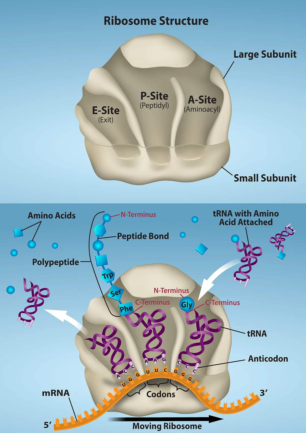
Extended description
Top panel, ‘Ribosome Structure’: a single tan ribosome shape has lines pointing to labels — ‘Large Subunit’ (upper right), ‘Small Subunit’ (lower right), and three shallow pockets across its front face labeled, left to right, ‘E-Site (Exit)’, ‘P-Site (Peptidyl)’, and ‘A-Site (Aminoacyl)’. Bottom panel: the same single ribosome rides an orange zig-zag mRNA strand, labeled ‘5′’ at its left end and ‘3′’ at its right end; where the strand passes under the ribosome, three groups of three orange nucleotide teeth (UGG, UUC, GGG) are bracketed and labeled ‘Codons’. Three purple helical tRNAs occupy the ribosome’s three pockets, their anticodons (ACC, AAG, CCC) paired to the codons below. The growing chain labeled ‘Polypeptide’ — blue shapes strung from a free ‘N-Terminus’ end, through a ‘Peptide Bond’ label, down through ‘Trp’ and ‘Ser’ to ‘Phe’ — hangs from the middle (P-site) tRNA, with ‘C-Terminus’ marked at ‘Phe’; the right (A-site) tRNA carries a single blue circle labeled ‘Gly’, marked with its own ‘N-Terminus’ and ‘C-Terminus’. A white arrow at the left shows a spent tRNA leaving the E-site. A cluster of separate blue squares, circles, and diamonds at the upper left is labeled ‘Amino Acids’, and at the upper right a purple tRNA carrying a blue circle is labeled ’tRNA with Amino Acid Attached’, arriving on a white arrow pointing into the ribosome. A black arrow below the strand, labeled ‘Moving Ribosome’, points right.
Aminoacyl tRNA Synthetases
The process of pre-tRNA synthesis by RNA polymerase III only creates the RNA portion of the adaptor molecule. The corresponding amino acid must be added later, once the tRNA is processed and exported to the cytoplasm. Through the process of tRNA “charging,” each tRNA molecule is linked to its correct amino acid by one of a group of enzymes called aminoacyl tRNA synthetases. At least one type of aminoacyl tRNA synthetase exists for each of the 20 amino acids; the exact number of aminoacyl tRNA synthetases varies by species. These enzymes first bind and hydrolyze ATP to catalyze a high-energy bond between an amino acid and adenosine monophosphate (AMP); a pyrophosphate molecule is expelled in this reaction. The activated amino acid is then transferred to the tRNA, and AMP is released. The term “charging” is appropriate, since the high-energy bond that attaches an amino acid to its tRNA is later used to drive the formation of the peptide bond. Each tRNA is named for its amino acid.
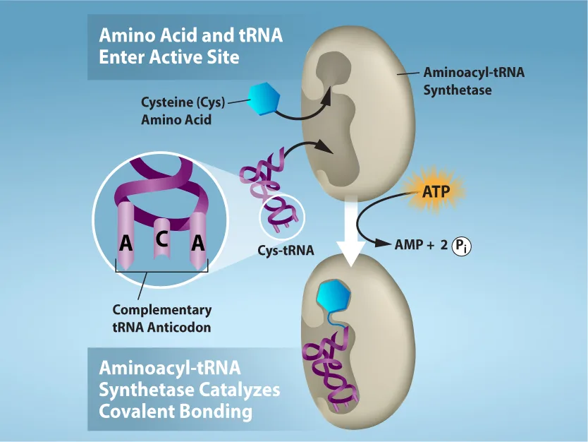
Extended description
A blue-gray background holds two dark banners. The upper banner reads ‘Amino Acid and tRNA Enter Active Site’ above a Cysteine (Cys) amino acid hexagon and a purple Cys-tRNA, both arrowing toward a tan kidney-shaped ‘Aminoacyl-tRNA Synthetase’ enzyme; an inset circle to the left zooms into the tRNA’s anticodon loop, showing three bases labeled A, C, A and captioned ‘Complementary tRNA Anticodon’; an orange starburst labeled ‘ATP’ arrows into the enzyme from the right. The lower banner reads ‘Aminoacyl-tRNA Synthetase Catalyzes Covalent Bonding’ above the same enzyme shape, now shown with the Cys-tRNA’s loop bonded to the blue amino acid at its tip; a white arrow leads down from the ATP starburst to the text ‘AMP + 2 Pᵢ’.
The Mechanism of Protein Synthesis
As with mRNA synthesis, protein synthesis can be divided into three phases: initiation, elongation, and termination. The process of translation is similar in prokaryotes and eukaryotes. Here we’ll explore how translation occurs in E. coli, a representative prokaryote, and specify any differences between prokaryotic and eukaryotic translation.
Initiation of Translation
Protein synthesis begins with the formation of an initiation complex. In E. coli, this complex involves the small 30S ribosome, the mRNA template, three initiation factors (IFs; IF-1, IF-2, and IF-3), and a special initiator tRNA, called tRNAfMet.
In E. coli mRNA, a sequence upstream of the first AUG codon, called the Shine-Dalgarno sequence (AGGAGG), interacts with the rRNA molecules that compose the ribosome. This interaction anchors the 30S ribosomal subunit at the correct location on the mRNA template. Guanosine triphosphate (GTP), which is a purine nucleotide triphosphate, acts as an energy source during translation—both at the start of elongation and during the ribosome’s translocation. Binding of the mRNA to the 30S ribosome also requires IF-3.
The initiator tRNA then interacts with the start codon AUG (or rarely, GUG). This tRNA carries the amino acid methionine, which is formylated after its attachment to the tRNA. The formylation creates a “faux” peptide bond between the formyl carboxyl group and the amino group of the methionine. Binding of the fMet-tRNAfMet is mediated by the initiation factor IF-2. The fMet begins every polypeptide chain synthesized by E. coli, but it is usually removed after translation is complete. When an in-frame AUG is encountered during translation elongation, a non-formylated methionine is inserted by a regular Met-tRNAMet. After the formation of the initiation complex, the 30S ribosomal subunit is joined by the 50S subunit to form the translation complex. In eukaryotes, a similar initiation complex forms, comprising mRNA, the 40S small ribosomal subunit, eukaryotic IFs, and nucleoside triphosphates (GTP and ATP). The methionine on the charged initiator tRNA, called Met-tRNAi, is not formylated. However, Met-tRNAi is distinct from other Met-tRNAs in that it can bind IFs.
Instead of depositing at the Shine-Dalgarno sequence, the eukaryotic initiation complex recognizes the 7-methylguanosine cap at the 5′ end of the mRNA. A cap-binding protein (CBP) and several other IFs assist the movement of the ribosome to the 5′ cap. Once at the cap, the initiation complex tracks along the mRNA in the 5′ to 3′ direction, searching for the AUG start codon. Many eukaryotic mRNAs are translated from the first AUG, but this is not always the case. According to Kozak’s rules, the nucleotides around the AUG indicate whether it is the correct start codon. Kozak’s rules state that the following consensus sequence must appear around the AUG of vertebrate genes: 5′-gccRccAUGG-3′. The R (for purine) indicates a site that can be either A or G, but cannot be C or U. Essentially, the closer the sequence is to this consensus, the higher the efficiency of translation.
Once the appropriate AUG is identified, the other proteins and CBP dissociate, and the 60S subunit binds to the complex of Met-tRNAi, mRNA, and the 40S subunit. This step completes the initiation of translation in eukaryotes.
Translation, Elongation, and Termination
In prokaryotes and eukaryotes, the basics of elongation are the same, so we will review elongation from the perspective of E. coli. When the translation complex is formed, the tRNA binding region of the ribosome consists of three compartments. The A (aminoacyl) site binds incoming charged aminoacyl tRNAs. The P (peptidyl) site binds charged tRNAs carrying amino acids that have formed peptide bonds with the growing polypeptide chain but have not yet dissociated from their corresponding tRNA. The E (exit) site releases dissociated tRNAs so that they can be recharged with free amino acids. The initiating methionyl-tRNA, however, occupies the P site at the beginning of the elongation phase of translation in both prokaryotes and eukaryotes.
During translation elongation, the mRNA template provides tRNA binding specificity. As the ribosome moves along the mRNA, each mRNA codon comes into register, and specific binding with the corresponding charged tRNA anticodon is ensured. If mRNA were not present in the elongation complex, the ribosome would bind tRNAs nonspecifically and randomly.
Elongation proceeds with charged tRNAs sequentially entering and leaving the ribosome as each new amino acid is added to the polypeptide chain. Movement of a tRNA from A to P to E site is induced by conformational changes that advance the ribosome by three bases in the 3′ direction. The energy for each step along the ribosome is donated by elongation factors that hydrolyze GTP. GTP energy is required both for the binding of a new aminoacyl-tRNA to the A site and for its translocation to the P site after formation of the peptide bond. Peptide bonds form between the amino group of the amino acid attached to the A-site tRNA and the carboxyl group of the amino acid attached to the P-site tRNA. The formation of each peptide bond is catalyzed by peptidyl transferase, an RNA-based enzyme that is integrated into the 50S ribosomal subunit. The energy for each peptide bond formation is derived from the high-energy bond linking each amino acid to its tRNA. After peptide bond formation, the A-site tRNA that now holds the growing peptide chain moves to the P site, and the P-site tRNA that is now empty moves to the E site and is expelled from the ribosome (see the figure below). Amazingly, the E. coli translation apparatus takes only 0.05 seconds to add each amino acid, meaning that a 200-amino-acid protein can be translated in just 10 seconds.
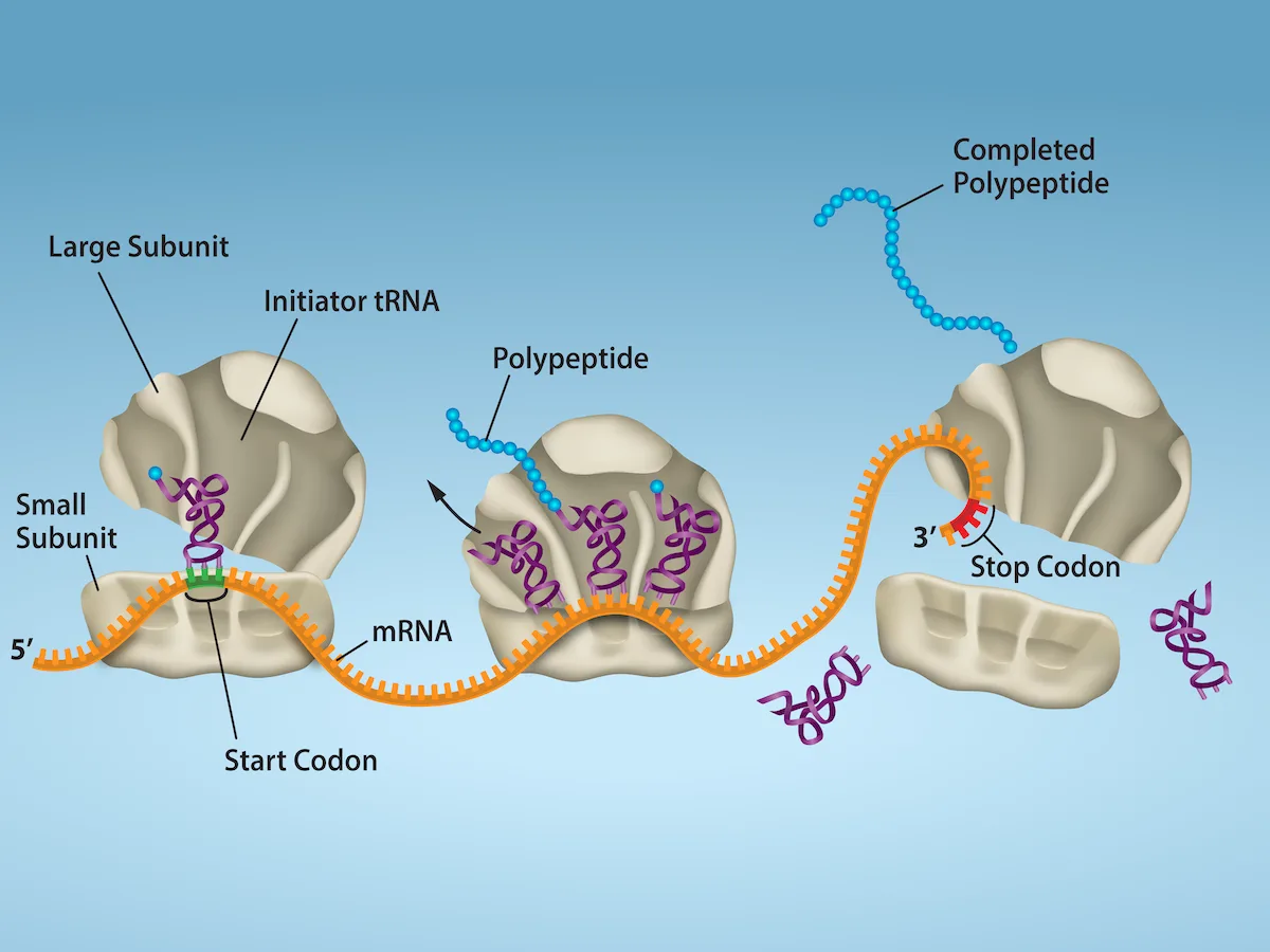
Extended description
Left to right along an orange mRNA strand labeled ‘5′’ at its left end: a small ribosomal subunit sits at a green-highlighted ‘Start Codon,’ with a purple ‘Initiator tRNA’ in place and a tan ‘Large Subunit’ arriving from above with an arrow; a ‘Small Subunit’ label points to the small ribosomal piece. Moving right, a complete ribosome holds three purple tRNAs bearing a growing blue ‘Polypeptide’ chain, with an arrow showing an empty tRNA leaving toward the upper left. Farther right, a fully formed blue chain labeled ‘Completed Polypeptide’ floats free at the top, with an empty purple tRNA drifting away below it. At the far right, a ribosome sits over a red-highlighted ‘Stop Codon’ near the mRNA’s ‘3′’ end, with its subunits shown separated above and below the strand.
Many antibiotics inhibit bacterial protein synthesis. For example, tetracycline blocks the A site on the bacterial ribosome, and chloramphenicol blocks peptidyl transfer.
Tetracycline blocks the A site on the bacterial ribosome. Which of the following processes would tetracycline directly affect?
The A site is where an incoming charged tRNA first binds the ribosome, before any peptide bond forms.Chloramphenicol blocks peptidyl transfer. Which of the following processes would chloramphenicol directly affect?
Peptidyl transferase catalyzes the peptide bonds that lengthen the polypeptide chain one amino acid at a time.Termination of translation occurs when a nonsense codon (UAA, UAG, or UGA) is encountered. Upon aligning with the A site, these nonsense codons are recognized by protein release factors that resemble tRNAs. The releasing factors in both prokaryotes and eukaryotes instruct peptidyl transferase to add a water molecule to the carboxyl end of the P-site amino acid. This reaction forces the P-site amino acid to detach from its tRNA, and the newly made protein is released. The small and large ribosomal subunits dissociate from the mRNA and from each other; they are recruited almost immediately into another translation initiation complex. After many ribosomes have completed translation, the mRNA is degraded so the nucleotides can be reused in another transcription reaction.
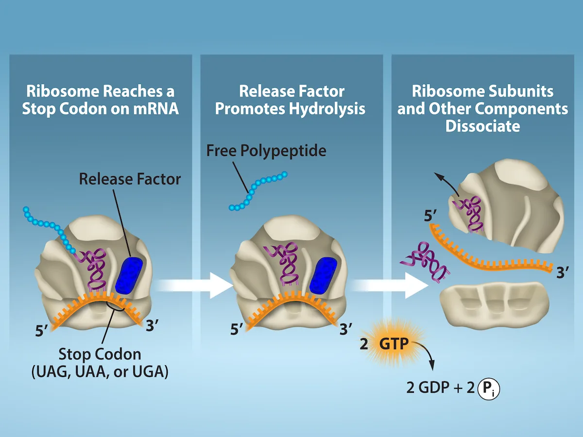
Extended description
Panel 1 (‘Ribosome Reaches a Stop Codon on mRNA’): a tan ribosome sits on an orange mRNA strand labeled 5′ at left and 3′ at right; a blue rectangular ‘Release Factor’ occupies the ribosome’s A site beside a purple tRNA carrying a short blue polypeptide, over a boxed label ‘Stop Codon (UAG, UAA, or UGA)’. Panel 2 (‘Release Factor Promotes Hydrolysis’): the same arrangement, with a short blue ‘Free Polypeptide’ chain now shown detached above the ribosome. Panel 3 (‘Ribosome Subunits and Other Components Dissociate’): the ribosome has split into an upper piece (still holding the purple tRNA) drifting up and away, and a lower piece with the mRNA strand (labeled 5′ and 3′) drifting apart from it; below the panel, an orange burst labeled ‘2 GTP’ arrows to the text ‘2 GDP + 2 Pᵢ’.
Protein Folding, Modification, and Targeting
During and after translation, individual amino acids may be chemically modified, signal sequences appended, and the new protein “folded” into a distinct three-dimensional structure as a result of intramolecular interactions. A signal sequence is a short sequence at the amino end of a protein that directs it to a specific cellular compartment. These sequences can be thought of as the protein’s “train ticket” to its ultimate destination, and are recognized by signal-recognition proteins that act as conductors. For instance, a specific signal sequence terminus will direct a protein to the mitochondria or chloroplasts (in plants). Once the protein reaches its cellular destination, the signal sequence is usually clipped off.
Many proteins fold spontaneously, but some proteins require helper molecules, called chaperones, to prevent them from aggregating during the complicated process of folding. Even if a protein is properly specified by its corresponding mRNA, it could take on a completely dysfunctional shape if abnormal temperature or pH conditions prevent it from folding correctly.
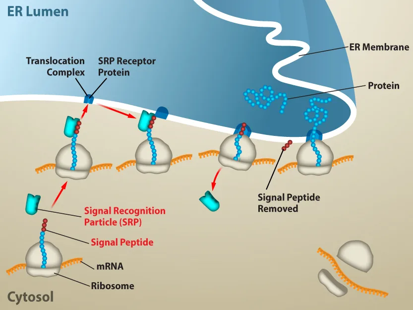
Extended description
Left to right, five ribosome-mRNA complexes trace the pathway through a region labeled ‘Cytosol,’ and a sixth ribosome at the lower right sits dissociated into its two separate subunits beside an mRNA strand. The first (bottom left) is labeled with ‘Ribosome’ and ‘mRNA’; from it rises a blue polypeptide topped with a red bead labeled ‘Signal Peptide,’ which a teal L-shaped ‘Signal Recognition Particle (SRP)’ clamps onto, shown with an upward arrow. The second complex shows this SRP-bound ribosome now positioned just under the ER membrane, with red arrows pointing from it to a small blue ‘Translocation Complex’ and ‘SRP Receptor Protein’ embedded in the wavy blue-shaded ER membrane at the top of the image, which separates the ‘Cytosol’ below from the ‘ER Lumen’ above. The third complex docks at the translocation complex while the teal SRP detaches and drifts away on a downward red arrow. The fourth shows the growing chain threading directly through the membrane into the ER Lumen, with a clipped-off run of red beads beside it labeled ‘Signal Peptide Removed.’ The fifth, rightmost complex shows the chain fully through the membrane with no signal peptide; inside the ER Lumen at the upper right, two tangled blue ‘Protein’ shapes are labeled, representing completed proteins released into the lumen.
Summary
The players in translation include the mRNA template, ribosomes, tRNAs, and various enzymatic factors. The small ribosomal subunit binds to the mRNA template either at the Shine-Dalgarno sequence (prokaryotes) or the 5′ cap (eukaryotes). Translation begins at the initiating AUG on the mRNA, specifying methionine. The formation of peptide bonds occurs between sequential amino acids matched to the mRNA template by their tRNAs according to the genetic code. Charged tRNAs enter the ribosomal A site, and their amino acid bonds with the amino acid at the P site. The entire mRNA is translated in three-nucleotide “steps” of the ribosome. When a nonsense codon is encountered, a release factor binds and dissociates the components and frees the new protein. Folding of the protein occurs during and after translation.
Key terms
- aminoacyl tRNA synthetase — enzyme that “charges” tRNA molecules by catalyzing a bond between the tRNA and a corresponding amino acid
- initiator tRNA — in prokaryotes, called tRNAfMet; in eukaryotes, called tRNAi; a tRNA that interacts with a start codon, binds directly to the ribosome P site, and links to a special methionine to begin a polypeptide chain
- Kozak’s rules — determines the correct initiation AUG in a eukaryotic mRNA; the following consensus sequence must appear around the AUG: 5′-GCC(purine)CCAUGG-3′; the bolded bases are most important
- peptidyl transferase — RNA-based enzyme that is integrated into the 50S ribosomal subunit and catalyzes the formation of peptide bonds
- polysome — mRNA molecule simultaneously being translated by many ribosomes all going in the same direction
- Shine-Dalgarno sequence — (AGGAGG); initiates prokaryotic translation by interacting with rRNA molecules comprising the 30S ribosome
- signal sequence — short tail of amino acids that directs a protein to a specific cellular compartment
- start codon — AUG (or rarely, GUG) on an mRNA from which translation begins; always specifies methionine
Practice
Describe the different steps in protein synthesis
In any given species, there are at least how many types of aminoacyl tRNA synthetases?
At least one synthetase exists for each amino acid used to build proteins.Transcribe and translate the following DNA sequence (nontemplate strand): 5′-ATGGCCGGTTATTAAGCA-3′
Show model answer
Did your answer mention:
Explain how single nucleotide changes can have vastly different effects on protein function.
Show model answer
Did your answer mention:
A normal mRNA that reads 5′-UGCCAUGGUAAUAACACAUGAGGCCUGAAC-3′ has an insertion mutation that changes the sequence to 5′-UGCCAUGGUUAAUAACACAUGAGGCCUGAAC-3′. Translate the original mRNA and the mutated mRNA, and explain how insertion mutations can have dramatic effects on proteins. (Hint: Be sure to find the initiation site.)
Show model answer
Did your answer mention:
The mRNA triplet from which translation begins, and which always specifies methionine, is called the ________.
It is usually AUG, though rarely GUG.Discuss the role of ribosomes in protein synthesis
The RNA components of ribosomes are synthesized in the ________.
This is the nuclear substructure the section says is completely specialized for the synthesis and assembly of rRNAs.A scientist introduces a mutation that makes the 60S ribosomal subunit nonfunctional in a human cell line. What would be the predicted effect on translation?
The 40S subunit alone can find the AUG start codon; recall which subunit joins afterward to complete the translation complex.The RNA-based enzyme integrated into the 50S ribosomal subunit that catalyzes the formation of peptide bonds is called ________.
It is built into the ribosome’s large subunit, not encoded by a separate free-floating protein.An mRNA molecule that is simultaneously being translated by many ribosomes, all moving in the same direction, is called a ________.
The complete mRNA/poly-ribosome structure gets this single name.The sequence (AGGAGG) upstream of the first AUG codon that interacts with rRNA to position the 30S ribosomal subunit on a prokaryotic mRNA is called the ________.
Eukaryotic ribosomes have no counterpart to it; they instead recognize the 5′ cap.This section is adapted from Biology 2e, Section 15.5: Ribosomes and Protein Synthesis by Mary Ann Clark, Jung Choi, Matthew Douglas, and OpenStax, © OpenStax, licensed under CC BY-NC-SA 4.0. Access the original for free at openstax.org. Changes: figures re-encoded as WebP with kind="diagram" set explicitly on all six (three of which — Figure_15_05_01, aminoandtrna, and Targeted_Protein_Formation-XZZ — the manifest’s file-extension guess called “photo”; all six are colored line-art illustrations, confirmed by inspection); source alts rewritten from the image for four figures whose source alt was a letter-spaced screen-reader spelling (“m R N A,” “t R N A,” “A T P,” “E R Membrane”): ribosomestructure, aminoandtrna, translationtermination, and Targeted_Protein_Formation-XZZ; the other two figures’ (Figure_15_05_01, Figure_15_05_02) alts also rewritten from the image to describe the drawing’s visible layout rather than restate the caption; a longdesc added to all six figures, since each is a labeled diagram whose full panel-by-panel content is not carried by its caption; two bare numbered cross-references (to the peptide-bond figure and to the visual-connection figure) changed to “(see the figure below)” since figures are not numbered here; primes normalized to Unicode ′ throughout the section and its Practice items, since the source mixes a straight apostrophe, a curly right-single-quote, and an en dash as its 5′/3′ notation; the glossary’s “initiator tRNA” entry’s sub/superscript notation (rendered in the source as MathML, “A” with a simultaneous subscript f and superscript Met) set to match the body’s own tRNAfMet / tRNAi glyphs for one consistent notation; the Visual Connection note’s two-part antibiotic question (keyed “Tetracycline: a; Chloramphenicol: c”) split into two separate multiple-choice components, kept in the body immediately after the figure, each restating its own antibiotic’s mechanism from the source stem so it stands alone; the interactive note rendered as a Link to Learning callout with descriptive link text (“this PBS interactive”) in place of the source’s own link text; the end-of-section Review Questions and Critical Thinking Questions adapted into the closing interactive Practice block (multiple choice and self-check respectively), with rubric checkpoints added to each self-check, decomposing its model answer (the source solution, lightly reformatted into complete sentences) into check-off clauses with no new claims; and four key-term recall items (start codon, peptidyl transferase, polysome, Shine-Dalgarno sequence) added from the glossary to bring both Practice groups to the book’s floor.