Eukaryotic Epigenetic Gene Regulation
By the end of this section, you will be able to:
- Explain how chromatin remodeling controls transcriptional access
- Describe how access to DNA is controlled by histone modification
- Describe how DNA methylation is related to epigenetic gene changes
Eukaryotic gene expression is more complex than prokaryotic gene expression because the processes of transcription and translation are physically separated. Unlike prokaryotic cells, eukaryotic cells can regulate gene expression at many different levels. Epigenetic changes are inheritable changes in gene expression that do not result from changes in the DNA sequence. Eukaryotic gene expression begins with control of access to the DNA. Transcriptional access to the DNA can be controlled in two general ways: chromatin remodeling and DNA methylation. Chromatin remodeling changes the way that DNA is associated with chromosomal histones. DNA methylation is associated with developmental changes and gene silencing.
Epigenetic Control: Regulating Access to Genes within the Chromosome
The human genome encodes over 20,000 genes, with hundreds to thousands of genes on each of the 23 human chromosomes. The DNA in the nucleus is precisely wound, folded, and compacted into chromosomes so that it will fit into the nucleus. It is also organized so that specific segments can be accessed as needed by a specific cell type.
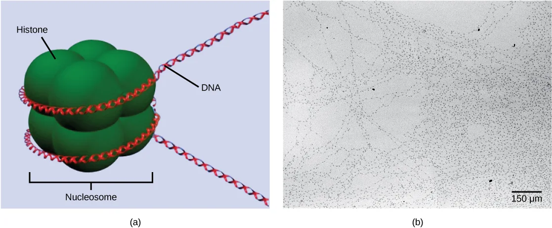
The first level of organization, or packing, is the winding of DNA strands around histone proteins. Histones package and order DNA into structural units called nucleosome complexes, which can control the access of proteins to the DNA regions (see the figure below, panel a). Under the electron microscope, this winding of DNA around histone proteins to form nucleosomes looks like small beads on a string (panel b).
Extended description
(a) A 3D rendering of a single nucleosome: a tightly fused cluster of large, rounded, dark-green histone lobes forms one compact globular shape. A red-and-white DNA double helix winds around the cluster twice; a line labeled ‘DNA’ points to the helix, a line labeled ‘Histone’ points to the green cluster, and a bracket labeled ‘Nucleosome’ spans the whole cluster. Free DNA helix continues beyond each side of the nucleosome. (b) A grayscale electron micrograph: many thin, thread-like DNA strands loop loosely across the field, each dotted with small dark round particles spaced along its length so that stretches of nucleosomes look like beads threaded loosely on a string; a scale bar in the lower right reads 150 µm.
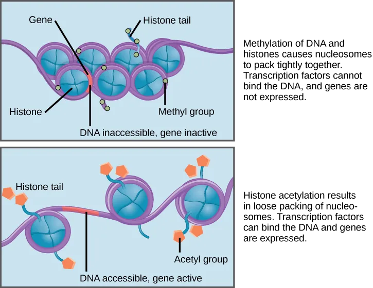
These beads (histone proteins) can move along the string (DNA) to expose different sections of the molecule. If DNA encoding a specific gene is to be transcribed into RNA, the nucleosomes surrounding that region of DNA can slide down the DNA to open that specific chromosomal region and allow for the transcriptional machinery (RNA polymerase) to initiate transcription (see the figure below).
Extended description
Top panel: seven blue-teal nucleosome circles sit packed in two interleaved rows — four behind and three in front — along a purple DNA strand. A line labeled ‘Gene’ points to a short red segment of exposed DNA between two of the nucleosomes, captioned ‘DNA inaccessible, gene inactive.’ A line labeled ‘Histone’ points to one of the circles; a line labeled ‘Histone tail’ points to a short projecting strand topped by a small pale-yellow dot; a line labeled ‘Methyl group’ points to another of those pale-yellow dots. Bottom panel: three blue-teal nucleosome circles sit spaced apart along the purple DNA strand, with visible stretches of exposed DNA between them, one highlighted red and captioned ‘DNA accessible, gene active.’ A line labeled ‘Histone tail’ points to a projecting strand; a line labeled ‘Acetyl group’ points to one of several small orange pentagons attached to the projecting histone tails.
In females, one of the two X chromosomes is inactivated during embryonic development because of epigenetic changes to the chromatin. What impact do you think these changes would have on nucleosome packing?
Show model answer
Did your answer mention:
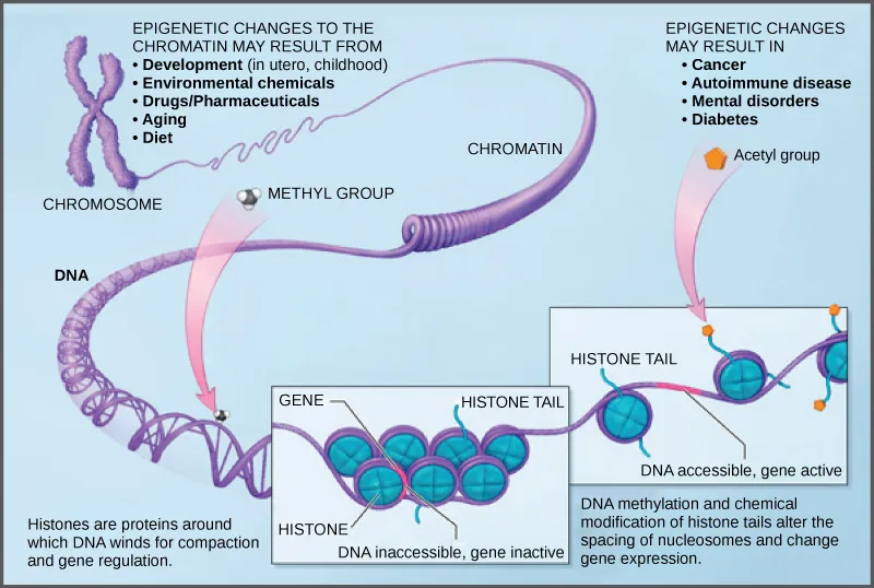
How closely the histone proteins associate with the DNA is regulated by signals found on both the histone proteins and on the DNA. These signals are functional groups added to histone proteins or to DNA and determine whether a chromosomal region should be open or closed (see the figure below, which depicts modifications to histone proteins and DNA). These tags are not permanent, but may be added or removed as needed. Some chemical groups (phosphate, methyl, or acetyl groups) are attached to specific amino acids in histone “tails” at the N-terminus of the protein. These groups do not alter the DNA base sequence, but they do alter how tightly wound the DNA is around the histone proteins. DNA is a negatively charged molecule and unmodified histones are positively charged; therefore, changes in the charge of the histone will change how tightly wound the DNA molecule will be. By adding chemical modifications like acetyl groups, the charge becomes less positive, and the binding of DNA to the histones is relaxed. Altering the location of nucleosomes and the tightness of histone binding opens some regions of chromatin to transcription and closes others.
The DNA molecule itself can also be modified by methylation. DNA methylation occurs within very specific regions called CpG islands. These are stretches with a high frequency of cytosine and guanine dinucleotide DNA pairs (CG) found in the promoter regions of genes. The cytosine member of the CG pair can be methylated (a methyl group is added). Methylated genes are usually silenced, although methylation may have other regulatory effects. In some cases, genes that are silenced during the development of the gametes of one parent are transmitted in their silenced condition to the offspring. Such genes are said to be imprinted. Parental diet or other environmental conditions may also affect the methylation patterns of genes, which in turn modifies gene expression. Changes in chromatin organization interact with DNA methylation. DNA methyltransferases appear to be attracted to chromatin regions with specific histone modifications. Highly methylated (hypermethylated) DNA regions with deacetylated histones are tightly coiled and transcriptionally inactive.
Extended description
A purple X-shaped chromosome at the upper left unravels rightward into a looser coiled fiber labeled ‘Chromatin,’ which continues down and left into a bare DNA double helix labeled ‘DNA.’ A bulleted list at the upper left, headed ‘Epigenetic changes to the chromatin may result from,’ names Development (in utero, childhood), Environmental chemicals, Drugs/Pharmaceuticals, Aging, and Diet. A bulleted list at the upper right, headed ‘Epigenetic changes may result in,’ names Cancer, Autoimmune disease, Mental disorders, and Diabetes. A pink arrow carries a gray sphere labeled ‘Methyl group’ down from the chromatin toward the DNA strand, pointing into a left inset box. A second pink arrow carries an orange pentagon labeled ‘Acetyl group’ down toward a right inset box. The left inset shows a dense cluster of teal nucleosome circles crowded tightly together along the DNA, too close to count individually, labeled ‘Gene,’ ‘Histone,’ and ‘Histone tail,’ captioned ‘DNA inaccessible, gene inactive,’ with the note ‘Histones are proteins around which DNA winds for compaction and gene regulation’ beneath it. The right inset shows three teal nucleosome circles spaced apart along the DNA with orange acetyl groups on projecting histone tails, labeled ‘Histone tail,’ captioned ‘DNA accessible, gene active,’ with the note ‘DNA methylation and chemical modification of histone tails alter the spacing of nucleosomes and change gene expression’ beneath it.
Epigenetic changes are not permanent, although they often persist through multiple rounds of cell division and may even cross generational lines. Chromatin remodeling alters the chromosomal structure (open or closed) as needed. If a gene is to be transcribed, the histone proteins and DNA in the chromosomal region encoding that gene are modified in a way that opens the promoter region to allow RNA polymerase and other proteins, called transcription factors, to bind and initiate transcription. If a gene is to remain turned off, or silenced, the histone proteins and DNA have different modifications that signal a closed chromosomal configuration. In this closed configuration, the RNA polymerase and transcription factors do not have access to the DNA and transcription cannot occur (see the figure above).
Summary
In eukaryotic cells, the first stage of gene-expression control occurs at the epigenetic level. Epigenetic mechanisms control access to the chromosomal region to allow genes to be turned on or off. Chromatin remodeling controls how DNA is packed into the nucleus by regulating how tightly the DNA is wound around histone proteins. The DNA itself may be methylated to selectively silence genes. The addition or removal of chemical modifications (or flags) to histone proteins or DNA signals the cell to open or close a chromosomal region. Therefore, eukaryotic cells can control whether a gene is expressed by controlling accessibility to the binding of RNA polymerase and its transcription factors.
Key terms
- transcription factor — protein that binds to the DNA at the promoter or enhancer region and that influences transcription of a gene
Practice
Explain how chromatin remodeling controls transcriptional access
What are epigenetic modifications?
Epigenetic changes never alter the DNA base sequence itself — think about what kind of change to histones and DNA can be added and later removed.Which of the following are true of epigenetic changes?
Check each of the first three statements against the section — if every one of them holds true, the last option follows.A protein that binds to the DNA at the promoter or enhancer region and that influences transcription of a gene is called a ________.
RNA polymerase needs this kind of protein’s help to bind the promoter or enhancer and initiate transcription.Describe how access to DNA is controlled by histone modification
Some autoimmune diseases show a positive correlation with dramatically decreased expression of histone deacetylase 9 (HDAC9, an enzyme that removes acetyl groups from histones). Why would the decreased expression of HDAC9 cause immune cells to produce inflammatory genes at inappropriate times?
Show model answer
Did your answer mention:
In cancer cells, alteration to epigenetic modifications turns off genes that are normally expressed. Hypothetically, how could you reverse this process to turn these genes back on?
Show model answer
Did your answer mention:
According to the section summary, chromatin remodeling controls how DNA is packed into the nucleus by regulating how tightly the DNA is wound around ________.
This is the type of protein that a nucleosome is built from.Describe how DNA methylation is related to epigenetic gene changes
A scientific study demonstrated that rat mothering behavior impacts the stress response in their pups. Rats that were born and grew up with attentive mothers showed low activation of stress-response genes later in life, while rats with inattentive mothers had high activation of stress-response genes in the same situation. An additional study that swapped the pups at birth (i.e., rats born to inattentive mothers grew up with attentive mothers and vice versa) showed the same positive effect of attentive mothering. How do genetics and/or epigenetics explain the results of this study?
Show model answer
Did your answer mention:
According to the section summary, the DNA itself may be methylated to selectively ________.
This is the same effect methylation is described as having on genes earlier in the section — think about what ‘usually silenced’ becomes as a verb phrase.DNA methylation occurs within very specific regions called ________.
These are stretches with a high frequency of cytosine and guanine dinucleotide pairs, found in the promoter regions of genes.This section is adapted from Biology 2e, Section 16.3: Eukaryotic Epigenetic Gene Regulation by Mary Ann Clark, Jung Choi, Matthew Douglas, and OpenStax, © OpenStax, licensed under CC BY-NC-SA 4.0. Access the original for free at openstax.org. Changes: figures re-encoded as WebP; Figure_16_03_01ab and Figure_16_03_03-6d14 re-kinded from the manifest’s file-extension guess of “photo” to “diagram” after inspection (both are illustrated diagrams, not photographs — Figure_16_03_01ab pairs a rendered diagram, panel (a), with a genuine electron micrograph, panel (b), and the labeled diagram carries the teaching); all three figures’ source alt text (which used letter-spaced screen-reader spellings such as “D N A”) rewritten from the images, with a longdesc added to each since none of the three figures’ full label content — histone/DNA/nucleosome labels, gene/methyl/acetyl-group labels, or the chromosome-to-chromatin-to-DNA unraveling with its two cause/effect bullet lists — is carried by its one- or two-sentence caption; the visual-connection note kept in the body immediately after its figure and rendered as a self-check, since the source keys it to a prose solution rather than a lettered option, with its one-sentence source solution kept verbatim as the model answer; the interactive note rendered as a Link to Learning callout with descriptive anchor text kept the source’s video URL; the end-of-section Review Questions and all three Critical Thinking Questions adapted into the closing interactive Practice block (multiple choice and self-check respectively); one key-term recall item (transcription factor) added from the glossary; rubric checkpoints added to each self-check, decomposing its model answer (the source solution) into check-off clauses with no new claims; and two summary-derived items added under the second and third objective groups (a select-the-term multiple choice on what DNA winds around, and a cloze recall on what selective methylation does to genes), plus one locally written text-recall item strictly from the section’s own sentence naming CpG islands, since no source exercise or glossary term in this section otherwise tests DNA methylation with an auto-graded item; the nucleosome micrograph (Figure_16_03_01ab, part b) prints a scale bar reading 150 µm while the source’s own alt text for the same image says 150 nm — the alt here describes the bar as printed, and the mismatch is reported as a source defect (erratum 318).