Eukaryotic Post-transcriptional Gene Regulation
By the end of this section, you will be able to:
- Understand RNA splicing and explain its role in regulating gene expression
- Describe the importance of RNA stability in gene regulation
RNA is transcribed, but must be processed into a mature form before translation can begin. This processing that takes place after an RNA molecule has been transcribed, but before it is translated into a protein, is called post-transcriptional modification. As with the epigenetic and transcriptional stages of processing, this post-transcriptional step can also be regulated to control gene expression in the cell. If the RNA is not processed, shuttled, or translated, then no protein will be synthesized.
RNA Splicing, the First Stage of Post-transcriptional Control
In eukaryotic cells, the RNA transcript often contains regions, called introns, that are removed prior to translation. The regions of RNA that code for protein are called exons (see the diagram below). After an RNA molecule has been transcribed, but prior to its departure from the nucleus to be translated, the RNA is processed and the introns are removed by splicing. Splicing is done by spliceosomes, ribonucleoprotein complexes that can recognize the two ends of the intron, cut the transcript at those two points, and bring the exons together for ligation.
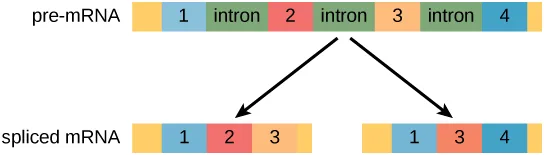
Extended description
A single row labeled ‘pre-mRNA’ shows, left to right, a plain colored end band, four numbered exon boxes (1, 2, 3, 4, each a different color), separated by three green boxes labeled ‘intron,’ and a plain colored end band. Two black arrows point down from this row to two shorter rows labeled ‘spliced mRNA’: the left one keeps exon boxes 1, 2, and 3 with the introns removed and the end bands unchanged; the right one keeps exon boxes 1, 3, and 4, omitting exon 2, again with the introns removed and the end bands unchanged.
Evolution Connection. Alternative RNA Splicing
In the 1970s, genes were first observed that exhibited alternative RNA splicing. Alternative RNA splicing is a mechanism that allows different protein products to be produced from one gene when different combinations of exons are combined to form the mRNA (see the diagram below). This alternative splicing can be haphazard, but more often it is controlled and acts as a mechanism of gene regulation, with the frequency of different splicing alternatives controlled by the cell as a way to control the production of different protein products in different cells or at different stages of development. Alternative splicing is now understood to be a common mechanism of gene regulation in eukaryotes; according to one estimate, 70 percent of genes in humans are expressed as multiple proteins through alternative splicing. Although there are multiple ways to alternatively splice RNA transcripts, the original 5′-3′ order of the exons is always conserved. That is, a transcript with exons 1 2 3 4 5 6 7 might be spliced 1 2 4 5 6 7 or 1 2 3 6 7, but never 1 2 5 4 3 6 7.
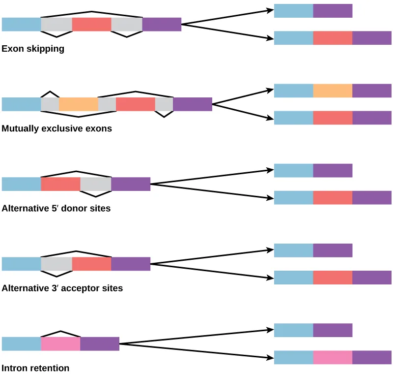
Extended description
Five rows, top to bottom, each showing a pre-mRNA on the left with two output arrows to spliced products on the right, labeled beneath: (1) Exon skipping — a blue exon, a red optional exon bounded by gray splice-choice shapes, and a purple exon; the top output skips the red exon (blue, purple only), the bottom output keeps it (blue, red, purple). (2) Mutually exclusive exons — a blue exon, an orange exon and a red exon shown as gray-bounded alternatives, and a purple exon; the top output keeps blue, orange, and purple, the bottom keeps blue, red, and purple. (3) Alternative 5′ donor sites — a blue exon, a gray splice-choice region of variable size, a red exon, and a purple exon; the top output keeps blue and purple only, the bottom keeps blue, red, and purple. (4) Alternative 3′ acceptor sites — the same three-exon layout with the gray region trailing the red exon instead of leading it; the top output again keeps blue and purple only, the bottom keeps all three. (5) Intron retention — a blue exon, a pink intron, and a purple exon; the top output keeps only blue and purple with the intron removed, the bottom keeps blue, the pink intron, and purple.
How could alternative splicing evolve? Introns have a beginning- and ending-recognition sequence; it is easy to imagine the failure of the splicing mechanism to identify the end of an intron and instead find the end of the next intron, thus removing two introns and the intervening exon. In fact, there are mechanisms in place to prevent such intron skipping, but mutations are likely to lead to their failure. Such “mistakes” would more than likely produce a nonfunctional protein. Indeed, the cause of many genetic diseases is abnormal splicing rather than mutations in a coding sequence. However, alternative splicing could possibly create a protein variant without the loss of the original protein, opening up possibilities for adaptation of the new variant to new functions. Gene duplication has played an important role in the evolution of new functions in a similar way by providing genes that may evolve without eliminating the original, functional protein.
Question: In the corn snake Pantherophis guttatus, there are several different color variants, including amelanistic snakes whose skin patterns display only red and yellow pigments. The cause of amelanism in these snakes was recently identified as the insertion of a transposable element into an intron in the OCA2 (oculocutaneous albinism) gene. How might the insertion of extra genetic material into an intron lead to a nonfunctional protein?
Control of RNA Stability
Before the mRNA leaves the nucleus, it is given two protective “caps” that prevent the ends of the strand from degrading during its journey. 5′ and 3′ exonucleases can degrade unprotected RNAs. The 5′ cap, which is placed on the 5′ end of the mRNA, is usually composed of a methylated guanosine triphosphate molecule (GTP). The GTP is placed “backward” on the 5′ end of the mRNA, so that the 5′ carbons of the GTP and the terminal nucleotide are linked through three phosphates. The poly-A tail, which is attached to the 3′ end, is usually composed of a long chain of adenine nucleotides. These changes protect the two ends of the RNA from exonuclease attack.
Once the RNA is transported to the cytoplasm, the length of time that the RNA resides there can be controlled. Each RNA molecule has a defined lifespan and decays at a specific rate. This rate of decay can influence how much protein is in the cell. If the decay rate is increased, the RNA will not exist in the cytoplasm as long, shortening the time available for translation of the mRNA to occur. Conversely, if the rate of decay is decreased, the mRNA molecule will reside in the cytoplasm longer and more protein can be translated. This rate of decay is referred to as the RNA stability. If the RNA is stable, it will be detected for longer periods of time in the cytoplasm.
Binding of proteins to the RNA can also influence its stability. Proteins called RNA-binding proteins, or RBPs, can bind to the regions of the mRNA just upstream or downstream of the protein-coding region. These regions in the RNA that are not translated into protein are called the untranslated regions, or UTRs. They are not introns (those have been removed in the nucleus). Rather, these are regions that regulate mRNA localization, stability, and protein translation. The region just before the protein-coding region is called the 5′ UTR, whereas the region after the coding region is called the 3′ UTR (see the diagram below). The binding of RBPs to these regions can increase or decrease the stability of an RNA molecule, depending on the specific RBP that binds.
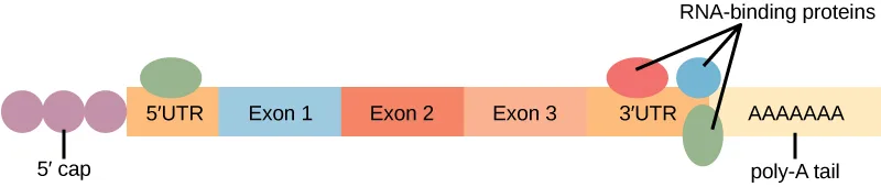
Extended description
Left to right: three small pink circles labeled ‘5′ cap’; an orange box labeled ‘5′ UTR’; three boxes labeled ‘Exon 1,’ ‘Exon 2,’ and ‘Exon 3’; an orange box labeled ‘3′ UTR’; and a row of A’s labeled ‘poly-A tail.’ A green oval sits above the 5′ UTR box. Above the 3′ UTR box, a red oval and a blue oval are pointed to by an arrow from the label ‘RNA-binding proteins’; a second green oval sits just below, between the 3′ UTR and the poly-A tail.
RNA Stability and microRNAs
In addition to RBPs that bind to and control (increase or decrease) RNA stability, other elements called microRNAs can bind to the RNA molecule. These microRNAs, or miRNAs, are short RNA molecules that are only 21 to 24 nucleotides in length. The miRNAs are made in the nucleus as longer pre-miRNAs. These pre-miRNAs are chopped into mature miRNAs by a protein called Dicer. Like transcription factors and RBPs, mature miRNAs recognize a specific sequence and bind to the RNA; however, miRNAs also associate with a ribonucleoprotein complex called the RNA-induced silencing complex (RISC). The RNA component of the RISC base-pairs with complementary sequences on an mRNA and either impede translation of the message or lead to the degradation of the mRNA.
Summary
Post-transcriptional control can occur at any stage after transcription, including RNA splicing and RNA stability. Once RNA is transcribed, it must be processed to create a mature RNA that is ready to be translated. This involves the removal of introns that do not code for protein. Spliceosomes bind to the signals that mark the exon/intron border to remove the introns and ligate the exons together. Once this occurs, the RNA is mature and can be translated. Alternative splicing can produce more than one mRNA from a given transcript. Different splicing variants may be produced under different conditions.
RNA is created and spliced in the nucleus, but needs to be transported to the cytoplasm to be translated. RNA is transported to the cytoplasm through the nuclear pore complex. Once the RNA is in the cytoplasm, the length of time it resides there before being degraded, called RNA stability, can also be altered to control the overall amount of protein that is synthesized. The RNA stability can be increased, leading to longer residency time in the cytoplasm, or decreased, leading to shortened time and less protein synthesis. RNA stability is controlled by RNA-binding proteins (RBPs) and microRNAs (miRNAs). These RBPs and miRNAs bind to the 5′ UTR or the 3′ UTR of the RNA to increase or decrease RNA stability. MicroRNAs associated with RISC complexes may repress translation or lead to mRNA breakdown.
Key terms
- 3′ UTR — 3′ untranslated region; region just downstream of the protein-coding region in an RNA molecule that is not translated
- 5′ cap — a methylated guanosine triphosphate (GTP) molecule that is attached to the 5′ end of a messenger RNA to protect the end from degradation
- 5′ UTR — 5′ untranslated region; region just upstream of the protein-coding region in an RNA molecule that is not translated
- Dicer — enzyme that chops the pre-miRNA into the mature form of the miRNA
- microRNA (miRNA) — small RNA molecules (approximately 21 nucleotides in length) that bind to RNA molecules to degrade them
- poly-A tail — a series of adenine nucleotides that are attached to the 3′ end of an mRNA to protect the end from degradation
- RNA-binding protein (RBP) — protein that binds to the 3′ or 5′ UTR to increase or decrease the RNA stability
- RNA stability — how long an RNA molecule will remain intact in the cytoplasm
- untranslated region — segment of the RNA molecule that is not translated into protein. These regions lie before (upstream or 5′) and after (downstream or 3′) the protein-coding region
- RISC — protein complex that binds along with the miRNA to the RNA to degrade it
Practice
Understand RNA splicing and explain its role in regulating gene expression
Which of the following are involved in post-transcriptional control?
Check whether the section treats each option as its own control point once the RNA has been made — each has its own subsection.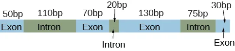
An unprocessed pre-mRNA has the structure shown in the diagram above. Which of the following is not a possible size, in base pairs, of the mature mRNA?
Add up different combinations of exon lengths that keep their original left-to-right order; one of the four options cannot be reached by any such combination.Alternative splicing has been estimated to occur in more than 95% of multi-exon genes. Which of the following is not an evolutionary advantage of alternative splicing?
Three of the four options describe how one gene can do more without changing anything about the genome or how the gene itself is expressed; one option describes a byproduct, not an advantage.Describe the importance of RNA stability in gene regulation
Binding of an RNA binding protein will ________ the stability of the RNA molecule.
The text says the specific RBP that binds determines the direction of the effect — re-read the last sentence of the RNA-binding-protein paragraph.Describe how RBPs can prevent miRNAs from degrading an RNA molecule.
Show model answer
Did your answer mention:
How can external stimuli alter post-transcriptional control of gene expression?
Show model answer
Did your answer mention:
A protein that binds just upstream or downstream of an mRNA’s protein-coding region to increase or decrease its stability is called a(n) ________.
This section abbreviates the term with three letters, always ending in P for protein.A segment of an RNA molecule that lies before or after the protein-coding region and is not itself translated into protein is called a(n) ________.
The section names two of these regions, one on each side of the protein-coding sequence.A series of adenine nucleotides attached to the 3′ end of an mRNA to protect it from degradation is called the ________.
It is a long chain of a single repeated nucleotide, attached opposite the end that gets the 5′ cap.Short RNA molecules only 21 to 24 nucleotides long that bind to RNA molecules and can lead to their degradation are called ________.
Dicer chops the longer pre-form of this molecule into its mature, functional form.This section is adapted from Biology 2e, Section 16.5: Eukaryotic Post-transcriptional Gene Regulation by Mary Ann Clark, Jung Choi, Matthew Douglas, and OpenStax, © OpenStax, licensed under CC BY-NC-SA 4.0. Access the original for free at openstax.org. Changes: figures re-encoded as WebP; Figure_16_05_03-becb and Figure_16_05_02 re-kinded from the manifest’s file-extension guess of “photo” to “diagram” after inspection (both are colored schematic illustrations, not photographs), and their source alts, which were letter-spaced text-to-speech spellings (“m R N A,” “5 prime”), rewritten from the images, with a full element-by-element walkthrough moved into a longdesc for each; Figure_15_04_02-0563 likewise re-kinded from “photo” to “diagram,” but its source alt was already an accurate, unspaced description — it was shortened to a one-line summary of the five splicing modes, and a panel-by-panel walkthrough written from the image — carrying colors and counts the source alt lacked — added as a longdesc; Figure_B16_Ass_01’s similarly letter-spaced source alt also rewritten from the image, without a longdesc, since its one-line structure is fully carried by the alt; the Evolution Connection and Link to Learning feature boxes rendered as callouts, the latter with descriptive anchor text in place of the source’s bare video link; the end-of-section Review Questions and Critical Thinking Questions adapted into the closing interactive Practice block; the mature-mRNA-length Review Question’s embedded diagram, shown in the source problem statement rather than in the body, added as its own mediafigure (with a locally written caption, since the source item carries none) immediately before the multiple choice that depends on it; four key-term recall items added from the glossary (RNA-binding protein, untranslated region, poly-A tail, microRNA); rubric checkpoints added to each self-check, decomposing its model answer (the source solution) into check-off clauses with no new claims; and the Section Summary’s twice-printed “RPBs” corrected to “RBPs” to match this same section’s own definition and every other use of the abbreviation — reported as a source defect.