Viral Evolution, Morphology, and Classification
By the end of this section, you will be able to:
- Describe how viruses were first discovered and how they are detected
- Discuss three hypotheses about how viruses evolved
- Describe the general structure of a virus
- Recognize the basic shapes of viruses
- Understand past and emerging classification systems for viruses
- Describe the basis for the Baltimore classification system
Viruses are diverse entities: They vary in structure, methods of replication, and the hosts they infect. Nearly all forms of life—from prokaryotic bacteria and archaeans, to eukaryotes such as plants, animals, and fungi—have viruses that infect them. While most biological diversity can be understood through evolutionary history (such as how species have adapted to changing environmental conditions and how different species are related to one another through common descent), much about virus origins and evolution remains unknown.
Discovery and Detection
Viruses were first discovered after the development of a porcelain filter—the Chamberland-Pasteur filter—that could remove all bacteria visible in the microscope from any liquid sample. In 1886, Adolf Mayer (Source note: the source prints “Adolph Meyer”; the 1886 tobacco-mosaic transmission work is Adolf Mayer’s, Die Landwirtschaftlichen Versuchs-Stationen 32:451.) demonstrated that a disease of tobacco plants—tobacco mosaic disease—could be transferred from a diseased plant to a healthy one via liquid plant extracts. In 1892, Dmitri Ivanowski showed that this disease could be transmitted in this way even after the Chamberland-Pasteur filter had removed all viable bacteria from the extract. Still, it was many years before it was proved that these “filterable” infectious agents were not simply very small bacteria but were a new type of very small, disease-causing particle.
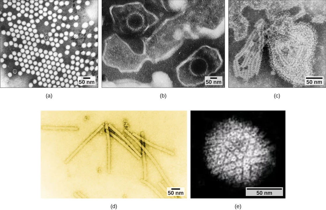
Most virions, or single virus particles, are very small, about 20 to 250 nanometers in diameter. However, some recently discovered viruses from amoebae range up to 1000 nm in diameter. With the exception of large virions, like the poxvirus and other large DNA viruses, viruses cannot be seen with a light microscope. It was not until the development of the electron microscope in the late 1930s that scientists got their first good view of the structure of the tobacco mosaic virus (TMV), discussed above, and other viruses, such as the ones shown below. The surface structure of virions can be observed by both scanning and transmission electron microscopy, whereas the internal structures of the virus can only be observed in images from a transmission electron microscope. The use of electron microscopy and other technologies has allowed for the discovery of many viruses of all types of living organisms.
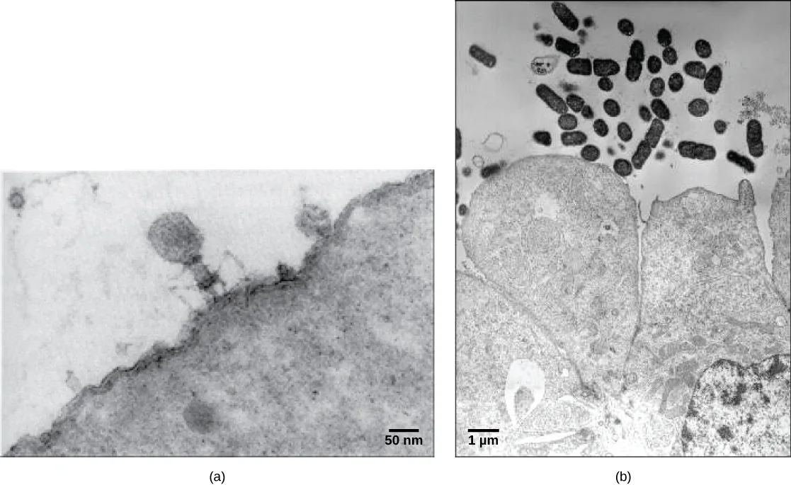
Evolution of Viruses
Although biologists have a significant amount of knowledge about how present-day viruses mutate and adapt, much less is known about how viruses originated in the first place. When exploring the evolutionary history of most organisms, scientists can look at fossil records and similar historic evidence. However, viruses do not fossilize, as far as we know, so researchers must extrapolate from investigations of how today’s viruses evolve and by using biochemical and genetic information to create speculative virus histories.
Most scholars agree that viruses don’t have a single common ancestor, nor is there a single reasonable hypothesis about virus origins. There are current evolutionary scenarios that may explain the origin of viruses. One such hypothesis, the “devolution” or the regressive hypothesis, suggests that viruses evolved from free-living cells, or from intracellular prokaryotic parasites. However, many components of how this process might have occurred remain a mystery. A second hypothesis, the escapist or the progressive hypothesis, suggests that viruses originated from RNA and DNA molecules, or self-replicating entities similar to transposons or other mobile genetic elements, that escaped from a host cell with the ability to enter another. A third hypothesis, the virus first hypothesis, suggests that viruses may have been the first self-replicating entities before the first cells. In all cases, viruses are probably continuing to evolve along with the cells on which they rely on as hosts.
As technology advances, scientists may develop and refine additional hypotheses to explain the origins of viruses. The emerging field called virus molecular systematics attempts to do just that through comparisons of sequenced genetic material. These researchers hope one day to better understand the origin of viruses—a discovery that could lead to advances in the treatments for the ailments they produce.
Viral Morphology
Viruses are noncellular, meaning they are biological entities that do not have a cellular structure. They therefore lack most of the components of cells, such as organelles, ribosomes, and the plasma membrane. A virion consists of a nucleic acid core, an outer protein coating or capsid, and sometimes an outer envelope made of protein and phospholipid membranes derived from the host cell. Viruses may also contain additional proteins, such as enzymes, within the capsid or attached to the viral genome. The most obvious difference between members of different viral families is the variation in their morphology, which is quite diverse. An interesting feature of viral complexity is that the complexity of the host does not necessarily correlate with the complexity of the virion. In fact, some of the most complex virion structures are found in the bacteriophages—viruses that infect the simplest living organisms, bacteria.
Morphology
Viruses come in many shapes and sizes, but these features are consistent for each viral family. As we have seen, all virions have a nucleic acid genome covered by a protective capsid. The proteins of the capsid are encoded in the viral genome, and are called capsomeres. Some viral capsids are simple helices or polyhedral “spheres,” whereas others are quite complex in structure.
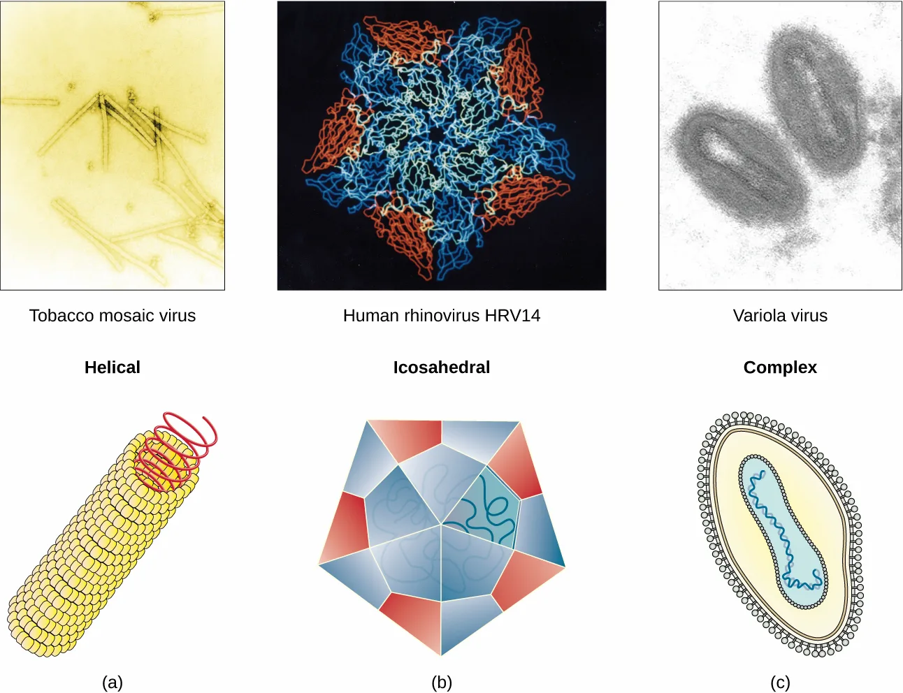
Extended description
Two rows of three panels, columns labeled (a), (b), (c) beneath the bottom row. Top row shows a real image of each virus: (a) a yellow-toned micrograph of tobacco mosaic virus as several long, thin rods; (b) a blue-and-red ribbon-diagram rendering of human rhinovirus HRV14, showing its protein subunits arranged with five-fold symmetry around a dark center; (c) a grey micrograph of three oval variola virus particles. Bottom row shows the matching schematic drawing for each: (a) ‘Helical’ — a red spiral coil wound around a cylinder built from stacked yellow protein subunits; (b) ‘Icosahedral’ — a many-faced polygon shell of red and blue triangular facets, with a tangled blue line representing genetic material inside; (c) ‘Complex’ — an oval shell with an outer ring of glycoprotein spikes, a middle protein layer, and a coiled blue-green line of genetic material at the center.
In general, the capsids of viruses are classified into four groups: helical, icosahedral, enveloped, and head-and-tail. Helical capsids are long and cylindrical. Many plant viruses are helical, including TMV. Icosahedral viruses have shapes that are roughly spherical, such as those of poliovirus or herpesviruses. Enveloped viruses have membranes derived from the host cell that surrounds the capsids. Animal viruses, such as HIV, are frequently enveloped. Head-and-tail viruses infect bacteria and have a head that is similar to icosahedral viruses and a tail shaped like helical viruses.
Many viruses use some sort of glycoprotein to attach to their host cells via molecules on the cell called viral receptors. For these viruses, attachment is required for later penetration of the cell membrane; only after penetration takes place can the virus complete its replication inside the cell. The receptors that viruses use are molecules that are normally found on cell surfaces and have their own physiological functions. It appears that viruses have simply evolved to make use of these molecules for their own replication. For example, HIV uses the CD4 molecule on T lymphocytes as one of its receptors. CD4 is a type of molecule called a cell adhesion molecule, which functions to keep different types of immune cells in close proximity to each other during the generation of a T lymphocyte immune response.
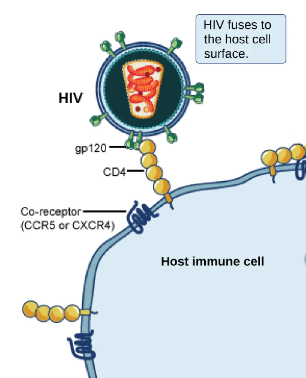
Extended description
At the top, a circular HIV particle labeled ‘HIV’ is studded with green Y-shaped spikes labeled ‘gp120’ and encloses a red coiled structure representing its genome. A light-blue caption box beside it reads ‘HIV fuses to the host cell surface.’ One gp120 spike reaches down to a chain of four yellow bead-shaped molecules labeled ‘CD4,’ embedded in the curving blue membrane of the ‘Host immune cell.’ Just to the left of that CD4 chain, a dark-blue coiled transmembrane protein is labeled ‘Co-receptor (CCR5 or CXCR4).’ A two more CD4-and-co-receptor pairs sit along the membrane, one at the lower left and one partly cropped at the upper right, not connected to the virus.
One of the most complex virions known, the T4 bacteriophage (which infects the Escherichia coli) bacterium, has a tail structure that the virus uses to attach to host cells and a head structure that houses its DNA.
Adenovirus, a non-enveloped animal virus that causes respiratory illnesses in humans, uses glycoprotein spikes protruding from its capsomeres to attach to host cells. Non-enveloped viruses also include those that cause polio (poliovirus), plantar warts (papillomavirus), and hepatitis A (hepatitis A virus).
Enveloped virions, such as the influenza virus, consist of nucleic acid (RNA in the case of influenza) and capsid proteins surrounded by a phospholipid bilayer envelope that contains virus-encoded proteins. Glycoproteins embedded in the viral envelope are used to attach to host cells. Other envelope proteins are the matrix proteins that stabilize the envelope and often play a role in the assembly of progeny virions. Chicken pox, HIV, and mumps are other examples of diseases caused by viruses with envelopes. Because of the fragility of the envelope, non-enveloped viruses are more resistant to changes in temperature, pH, and some disinfectants than enveloped viruses.
Overall, the shape of the virion and the presence or absence of an envelope tell us little about what disease the virus may cause or what species it might infect, but they are still useful means to begin viral classification.
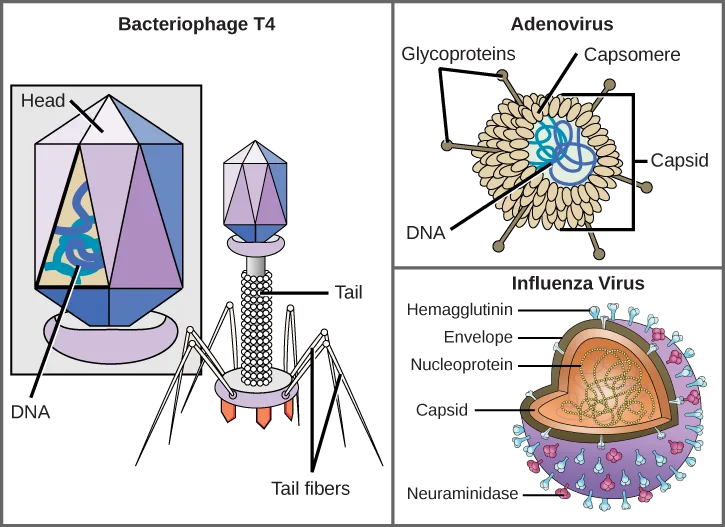
Extended description
Three side-by-side labeled diagrams under a shared header. Left panel, ‘Bacteriophage T4’: a hexagonal head (labeled Head), outlined in blue and purple facets, encloses a tangled blue line labeled DNA; below the head a cylindrical structure (labeled Tail) leads to a base ringed by six bent, spider-leg-like tail fibers (labeled Tail fibers). Middle panel, ‘Adenovirus’: a round capsid made of many small rounded subunits (labeled Capsomere, with a bracket also labeling the whole cluster Capsid) encloses a tangled blue line labeled DNA; several thin spikes (labeled Glycoproteins) project outward from the capsid surface. Right panel, ‘Influenza Virus’: a heart-shaped orange capsid (labeled Capsid) encloses a tangled dotted line (labeled Nucleoprotein); a purple spherical envelope (labeled Envelope) surrounds the capsid, and its outer surface is studded with two kinds of spike, labeled Hemagglutinin and Neuraminidase.
Which of the following statements about virus structure is true?
Re-read what the section says the glycoproteins on an envelope or capsid do when a virus meets a host cell.Types of Nucleic Acid
Unlike nearly all living organisms that use DNA as their genetic material, viruses may use either DNA or RNA. The virus core contains the genome—the total genetic content of the virus. Viral genomes tend to be small, containing only those genes that encode proteins which the virus cannot get from the host cell. This genetic material may be single- or double-stranded. It may also be linear or circular. While most viruses contain a single nucleic acid, others have genomes divided into several segments. The RNA genome of the influenza virus is segmented, which contributes to its variability and continuous evolution, and explains why it is difficult to develop a vaccine against it.
In DNA viruses, the viral DNA directs the host cell’s replication proteins to synthesize new copies of the viral genome and to transcribe and translate that genome into viral proteins. Human diseases caused by DNA viruses include chickenpox, hepatitis B, and adenoviruses. Sexually transmitted DNA viruses include the herpes virus and the human papilloma virus (HPV), which has been associated with cervical cancer and genital warts.
RNA viruses contain only RNA as their genetic material. To replicate their genomes in the host cell, the RNA viruses must encode their own enzymes that can replicate RNA into RNA or, in the retroviruses, into DNA. These RNA polymerase enzymes are more likely to make copying errors than DNA polymerases, and therefore often make mistakes during transcription. For this reason, mutations in RNA viruses occur more frequently than in DNA viruses. This causes them to change and adapt more rapidly to their host. Human diseases caused by RNA viruses include influenza, hepatitis C, measles, and rabies. The HIV virus, which is sexually transmitted, is an RNA retrovirus.
The Challenge of Virus Classification
Because most viruses probably evolved from different ancestors, the systematic methods that scientists have used to classify prokaryotic and eukaryotic cells are not very useful. If viruses represent “remnants” of different organisms, then even genomic or protein analysis is not useful. Why? Because viruses have no common genomic sequence that they all share. For example, the 16S rRNA sequence so useful for constructing prokaryote phylogenies is of no use for a creature with no ribosomes! Biologists have used several classification systems in the past. Viruses were initially grouped by shared morphology. Later, groups of viruses were classified by the type of nucleic acid they contained, DNA or RNA, and whether their nucleic acid was single- or double-stranded. However, these earlier classification methods grouped viruses differently, because they were based on different sets of characters of the virus. The most commonly used classification method today is called the Baltimore classification scheme, and is based on how messenger RNA (mRNA) is generated in each particular type of virus.
Past Systems of Classification
Viruses contain only a few elements by which they can be classified: the viral genome, the type of capsid, and the envelope structure for the enveloped viruses. All of these elements have been used in the past for viral classification. Viral genomes may vary in the type of genetic material (DNA or RNA) and its organization (single- or double-stranded, linear or circular, and segmented or non-segmented). In some viruses, additional proteins needed for replication are associated directly with the genome or contained within the viral capsid.
Virus Classification by Genome Structure
| Genome Structure | Examples |
|---|---|
| RNA | Rabies virus, retroviruses |
| DNA | Herpesviruses, smallpox virus |
| Single-stranded | Rabies virus, retroviruses |
| Double-stranded | Herpesviruses, smallpox virus |
| Linear | Rabies virus, retroviruses, herpesviruses, smallpox virus |
| Circular | Papillomaviruses, many bacteriophages |
| Non-segmented: genome consists of a single segment of genetic material | Parainfluenza viruses |
| Segmented: genome is divided into multiple segments | Influenza viruses |
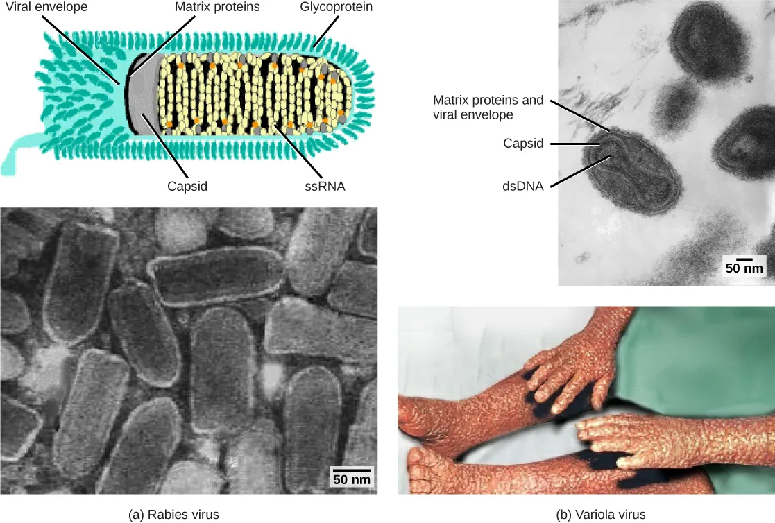
Extended description
Two columns, (a) Rabies virus on the left and (b) Variola virus on the right, each with a top panel and a bottom panel. Top-left is a colored schematic of a single bullet-shaped rabies virion: an outer teal-green fringe labeled Glycoprotein surrounds a grey shell labeled Matrix proteins, which encases a pale interior packed with yellow lipid-bilayer beads and a red coiled strand labeled ssRNA at the core, with a line pointing to a grey inner cylinder labeled Capsid, and the whole outer fringe-and-shell layer also labeled Viral envelope. Bottom-left is a black-and-white electron micrograph of roughly a dozen bullet-shaped rabies virus particles clustered together, with a 50 nm scale bar. Top-right is a black-and-white electron micrograph of two dark oval variola virus particles, each with an inner dark core and outer shell, with lines labeled ‘Matrix proteins and viral envelope,’ ‘Capsid,’ and ‘dsDNA’ pointing to the corresponding layers, with a 50 nm scale bar. Bottom-right is a color photo of a person’s forearms and lower legs covered in raised, irregular, bumpy pox lesions.
Viruses can also be classified by the design of their capsids. Capsids are classified as naked icosahedral, enveloped icosahedral, enveloped helical, naked helical, and complex. The type of genetic material (DNA or RNA) and its structure (single- or double-stranded, linear or circular, and segmented or non-segmented) are used to classify the virus core structures.
Virus Classification by Capsid Structure
| Capsid Classification | Examples |
|---|---|
| Naked icosahedral | Hepatitis A virus, polioviruses |
| Enveloped icosahedral | Epstein-Barr virus, herpes simplex virus, rubella virus, yellow fever virus, HIV-1 |
| Enveloped helical | Influenza viruses, mumps virus, measles virus, rabies virus |
| Naked helical | Tobacco mosaic virus |
| Complex with many proteins; some have combinations of icosahedral and helical capsid structures | Herpesviruses, smallpox virus, hepatitis B virus, T4 bacteriophage |
Baltimore Classification
The most commonly and currently used system of virus classification was first developed by Nobel Prize-winning biologist David Baltimore in the early 1970s. In addition to the differences in morphology and genetics mentioned above, the Baltimore classification scheme groups viruses according to how the mRNA is produced during the replicative cycle of the virus.
Group I viruses contain double-stranded DNA (dsDNA) as their genome. Their mRNA is produced by transcription in much the same way as with cellular DNA, using the enzymes of the host cell.
Group II viruses have single-stranded DNA (ssDNA) as their genome. They convert their single-stranded genomes into a dsDNA intermediate before transcription to mRNA can occur.
Group III viruses use dsRNA as their genome. The strands separate, and one of them is used as a template for the generation of mRNA using the RNA-dependent RNA polymerase encoded by the virus.
Group IV viruses have ssRNA as their genome with a positive polarity, which means that the genomic RNA can serve directly as mRNA. Intermediates of dsRNA, called replicative intermediates, are made in the process of copying the genomic RNA. Multiple, full-length RNA strands of negative polarity (complementary to the positive-stranded genomic RNA) are formed from these intermediates, which may then serve as templates for the production of RNA with positive polarity, including both full-length genomic RNA and shorter viral mRNAs.
Group V viruses contain ssRNA genomes with a negative polarity, meaning that their sequence is complementary to the mRNA. As with Group IV viruses, dsRNA intermediates are used to make copies of the genome and produce mRNA. In this case, the negative-stranded genome can be converted directly to mRNA. Additionally, full-length positive RNA strands are made to serve as templates for the production of the negative-stranded genome.
Group VI viruses have diploid (two copies) ssRNA genomes that must be converted, using the enzyme reverse transcriptase, to dsDNA; the dsDNA is then transported to the nucleus of the host cell and inserted into the host genome. Then, mRNA can be produced by transcription of the viral DNA that was integrated into the host genome.
Group VII viruses have partial dsDNA genomes and make ssRNA intermediates that act as mRNA, but are also converted back into dsDNA genomes by reverse transcriptase, necessary for genome replication.
The characteristics of each group in the Baltimore classification are summarized below, with examples of each group.
Baltimore Classification
| Group | Characteristics | Mode of mRNA Production | Example |
|---|---|---|---|
| I | Double-stranded DNA | mRNA is transcribed directly from the DNA template | Herpes simplex (herpesvirus) |
| II | Single-stranded DNA | DNA is converted to double-stranded form before RNA is transcribed | Canine parvovirus (parvovirus) |
| III | Double-stranded RNA | mRNA is transcribed from the RNA genome | Childhood gastroenteritis (rotavirus) |
| IV | Single stranded RNA (+) | Genome functions as mRNA | Common cold (picornavirus) |
| V | Single stranded RNA (-) | mRNA is transcribed from the RNA genome | Rabies (rhabdovirus) |
| VI | Single stranded RNA viruses with reverse transcriptase | Reverse transcriptase makes DNA from the RNA genome; DNA is then incorporated in the host genome; mRNA is transcribed from the incorporated DNA | Human immunodeficiency virus (HIV) |
| VII | Double stranded DNA viruses with reverse transcriptase | The viral genome is double-stranded DNA, but viral DNA is replicated through an RNA intermediate; the RNA may serve directly as mRNA or as a template to make mRNA | Hepatitis B virus (hepadnavirus) |
Summary
Viruses are tiny, noncellular entities that usually can be seen only with an electron microscope. Their genomes contain either DNA or RNA—never both—and they replicate either by using the replication proteins of a host cell or by using proteins encoded in the viral genome. Viruses are diverse, infecting archaea, bacteria, fungi, plants, and animals. Viruses consist of a nucleic acid core surrounded by a protein capsid with or without an outer lipid envelope. The capsid shape, presence of an envelope, and core composition dictate some elements of the classification of viruses. The most commonly used classification method, the Baltimore classification, categorizes viruses based on how they produce their mRNA.
Key terms
- acellular — lacking cells
- capsid — protein coating of the viral core
- capsomere — protein subunit that makes up the capsid
- envelope — lipid bilayer that encircles some viruses
- group I virus — virus with a dsDNA genome
- group II virus — virus with an ssDNA genome
- group III virus — virus with a dsRNA genome
- group IV virus — virus with an ssRNA genome with positive polarity
- group V virus — virus with an ssRNA genome with negative polarity
- group VI virus — virus with an ssRNA genome converted into dsDNA by reverse transcriptase
- group VII virus — virus with a single-stranded mRNA converted into dsDNA for genome replication
- matrix protein — envelope protein that stabilizes the envelope and often plays a role in the assembly of progeny virions
- negative polarity — ssRNA viruses with genomes complementary to their mRNA
- positive polarity — ssRNA virus with a genome that contains the same base sequences and codons found in their mRNA
- replicative intermediate — dsRNA intermediate made in the process of copying genomic RNA
- reverse transcriptase — enzyme found in Baltimore groups VI and VII that converts single-stranded RNA into double-stranded DNA
- viral receptor — glycoprotein used to attach a virus to host cells via molecules on the cell
- virion — individual virus particle outside a host cell
- virus core — contains the virus genome
Practice
Describe how viruses were first discovered and how they are detected
The first electron micrograph of a virus (tobacco mosaic virus) was produced in 1939. Before that time, how did scientists know that viruses existed if they could not see them? (Hint: Early scientists called viruses “filterable agents.”)
Show model answer
Did your answer mention:
An individual virus particle outside a host cell is called a ________.
The section’s first paragraph on detection calls a single virus particle by this name.Viruses are tiny, noncellular entities that usually can be seen only with a(n) ________.
This instrument was developed in the late 1930s and gave scientists their first good view of viruses like TMV.Discuss three hypotheses about how viruses evolved
The observation that the bacteria genus Chlamydia contains species that can only survive as intracellular parasites supports which viral origin hypothesis?
A parasite that can only survive inside a host cell fits the hypothesis that viruses evolved FROM free-living or intracellular organisms, not the hypothesis that they escaped from one.According to this section, what does the progressive hypothesis suggest about how viruses originated?
Show model answer
Did your answer mention:
The hypothesis suggesting that viruses may have been the earliest self-replicating entities, predating cells, is called the ________ hypothesis.
This hypothesis puts the virus before the cell, rather than the cell before the virus.Describe the general structure of a virus
Which statement is true?
A virion lacks organelles, ribosomes, and a plasma membrane — the components of a true cell.The viral ________ play(s) a role in attaching a virion to the host cell.
Re-read what the capsid and the envelope do for a virus that must attach before it can penetrate the cell membrane.Varicella-zoster virus is a double-stranded DNA virus that causes chickenpox. How does its genome structure provide an evolutionary advantage over a single-stranded DNA virus?
Show model answer
Did your answer mention:
Recognize the basic shapes of viruses
Viruses_______.
The section opens by comparing helical, icosahedral, enveloped, and head-and-tail capsids.According to the capsid-classification table, which capsid design describes tobacco mosaic virus?
TMV’s capsid is a hollow tube with no surrounding envelope.A capsid that is long and cylindrical, such as that of tobacco mosaic virus, is described as ________.
This shape describes a hollow tube, not a roughly spherical shell.Understand past and emerging classification systems for viruses
A scientist discovers a new virus with a linear, RNA genome surrounded by a helical capsid. The virus is most likely a member of which family based on structure classification?
Two of these options share the enveloped-helical row of the capsid classification table; the genome-structure table’s segmented row rules one of them out.According to the genome-structure classification table, which examples have a segmented genome?
Check the genome-structure table’s row for a genome divided into multiple segments.The part of a virion that contains the genome is called the ________.
It is one of the few elements, along with the capsid and envelope structure, used to classify a virus.Describe the basis for the Baltimore classification system
Classify the Rabies virus (a rhabdovirus family member) and HIV-1 with both the Baltimore and genomic structure systems. Compare your results. What conclusions can be made about these two different methods?
Show model answer
Did your answer mention:
A virus whose genome is double-stranded DNA (dsDNA) belongs to Baltimore ________.
Its mRNA is produced by transcription the same way cellular DNA is transcribed, using the host cell’s own enzymes.The enzyme found in Baltimore groups VI and VII that converts single-stranded RNA into double-stranded DNA is called ________.
HIV, a Group VI virus, depends on this enzyme to make a DNA copy of its RNA genome before integrating it into the host genome.This section is adapted from Biology 2e, Section 21.1: Viral Evolution, Morphology, and Classification by Mary Ann Clark, Jung Choi, Matthew Douglas, and OpenStax, © OpenStax, licensed under CC BY-NC-SA 4.0. Access the original for free at openstax.org. Changes: figures re-encoded as WebP; three figures (Figure_B21_01_02, Figure_B21_03_03a, Figure_21_01_04ab) re-kinded from the manifest’s file-extension guess of “photo” to “diagram,” since each pairs a micrograph or photo with a labeled schematic drawing that carries the teaching; Figure_21_01_01ab and Figure_21_01_06abcde kept as “photo” (pure micrograph panels); Figure_B21_02_01 kept as the manifest’s “diagram” guess, and its influenza panel’s capsid described as heart-shaped, as drawn, where the source alt calls it bullet-shaped (reported as a source defect); the letter-spaced screen-reader alts on Figure_B21_03_03a (“H I V,” “C D 4”), Figure_B21_02_01 (“D N A,” “R N A”), and Figure_21_01_04ab (“R N A,” “D N A”) rewritten as plain prose, and a longdesc added to those three plus Figure_B21_01_02, since none of the four diagrams’ labels and panel-by-panel content are fully carried by their captions; two cross-references to figures outside this section (the tobacco mosaic virus figure from the chapter introduction, and internal print figure numbers) rewritten as prose description, since figures are not numbered here; the note wrapping the Visual Connection question rendered as its figure plus a multiple choice kept in the body, keyed to the source answer; the genome-structure table’s bulleted cells (Table 21.1) reshaped to one Markdown row per bullet, pairing each genome-structure bullet with its matching example bullet in source order — compared against the printed table (p. 536) with no contradiction found; the end-of-section Review Questions and Critical Thinking Questions adapted into the closing interactive Practice block (multiple choice and self-check respectively); rubric checkpoints added to each self-check, decomposing its model answer (the source solution) into check-off clauses with no new claims; four glossary key-term recall items added (virion, virus core, Group I, reverse transcriptase) and one summary-derived cloze recall item added (electron microscope); and, where the source’s keyed exercises and the glossary left an objective’s Practice group thin, five locally written items added strictly from the section’s own sentences and tables, with no new claim — under “Discuss three hypotheses about how viruses evolved,” a self-check on the progressive hypothesis and a recall item on the virus first hypothesis; under “Recognize the basic shapes of viruses,” a multiple choice built from the capsid-structure table (naked helical → tobacco mosaic virus) and a recall item on the helical capsid shape; and under “Understand past and emerging classification systems for viruses,” a multiple choice built from the genome-structure table (segmented genome → influenza viruses). One name is corrected with a visible Source note: Adolf Mayer, the agricultural chemist of the 1886 tobacco-mosaic experiments, where the source prints “Adolph Meyer” (erratum 412).