Virus Infections and Hosts
By the end of this section, you will be able to:
- List the steps of replication and explain what occurs at each step
- Describe the lytic and lysogenic cycles of virus replication
- Explain the transmission of plant and animal viruses
- Discuss some of the diseases caused by plant and animal viruses
- Discuss the economic impact of plant and animal viruses
Viruses are obligate, intracellular parasites. A virus must first recognize and attach to a specific living cell prior to entering it. After penetration, the invading virus must copy its genome and manufacture its own proteins. Finally, the progeny virions must escape the host cell so that they can infect other cells. Viruses can infect only certain species of hosts and only certain cells within that host. Specific host cells that a virus must occupy and use to replicate are called permissive. In most cases, the molecular basis for this specificity is due to a particular surface molecule known as the viral receptor on the host cell surface. A specific viral receptor is required for the virus to attach. In addition, differences in metabolism and host-cell immune responses (based on differential gene expression) are a likely factor in determining which cells a virus may target for replication.
Steps of Virus Infections
A virus must use its host-cell processes to replicate. The viral replication cycle can produce dramatic biochemical and structural changes in the host cell, which may cause cell damage. These changes, called cytopathic effects, can change cell functions or even destroy the cell. Some infected cells, such as those infected by the common cold virus known as rhinovirus, die through lysis (bursting) or apoptosis (programmed cell death or “cell suicide”), releasing all progeny virions at once. The symptoms of viral diseases result both from such cell damage caused by the virus and from the immune response to the virus, which attempts to control and eliminate the virus from the body.
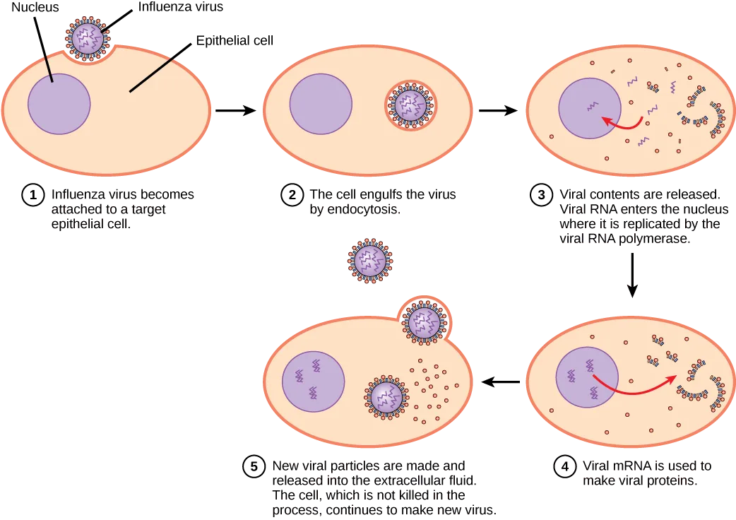
Many animal viruses, such as HIV (human immunodeficiency virus), leave the infected cells of the immune system by a process known as budding, where virions leave the cell individually. During the budding process, the cell does not undergo lysis and is not immediately killed. However, the damage to the cells that the virus infects may make it impossible for the cells to function normally, even though the cells remain alive for a period of time. Most productive viral infections follow similar steps in the virus replication cycle: attachment, penetration, uncoating, replication, assembly, and release (see the diagram below).
Attachment
A virus attaches to a specific receptor site on the host cell membrane through attachment proteins in the capsid or via glycoproteins embedded in the viral envelope. The specificity of this interaction determines the host—and the cells within the host—that can be infected by a particular virus. This can be illustrated by thinking of several keys and several locks, where each key will fit only one specific lock.
Entry
Viruses may enter a host cell either with or without the viral capsid. The nucleic acid of bacteriophages enters the host cell “naked,” leaving the capsid outside the cell. Plant and animal viruses can enter through endocytosis (as you may recall, the cell membrane surrounds and engulfs the entire virus). Some enveloped viruses enter the cell when the viral envelope fuses directly with the cell membrane. Once inside the cell, the viral capsid degrades, and then the viral nucleic acid is released and becomes available for replication and transcription.
Replication and Assembly
The replication mechanism depends on the viral genome. DNA viruses usually use host-cell proteins and enzymes to replicate the viral DNA and to transcribe viral mRNA, which is then used to direct viral protein synthesis. RNA viruses usually use the RNA core as a template for synthesis of viral genomic RNA and mRNA. The viral mRNA directs the host cell to synthesize viral enzymes and capsid proteins, and assemble new virions.
Of course, there are exceptions to this pattern. If a host cell does not provide the enzymes necessary for viral replication, viral genes supply the information to direct synthesis of the missing proteins. Retroviruses, such as HIV (group VI of the Baltimore classification scheme), have an RNA genome that must be reverse transcribed into DNA, which then is incorporated into the host cell genome. To convert RNA into DNA, retroviruses must contain genes that encode the virus-specific enzyme reverse transcriptase that transcribes an RNA template to DNA. Reverse transcription of a viral genome does not occur in uninfected host cells—the viral enzyme reverse transcriptase is derived only from the expression of viral genes within the infected host cells. (Source note: the source says reverse transcription “never occurs in uninfected host cells”; this book’s DNA Replication in Eukaryotes section describes telomerase, a cellular enzyme active in germ and stem cells, copying its built-in RNA template into DNA, so the claim is scoped here to the viral enzyme.) The fact that HIV produces some of its own enzymes not found in the host has allowed researchers to develop drugs that inhibit these enzymes without affecting the host’s metabolism.
This approach has led to the development of a variety of drugs used to treat HIV and has been effective at reducing the number of infectious virions (copies of viral RNA) in the blood to non-detectable levels in many HIV-infected individuals.
Egress
The last stage of viral replication is the release of the new virions produced in the host organism, where they are able to infect adjacent cells and repeat the replication cycle. As you’ve learned, some viruses are released when the host cell dies, and other viruses can leave infected cells by budding through the membrane without directly killing the cell.
Extended description
Five numbered panels show an oval epithelial cell with a purple nucleus. Step 1: an influenza virus, drawn as a small circle ringed with red spike proteins around a coiled RNA core, attaches to the cell’s outer membrane. Step 2: the cell engulfs the virus by endocytosis, enclosing it in a membrane-bound vesicle beside the nucleus. Step 3: the vesicle membrane dissolves, releasing viral RNA (red zigzag strands) and other viral components into the cytoplasm; a red arrow shows viral RNA moving into the nucleus, where it is copied by viral RNA polymerase. Step 4: viral mRNA exits the nucleus to the cytoplasm, where it directs synthesis of viral proteins, drawn as small dots and curved chains. Step 5: new virus particles assemble at the cell membrane and bud outward into the surrounding fluid; the cell’s outline and nucleus remain visible and intact.
Influenza virus is packaged in a viral envelope that fuses with the plasma membrane. This way, the virus can exit the host cell without killing it. What advantage does the virus gain by keeping the host cell alive?
Show model answer
Did your answer mention:
Different Hosts and Their Viruses
As you’ve learned, viruses often infect very specific hosts, as well as specific cells within the host. This feature of a virus makes it specific to one or a few species of life on Earth. On the other hand, so many different types of viruses exist on Earth that nearly every living organism has its own set of viruses trying to infect its cells. Even prokaryotes, the smallest and simplest of cells, may be attacked by specific types of viruses. In the following section, we will look at some of the features of viral infection of prokaryotic cells. As we have learned, viruses that infect bacteria are called bacteriophages (see the micrograph below). Archaea have their own similar viruses.
Bacteriophages
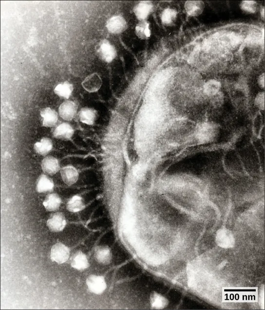
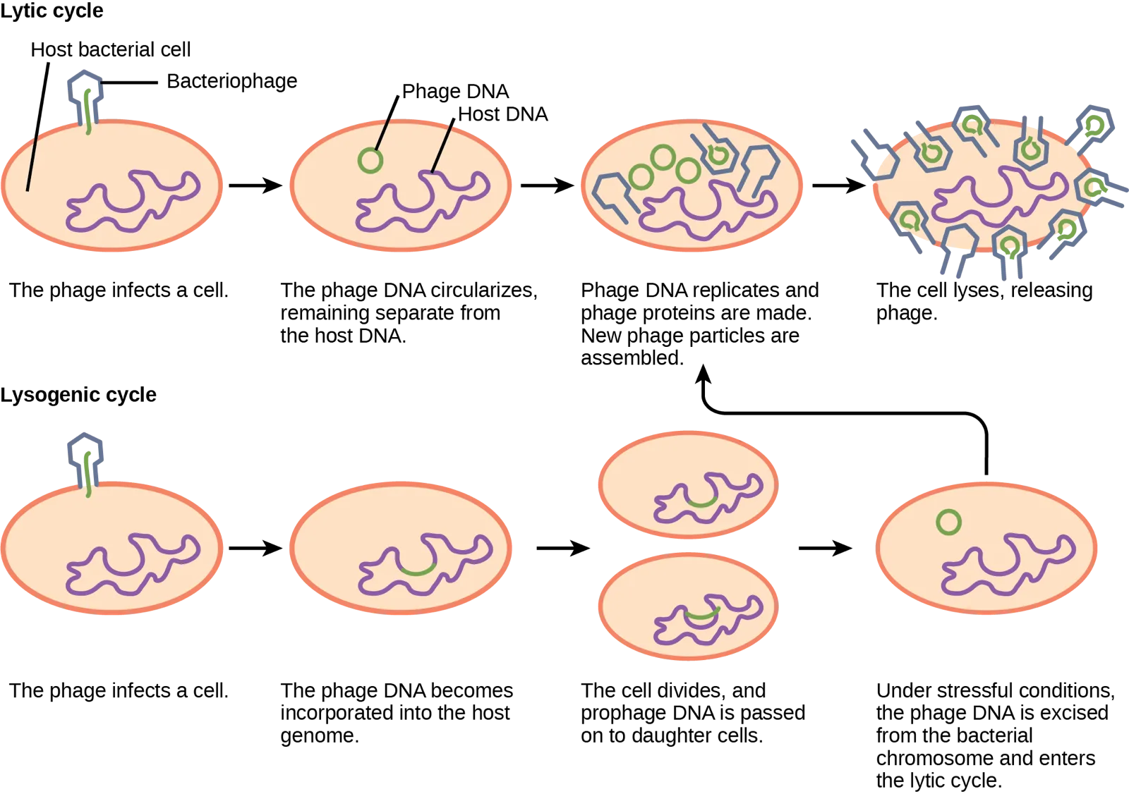
Most bacteriophages are dsDNA viruses, which use host enzymes for DNA replication and RNA transcription. Phage particles must bind to specific surface receptors and actively insert the genome into the host cell. (The complex tail structures seen in many bacteriophages are actively involved in getting the viral genome across the prokaryotic cell wall.) When infection of a cell by a bacteriophage results in the production of new virions, the infection is said to be productive. If the virions are released by bursting the cell, the virus replicates by means of a lytic cycle (illustrated below). An example of a lytic bacteriophage is T4, which infects Escherichia coli found in the human intestinal tract. Sometimes, however, a virus can remain within the cell without being released. For example, when a temperate bacteriophage infects a bacterial cell, it replicates by means of a lysogenic cycle (also illustrated below), and the viral genome is incorporated into the genome of the host cell. When the phage DNA is incorporated into the host-cell genome, it is called a prophage. An example of a lysogenic bacteriophage is the λ (lambda) virus, which also infects the E. coli bacterium. Viruses that infect plant or animal cells may sometimes undergo infections where they are not producing virions for long periods. An example is the animal herpesviruses, including herpes simplex viruses, the cause of oral and genital herpes in humans. In a process called latency, these viruses can exist in nervous tissue for long periods of time without producing new virions, only to leave latency periodically and cause lesions in the skin where the virus replicates. Even though there are similarities between lysogeny and latency, the term lysogenic cycle is usually reserved to describe bacteriophages. Latency will be described in more detail in the next section.
Extended description
Top row, labeled ‘Lytic cycle’: panel 1 shows a bacteriophage — a hexagonal head on a straight tail — attached to an oval host bacterial cell containing a purple, tangled host-DNA loop, with the phage’s straight green DNA inside its head. Panel 2 shows a small green circle labeled ‘Phage DNA’ inside the cell, separate from the purple ‘Host DNA’ loop. Panel 3 shows several green phage-DNA circles and assembled phage particles (hexagonal heads with tails) forming inside the cell. Panel 4 shows the cell’s outline broken open, releasing many phage particles, labeled ‘The cell lyses, releasing phage.’ Bottom row, labeled ‘Lysogenic cycle’: panel 1 shows the same phage attaching to a host cell. Panel 2 shows the green phage DNA now incorporated as a segment within the purple host-DNA loop. Panel 3 shows the cell having divided into two daughter cells, each still carrying the green phage-DNA segment within its host DNA. Panel 4 shows a single cell with the green phage DNA now excised back into a separate circle, labeled ‘Under stressful conditions, the phage DNA is excised from the bacterial chromosome and enters the lytic cycle,’ with a curved arrow leading back up to panel 3 of the lytic-cycle row.
Which of the following statements is false?
Re-read the diagram’s last lysogenic-cycle panel — a stressor triggers excision into which cycle, not which one?Plant Viruses
Most plant viruses, like the tobacco mosaic virus, have single-stranded (+) RNA genomes. However, there are also plant viruses in most other virus categories. Unlike bacteriophages, plant viruses do not have active mechanisms for delivering the viral genome across the protective cell wall. For a plant virus to enter a new host plant, some type of mechanical damage must occur. This damage is often caused by weather, insects, animals, fire, or human activities like farming or landscaping. Movement from cell to cell within a plant can be facilitated by viral modification of plasmodesmata (cytoplasmic threads that pass from one plant cell to the next). Additionally, plant offspring may inherit viral diseases from parent plants. Plant viruses can be transmitted by a variety of vectors, through contact with an infected plant’s sap, by living organisms such as insects and nematodes, and through pollen. The transfer of a virus from one plant to another is known as horizontal transmission, whereas the inheritance of a virus from a parent is called vertical transmission.
Symptoms of viral diseases vary according to the virus and its host (see the table below). One common symptom is hyperplasia, the abnormal proliferation of cells that causes the appearance of plant tumors known as galls. Other viruses induce hypoplasia, or decreased cell growth, in the leaves of plants, causing thin, yellow areas to appear. Still other viruses affect the plant by directly killing plant cells, a process known as cell necrosis. Other symptoms of plant viruses include malformed leaves; black streaks on the stems of the plants; altered growth of stems, leaves, or fruits; and ring spots, which are circular or linear areas of discoloration found in a leaf.
Some Common Symptoms of Plant Viral Diseases
| Symptom | Appears as |
|---|---|
| Hyperplasia | Galls (tumors) |
| Hypoplasia | Thinned, yellow splotches on leaves |
| Cell necrosis | Dead, blackened stems, leaves, or fruit |
| Abnormal growth patterns | Malformed stems, leaves, or fruit |
| Discoloration | Yellow, red, or black lines, or rings in stems, leaves, or fruit |
Plant viruses can seriously disrupt crop growth and development, significantly affecting our food supply. They are responsible for poor crop quality and quantity globally, and can bring about huge economic losses annually. Other viruses may damage plants used in landscaping. Some viruses that infect agricultural food plants include the name of the plant they infect, such as tomato spotted wilt virus, bean common mosaic virus, and cucumber mosaic virus. In plants used for landscaping, two of the most common viruses are peony ring spot and rose mosaic virus. There are far too many plant viruses to discuss each in detail, but symptoms of bean common mosaic virus result in lowered bean production and stunted, unproductive plants. In the ornamental rose, the rose mosaic disease causes wavy yellow lines and colored splotches on the leaves of the plant.
Animal Viruses
Animal viruses, unlike the viruses of plants and bacteria, do not have to penetrate a cell wall to gain access to the host cell. The virus may even induce the host cell to cooperate in the infection process. Non-enveloped or “naked” animal viruses may enter cells in two different ways. As a protein in the viral capsid binds to its receptor on the host cell, the virus may be taken inside the cell via a vesicle during the normal cell process of receptor-mediated endocytosis. An alternative method of cell penetration used by non-enveloped viruses is for capsid proteins to undergo shape changes after binding to the receptor, creating channels in the host cell membrane. The viral genome is then “injected” into the host cell through these channels in a manner analogous to that used by many bacteriophages.
Enveloped viruses also have two ways of entering cells after binding to their receptors: receptor-mediated endocytosis, or fusion. Many enveloped viruses enter the cell by receptor-mediated endocytosis in a fashion similar to that seen in some non-enveloped viruses. On the other hand, fusion only occurs with enveloped virions. These viruses, which include HIV among others, use special fusion proteins in their envelopes to cause the envelope to fuse with the plasma membrane of the cell, thus releasing the genome and capsid of the virus into the cell cytoplasm.
After making their proteins and copying their genomes, animal viruses complete the assembly of new virions and exit the cell. As we have already discussed using the example the influenza virus, enveloped animal viruses may bud from the cell membrane as they assemble themselves, taking a piece of the cell’s plasma membrane in the process. On the other hand, non-enveloped viral progeny, such as rhinoviruses, accumulate in infected cells until there is a signal for lysis or apoptosis, and all virions are released together.
As you will learn in the next module, animal viruses are associated with a variety of human diseases. Some of them follow the classic pattern of acute disease, where symptoms get increasingly worse for a short period followed by the elimination of the virus from the body by the immune system and eventual recovery from the infection. Examples of acute viral diseases are the common cold and influenza. Other viruses cause long-term chronic infections, such as the virus causing hepatitis C, whereas others, like herpes simplex virus, only cause intermittent symptoms. Still other viruses, such as human herpesviruses 6 and 7, which in some cases can cause the minor childhood disease roseola, often successfully cause productive infections without causing any symptoms at all in the host, and thus we say these patients have an asymptomatic infection.
In hepatitis C infections, the virus grows and reproduces in liver cells, causing low levels of liver damage. The damage is so low that infected individuals are often unaware that they are infected, and many infections are detected only by routine blood work on patients with risk factors such as intravenous drug use. On the other hand, since many of the symptoms of viral diseases are caused by immune responses, a lack of symptoms is an indication of a weak immune response to the virus. This allows the virus to escape elimination by the immune system and persist in individuals for years, all the while producing low levels of progeny virions in what is known as a chronic viral disease. Chronic infection of the liver by this virus leads to a much greater chance of developing liver cancer, sometimes as much as 30 years after the initial infection.
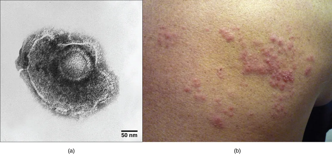
As already discussed, herpes simplex virus can remain in a state of latency in nervous tissue for months, even years. As the virus “hides” in the tissue and makes few if any viral proteins, there is nothing for the immune response to act against, and immunity to the virus slowly declines. Under certain conditions, including various types of physical and psychological stress, the latent herpes simplex virus may be reactivated and undergo a lytic replication cycle in the skin, causing the lesions associated with the disease. Once virions are produced in the skin and viral proteins are synthesized, the immune response is again stimulated and resolves the skin lesions in a few days or weeks by destroying viruses in the skin. As a result of this type of replicative cycle, appearances of cold sores and genital herpes outbreaks only occur intermittently, even though the viruses remain in the nervous tissue for life. Latent infections are common with other herpesviruses as well, including the varicella-zoster virus that causes chickenpox. After having a chickenpox infection in childhood, the varicella-zoster virus can remain latent for many years and reactivate in adults to cause the painful condition known as “shingles” (pictured below).
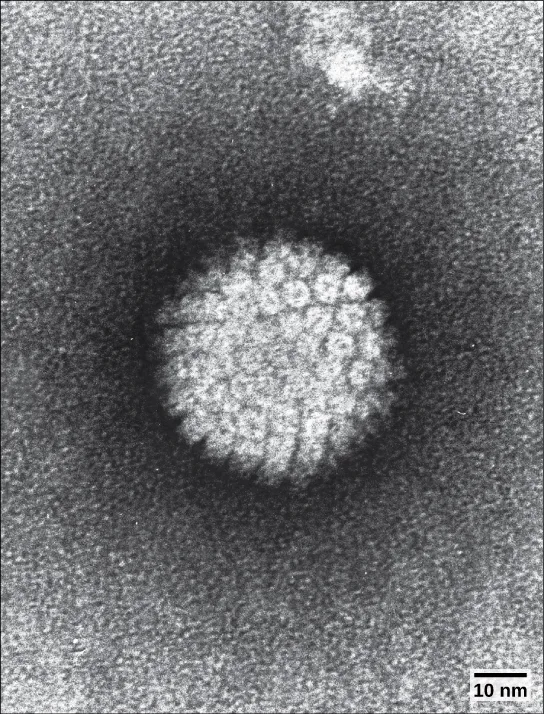
Some animal-infecting viruses, including the hepatitis C virus discussed above, are known as oncogenic viruses: They have the ability to cause cancer. These viruses interfere with the normal regulation of the host cell cycle either by introducing genes that stimulate unregulated cell growth (oncogenes) or by interfering with the expression of genes that inhibit cell growth. Oncogenic viruses can be either DNA or RNA viruses. Cancers known to be associated with viral infections include cervical cancer, caused by human papillomavirus (HPV) (pictured below), liver cancer caused by hepatitis C virus, T-cell leukemia, and several types of lymphoma.
Summary
Plant viruses may be transmitted either vertically from parent reproductive cells or horizontally through damaged plant tissues. Viruses of plants are responsible for significant economic damage in both crop plants and plants used for ornamentation. Animal viruses enter their hosts through several types of virus-host cell interactions and cause a variety of infections. Viral infections can be either acute, with a brief period of infection terminated by host immune responses, or chronic, in which the infection persists. Persistent infections may cause chronic symptoms (hepatitis C), intermittent symptoms (latent viruses such a herpes simplex virus 1), or even be effectively asymptomatic (human herpesviruses 6 and 7). Oncogenic viruses in animals have the ability to cause cancer by interfering with the regulation of the host cell cycle.
Key terms
- acute disease — disease where the symptoms rise and fall within a short period of time
- asymptomatic disease — disease where there are no symptoms and the individual is unaware of being infected unless lab tests are performed
- AZT — anti-HIV drug that inhibits the viral enzyme reverse transcriptase
- bacteriophage — virus that infects bacteria
- budding — method of exit from the cell used in certain animal viruses, where virions leave the cell individually by capturing a piece of the host plasma membrane
- cell necrosis — cell death
- chronic infection — describes when the virus persists in the body for a long period of time
- cytopathic — causing cell damage
- fusion — method of entry by some enveloped viruses, where the viral envelope fuses with the plasma membrane of the host cell
- gall — appearance of a plant tumor
- horizontal transmission — transmission of a disease between unrelated individuals
- hyperplasia — abnormally high cell growth and division
- hypoplasia — abnormally low cell growth and division
- intermittent symptom — symptom that occurs periodically
- latency — virus that remains in the body for a long period of time but only causes intermittent symptoms
- lysis — bursting of a cell
- lytic cycle — type of virus replication in which virions are released through lysis, or bursting, of the cell
- lysogenic cycle — type of virus replication in which the viral genome is incorporated into the genome of the host cell
- oncogenic virus — virus that has the ability to cause cancer
- permissive — cell type that is able to support productive replication of a virus
- productive — viral infection that leads to the production of new virions
- prophage — phage DNA that is incorporated into the host cell genome
- vertical transmission — transmission of disease from parent to offspring
Practice
List the steps of replication and explain what occurs at each step
Which statement is true of viral replication?
Re-read the Attachment subsection’s opening sentence about where on the host cell membrane a virus binds.Which statement is true of reverse transcriptase?
This enzyme lets a retrovirus like HIV convert its RNA genome into a form the host genome can incorporate.Which is true of DNA viruses?
Re-read the Replication and Assembly subsection’s first sentence about what DNA viruses use to replicate their DNA.People with the CCR5Δ32 mutation of a T-cell surface protein can be exposed to some strains of HIV-1 without becoming sick. What step of the virus life cycle is likely to be inhibited with this mutation?
A mutated surface protein most directly affects the step where a virus first binds its specific receptor on the host cell.Why can’t dogs catch the measles?
Show model answer
Did your answer mention:
One of the first and most important targets for drugs to fight infection with HIV (a retrovirus) is the reverse transcriptase enzyme. Why?
Show model answer
Did your answer mention:
Describe the lytic and lysogenic cycles of virus replication
Which statement is not true of viral replication?
Re-read the section’s second paragraph about cytopathic effects — what does it say happens to host cell function during viral replication?A bacteriophage can infect ________.
The word’s own root names the group of organisms these viruses attack.A bacteriophage with a lytic life cycle develops a mutation that allows it to now also go through the lysogenic cycle. How would this provide an evolutionary advantage over the other bacteriophages that can only spread through lytic cycles?
Show model answer
Did your answer mention:
Phage DNA that has become incorporated into the host cell’s genome is called a ________.
It forms during the lysogenic cycle, when the phage genome joins the bacterial chromosome.Explain the transmission of plant and animal viruses
An apple grower notices that several of his apple trees with fungi growing on their trunks have developed necrotic ring spots, while other trees in the orchard that lack fungi appear healthy. What is the most likely conclusion the farmer can make about the virus infecting his apple trees?
Only the trees carrying fungi got sick, so the fungi are the likely vector between unrelated trees, not something inherited from a parent plant.The transfer of a plant virus from one unrelated plant to another is called ________.
Insects, nematodes, pollen, sap contact, and mechanical damage all spread a virus this way, plant to plant.The inheritance of a virus by offspring from a parent plant is called ________.
This is the opposite of the plant-to-plant route named just before it in the text.Discuss some of the diseases caused by plant and animal viruses
Oncogenic virus cores can be ________.
The section states oncogenic viruses can belong to either major type of genome.A viral illness such as hepatitis C that persists in the body for a long period of time is called a ________.
Its opposite in this section is the short, sharp pattern called an acute disease.Abnormally high plant-cell growth and division is known as ________.
This symptom is what causes the plant tumors described just after it in the text.A plant tumor caused by a virus is known as a ________.
This symptom appears when a virus causes abnormally high cell growth and division in the plant.Discuss the economic impact of plant and animal viruses
Although plant viruses cannot infect humans, what are some of the ways in which they affect humans?
Show model answer
Did your answer mention:
Viruses of plants are responsible for significant ________ in both crop plants and plants used for ornamentation.
Crop failures and losses to ornamental plants both count as this kind of significant financial harm.According to this section, which of the following viruses affects plants used in landscaping rather than food crops?
This one is named in the same sentence as another ornamental-rose virus, right after the section lists viruses of tomato, bean, and cucumber.This section is adapted from Biology 2e, Section 21.2: Virus Infections and Hosts by Mary Ann Clark, Jung Choi, Matthew Douglas, and OpenStax, © OpenStax, licensed under CC BY-NC-SA 4.0. Access the original for free at openstax.org. Changes: figures re-encoded as WebP (all five kind guesses — diagram for Figure_21_02_01 and Figure_B21_03_01, photo for Figure_21_02_02-545c, Figure_21_02_04ab, and Figure_21_02_05-f075 — confirmed correct after inspection, no re-kinding needed); a longdesc added for Figure_21_02_01 (the five numbered steps are not fully carried by the one-sentence caption) and Figure_B21_03_01 (the panel-by-panel lytic/lysogenic sequence is not in the caption); the letter-spaced source alt for Figure_21_02_01 (“m R N A”, “R N A”) rewritten as “mRNA”/“RNA” and reworded, alongside its longdesc, to describe what the final panel draws rather than restate the adjoining Visual Connection question’s own answer; the over-600-character source alt for Figure_B21_03_01 shortened to a concise overview with the full step-by-step detail moved into longdesc; the source alt for Figure_21_02_04ab corrected from “a person’s face” to “a person’s shoulder and upper back,” matching the image (reported as a source defect); the source’s forward figure and table references (bracketed autonumber links) reworded as “see the diagram below,” “see the micrograph below,” “see the table below,” “illustrated below,” and “pictured below,” since figures are not numbered here; the three interactive notes rendered as Link to Learning callouts with descriptive link text in place of the source’s bare “video”; the two visual-connection notes rendered as their mediafigure followed by, respectively, a self-check (the prose-keyed influenza question) and a multiple choice (the lettered-keyed lytic/lysogenic question) kept in the body; the plant-viral-disease-symptoms table (tab-ch21_02_01) transcribed complete as a Markdown table, its spanning title row set as a bold line above it; one Review Question (the “not true of viral replication” item) re-keyed from the source’s “Newly released virions can infect adjacent cells” — which the section’s own Egress paragraph states is true — to “Viral replication does not affect host cell function,” which the section’s own cytopathic-effects sentence directly contradicts, and, because the source’s first option (“A lysogenic cycle kills the host cell”) is also false by the section’s own account of lysogeny, that option replaced with the section’s own sentence “In the lysogenic cycle, phage DNA is incorporated into the host genome” so that exactly one option is untrue (reported as a source defect); the end-of-section Review Questions and Critical Thinking Questions adapted into the closing interactive Practice block (multiple choice and self-check respectively), with one Critical Thinking Question omitted because its source solution is open-ended (“Answer is open and will vary”) and has no fixed content to check; rubric checkpoints added to each self-check, decomposing its model answer (the source solution) into check-off clauses with no new claims; six key-term recall items (prophage, horizontal transmission, vertical transmission, chronic infection, hyperplasia, gall) added from the glossary; one summary-derived cloze recall item (economic damage) added; and one multiple choice written locally, strictly from the section’s own sentence naming peony ring spot and rose mosaic virus as ornamental-plant viruses, since the module’s exercise sets left the fifth objective (“Discuss the economic impact of plant and animal viruses”) with only one source item. One claim is narrowed with a visible Source note: reverse transcription by the viral enzyme does not occur in uninfected cells, since this book’s own telomerase description is RNA-templated DNA synthesis in uninfected cells (erratum 411).