Structure of Prokaryotes: Bacteria and Archaea
By the end of this section, you will be able to:
- Describe the basic structure of a typical prokaryote
- Describe important differences in structure between Archaea and Bacteria
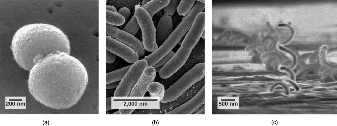
There are many differences between prokaryotic and eukaryotic cells. The name “prokaryote” suggests that prokaryotes are defined by exclusion—they are not eukaryotes, or organisms whose cells contain a nucleus and other internal membrane-bound organelles. However, all cells have four common structures: the plasma membrane, which functions as a barrier for the cell and separates the cell from its environment; the cytoplasm, a complex solution of organic molecules and salts inside the cell; a double-stranded DNA genome, the informational archive of the cell; and ribosomes, where protein synthesis takes place. Prokaryotes come in various shapes, but many fall into three categories: cocci (spherical), bacilli (rod-shaped), and spirilli (spiral-shaped), shown in the micrographs below.
The Prokaryotic Cell
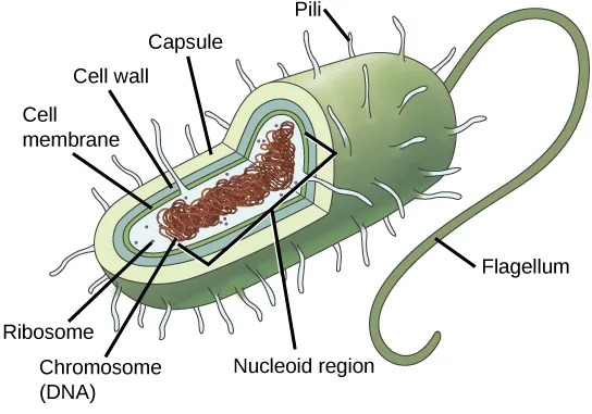
Recall that prokaryotes are unicellular organisms that lack membrane-bound organelles or other internal membrane-bound structures, illustrated in the diagram below. Their chromosome—usually single—consists of a piece of circular, double-stranded DNA located in an area of the cell called the nucleoid. Most prokaryotes have a cell wall outside the plasma membrane. The cell wall functions as a protective layer, and it is responsible for the organism’s shape. Some bacterial species have a capsule outside the cell wall. The capsule enables the organism to attach to surfaces, protects it from dehydration and attack by phagocytic cells, and makes pathogens more resistant to our immune responses. Some species also have flagella (singular, flagellum) used for locomotion, and pili (singular, pilus) used for attachment to surfaces including the surfaces of other cells. Plasmids, which consist of extra-chromosomal DNA, are also present in many species of bacteria and archaea.
Extended description
A green, rod-shaped cell is drawn in cutaway. Short, hair-like pili project from all around the cell surface, and one long, curved flagellum extends from one narrow end. Leader lines at the upper left point, from outside in, to the capsule (the outermost pale layer), the cell wall, and the cell membrane. Inside the cytoplasm, small circles labeled ribosomes are scattered throughout, and leader lines at lower left and center point to the chromosome (DNA) — a tangled reddish loop — and to the nucleoid region, the lighter area of the cytoplasm where that loop sits.
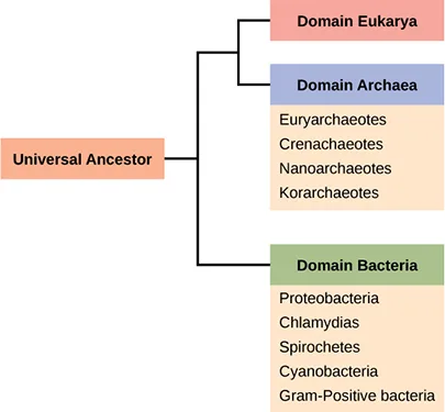
Recall that prokaryotes are divided into two different domains, Bacteria and Archaea, which together with Eukarya, comprise the three domains of life, illustrated below.
Extended description
A box labeled “Universal Ancestor” sits at the left. A line runs right from it to a branch point. The lower branch runs to a green box labeled “Domain Bacteria,” beneath which an orange list names its major phyla: Proteobacteria, Chlamydias, Spirochetes, Cyanobacteria, and Gram-Positive bacteria. The upper branch runs to a second branch point, which splits into a pink box labeled “Domain Eukarya” (top, with no phyla listed) and a purple box labeled “Domain Archaea” (below it), beneath which an orange list names its major phyla: Euryarchaeotes, Crenarchaeotes, Nanoarchaeotes, and Korarchaeotes.
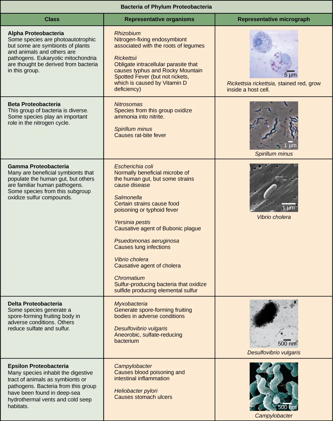
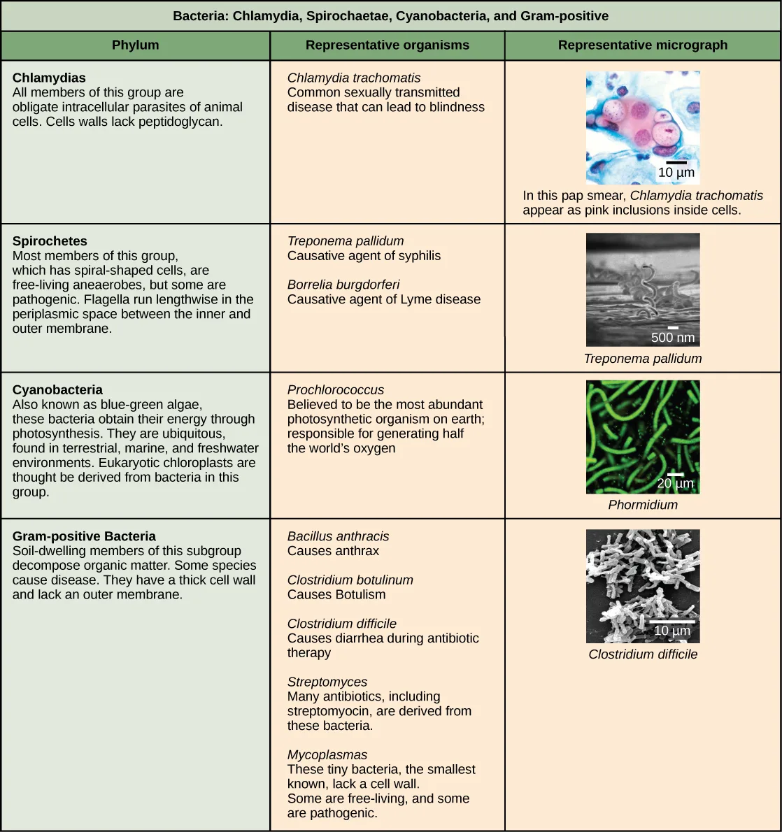
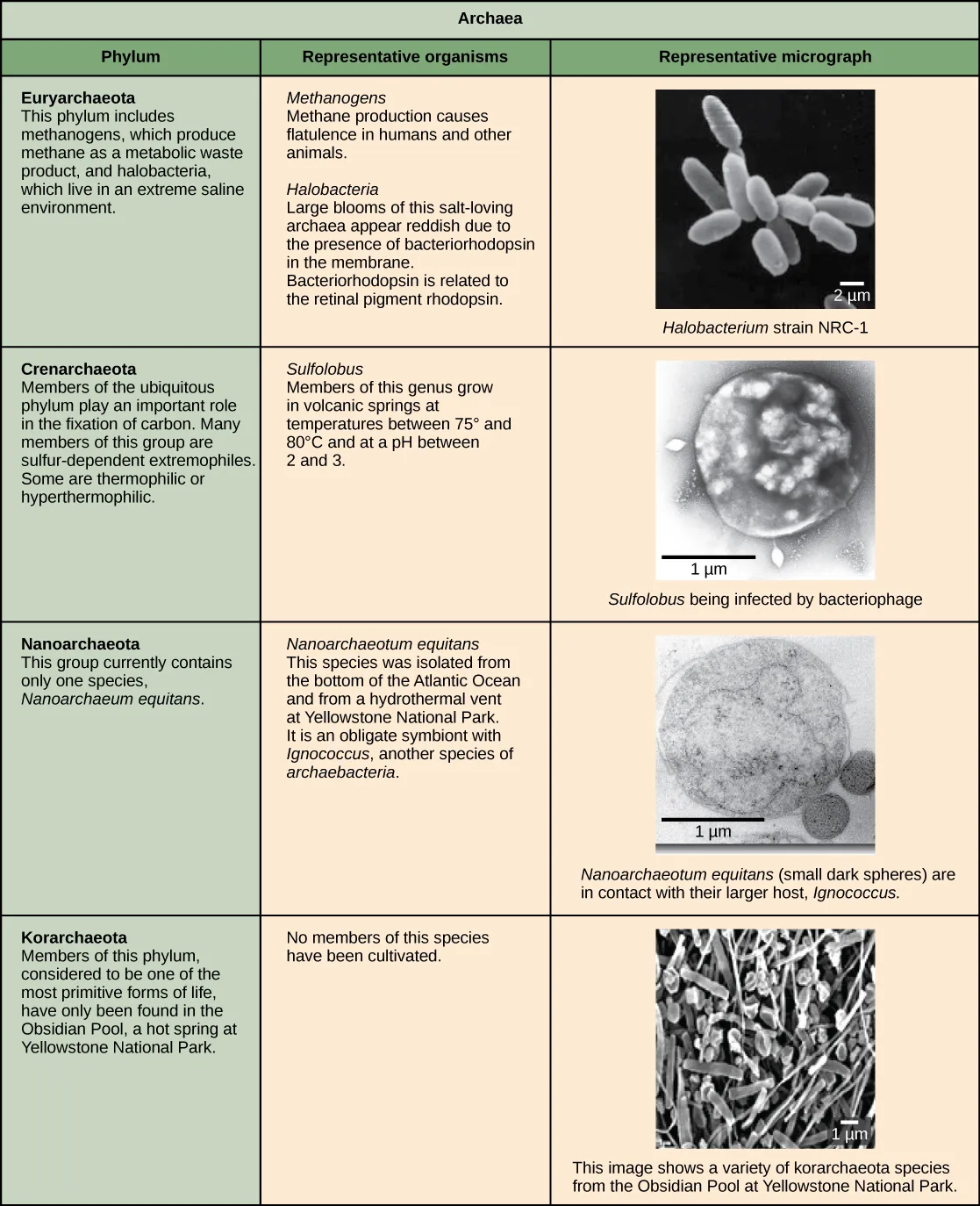
Characteristics of bacterial phyla are described in the tables below. Major bacterial phyla include the Proteobacteria, the Chlamydias, the Spirochaetes, the photosynthetic Cyanobacteria, and the Gram-positive bacteria. The Proteobacteria are in turn subdivided into several classes, from the Alpha- to the Epsilon proteobacteria. Eukaryotic mitochondria are thought to be the descendants of alphaproteobacteria, while eukaryotic chloroplasts are derived from cyanobacteria. Archaeal phyla are described in the archaea table below.
Extended description
The table has three columns — Class, Representative organisms, and Representative micrograph — and five rows, one per class. Alpha Proteobacteria: some species are photoautotrophic, some are symbionts of plants and animals, and others are pathogens; eukaryotic mitochondria are thought to be derived from bacteria in this group. Representative organisms: Rhizobium, a nitrogen-fixing endosymbiont associated with legume roots, and Rickettsia rickettsii, an obligate intracellular parasite; species of Rickettsia cause typhus and Rocky Mountain spotted fever (but not rickets, which is caused by vitamin D deficiency). The micrograph shows Rickettsia rickettsii, stained red, growing inside a host cell, with a 5 µm scale bar. Beta Proteobacteria: a diverse group, some of whose species play an important role in the nitrogen cycle. Representative organisms: Nitrosomonas, which oxidizes ammonia into nitrite, and Spirillum minus, which causes rat-bite fever. The micrograph shows spiral-shaped Spirillum minus cells, with a 1 µm scale bar. Gamma Proteobacteria: many are beneficial symbionts of the human gut, others are familiar human pathogens, and some oxidize sulfur compounds. Representative organisms: Escherichia coli, normally a beneficial gut microbe though some strains cause disease; Salmonella, some strains of which cause food poisoning or typhoid fever; Yersinia pestis, the causative agent of bubonic plague; Pseudomonas aeruginosa, which causes lung infections; Vibrio cholerae, the causative agent of cholera; and Chromatium, sulfur-producing bacteria that oxidize sulfide to elemental sulfur. The micrograph shows rod-shaped Vibrio cholerae. Delta Proteobacteria: some species generate a spore-forming fruiting body in adverse conditions, others reduce sulfate and sulfur. Representative organisms: Myxobacteria, which generate spore-forming fruiting bodies in adverse conditions, and Desulfovibrio vulgaris, an anaerobic, sulfate-reducing bacterium. The micrograph shows a bent, rod-shaped Desulfovibrio vulgaris cell, with a 500 nm scale bar. Epsilon Proteobacteria: many species inhabit the digestive tract of animals as symbionts or pathogens, and members of this group have been found in deep-sea hydrothermal vents and cold seep habitats. Representative organisms: Campylobacter, which causes blood poisoning and intestinal inflammation, and Helicobacter pylori, which causes stomach ulcers. The micrograph shows short, spiral-shaped Campylobacter cells, with a 500 nm scale bar.
Extended description
The table has three columns — Phylum, Representative organisms, and Representative micrograph — and four rows. Chlamydias: all members are obligate intracellular parasites of animal cells, and their cell walls lack peptidoglycan. Representative organism: Chlamydia trachomatis, which causes a common sexually transmitted disease that can lead to blindness. The micrograph is a Pap smear showing Chlamydia trachomatis as pink inclusions inside cells, with a 10 µm scale bar. Spirochetes: most members, which have spiral-shaped cells, are free-living anaerobes, though some are pathogenic; their flagella run lengthwise in the periplasmic space between the inner and outer membrane. Representative organisms: Treponema pallidum, the causative agent of syphilis, and Borrelia burgdorferi, the causative agent of Lyme disease. The micrograph shows a corkscrew-shaped Treponema pallidum cell, with a 500 nm scale bar. Cyanobacteria: also known as blue-green algae, these bacteria obtain their energy through photosynthesis and are found in terrestrial, marine, and freshwater environments; eukaryotic chloroplasts are thought to be derived from bacteria in this group. Representative organism: Prochlorococcus, believed to be the most abundant photosynthetic organism on Earth, responsible for generating half the world’s oxygen. The micrograph shows thin, thread-like Phormidium filaments, with a 20 µm scale bar. Gram-positive Bacteria: soil-dwelling members of this group decompose organic matter, and some species cause disease; they have a thick cell wall and lack an outer membrane. Representative organisms: Bacillus anthracis, which causes anthrax; Clostridium botulinum, which causes botulism; Clostridium difficile, which causes diarrhea during antibiotic therapy; Streptomyces, from which many antibiotics, including streptomycin, are derived; and Mycoplasma, the smallest known bacteria, which lack a cell wall and may be free-living or pathogenic. The micrograph shows rod-shaped Clostridium difficile cells, with a 10 µm scale bar.
Extended description
The table has three columns — Phylum, Representative organisms, and Representative micrograph — and four rows. Euryarchaeota: includes methanogens, which produce methane as a metabolic waste product, and halobacteria, which live in an extreme saline environment; large blooms of halobacteria appear reddish from the pigment bacteriorhodopsin, which is related to the retinal pigment rhodopsin. The micrograph shows several rod-shaped Halobacterium cells, with a 2 µm scale bar. Crenarchaeota: members of this ubiquitous phylum play an important role in carbon fixation; many are sulfur-dependent extremophiles, and some are thermophilic or hyperthermophilic. Representative organism: Sulfolobus, which grows in volcanic springs between 75 and 80 °C and at a pH between 2 and 3. The micrograph shows a single round Sulfolobus cell being infected by bacteriophage, with a 1 µm scale bar. Nanoarchaeota: this phylum currently contains only one species, Nanoarchaeum equitans, isolated from the bottom of the Atlantic Ocean and from a hydrothermal vent at Yellowstone National Park; it is an obligate symbiont of Ignicoccus, another archaeal species. The micrograph shows two small, round Nanoarchaeum equitans cells attached to a larger Ignicoccus cell, with a 1 µm scale bar. Korarchaeota: considered one of the most primitive forms of life, members of this phylum have so far only been found in the Obsidian Pool, a hot spring at Yellowstone National Park; no members of this phylum have been cultivated. The micrograph shows a variety of korarchaeote cells of different shapes from the Obsidian Pool, with a 1 µm scale bar.
The Plasma Membrane of Prokaryotes
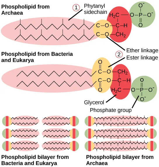
The prokaryotic plasma membrane is a thin lipid bilayer (6 to 8 nanometers) that completely surrounds the cell and separates the inside from the outside. Its selectively permeable nature keeps ions, proteins, and other molecules within the cell and prevents them from diffusing into the extracellular environment, while other molecules may move through the membrane. Recall that the general structure of a cell membrane is a phospholipid bilayer composed of two layers of lipid molecules. In archaeal cell membranes, isoprene (phytanyl) chains linked to glycerol replace the fatty acids linked to glycerol in bacterial membranes. Some archaeal membranes are lipid monolayers instead of bilayers, illustrated below.
Extended description
Two individual phospholipids are drawn stacked at the top. The upper one, labeled “Phospholipid from Archaea,” has a branched, zigzag phytanyl sidechain (numbered 1) attached through an ether linkage to a glycerol backbone, which is in turn attached to a phosphate group. The lower one, labeled “Phospholipid from Bacteria and Eukarya,” has a straight, unbranched fatty acid chain attached through an ester linkage (numbered 2) to the same glycerol-and-phosphate backbone. Below these, two rows of three phospholipids each are drawn tail-to-tail as bilayers: the left bilayer, labeled “Phospholipid bilayer from Bacteria and Eukarya,” uses the straight-chain lipid; the right bilayer, labeled “Phospholipid bilayer from Archaea,” uses the branched-chain lipid; the two bilayers otherwise look alike.
The Cell Wall of Prokaryotes
The cytoplasm of prokaryotic cells has a high concentration of dissolved solutes. Therefore, the osmotic pressure within the cell is relatively high. The cell wall is a protective layer that surrounds some cells and gives them shape and rigidity. It is located outside the cell membrane and prevents osmotic lysis (bursting due to increasing volume). The chemical composition of the cell wall varies between Archaea and Bacteria, and also varies between bacterial species.
Bacterial cell walls contain peptidoglycan, composed of polysaccharide chains that are cross-linked by unusual peptides containing both L- and D-amino acids including D-glutamic acid and D-alanine. (Proteins normally have only L-amino acids; as a consequence, many of our antibiotics work by mimicking D-amino acids and therefore have specific effects on bacterial cell-wall development.) There are more than 100 different forms of peptidoglycan. S-layer (surface layer) proteins are also present on the outside of cell walls of both Archaea and Bacteria.
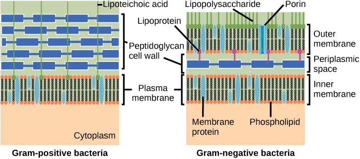
Bacteria are divided into two major groups: Gram positive and Gram negative, based on their reaction to Gram staining. Note that all Gram-positive bacteria belong to two phyla (Firmicutes and Actinobacteria); bacteria in the other phyla (Proteobacteria, Chlamydias, Spirochetes, Cyanobacteria, and others) are Gram-negative. The Gram staining method is named after its inventor, Danish scientist Hans Christian Gram (1853–1938). The different bacterial responses to the staining procedure are ultimately due to cell wall structure. Gram-positive organisms typically lack the outer membrane found in Gram-negative organisms, illustrated below. Up to 90 percent of the cell wall in Gram-positive bacteria is composed of peptidoglycan, and most of the rest is composed of acidic substances called teichoic acids. Teichoic acids may be covalently linked to lipids in the plasma membrane to form lipoteichoic acids. Lipoteichoic acids anchor the cell wall to the cell membrane. Gram-negative bacteria have a relatively thin cell wall composed of a few layers of peptidoglycan (only 10 percent of the total cell wall), surrounded by an outer envelope containing lipopolysaccharides (LPS) and lipoproteins. This outer envelope is sometimes referred to as a second lipid bilayer. The chemistry of this outer envelope is very different, however, from that of the typical lipid bilayer that forms plasma membranes.
Extended description
Two cutaway diagrams are shown side by side. Left, “Gram-positive bacteria”: from top to bottom, a green layer labeled lipoteichoic acid (with vertical strands reaching down through the wall), a thick layer of horizontal blue bars labeled peptidoglycan cell wall, and a light blue lipid-bilayer strip labeled plasma membrane, above an orange cytoplasm. Right, “Gram-negative bacteria”: from top to bottom, a green layer labeled lipopolysaccharide with vertical strands and blue upright shapes labeled porin, a lipid-bilayer strip labeled outer membrane, a thin single row of blue bars labeled peptidoglycan cell wall sitting in a gap labeled periplasmic space, purple connectors labeled lipoprotein linking the wall to the outer membrane, and a second lipid-bilayer strip labeled inner membrane with upright shapes labeled membrane protein and phospholipid, above the same orange cytoplasm.
Which of the following statements is true?
Check each statement against the diagram above: how many membranes does each cell type have, and which side does lipoteichoic acid attach to?Archaean cell walls do not have peptidoglycan. There are four different types of archaean cell walls. One type is composed of pseudopeptidoglycan, which is similar to peptidoglycan in morphology but contains different sugars in the polysaccharide chain. The other three types of cell walls are composed of polysaccharides, glycoproteins, or pure protein. Other differences between Bacteria and Archaea are seen in the table below. Note that features related to DNA replication, transcription, and translation in Archaea are similar to those seen in eukaryotes.
Differences and Similarities between Bacteria and Archaea
| Structural Characteristic | Bacteria | Archaea |
|---|---|---|
| Cell type | Prokaryotic | Prokaryotic |
| Cell morphology | Variable | Variable |
| Cell wall | Contains peptidoglycan | Does not contain peptidoglycan |
| Cell membrane type | Lipid bilayer | Lipid bilayer or lipid monolayer |
| Plasma membrane lipids | Fatty acids-glycerol ester | Phytanyl-glycerol ethers |
| Chromosome | Typically circular | Typically circular |
| Replication origins | Single | Multiple |
| RNA polymerase | Single | Multiple |
| Initiator tRNA | Formyl-methionine | Methionine |
| Streptomycin inhibition | Sensitive | Resistant |
| Calvin cycle | Yes | No |
Reproduction
Reproduction in prokaryotes is asexual and usually takes place by binary fission. (Recall that the DNA of a prokaryote is a single, circular chromosome.) Prokaryotes do not undergo mitosis; instead, the chromosome is replicated and the two resulting copies separate from one another, due to the growth of the cell. The prokaryote, now enlarged, is pinched inward at its equator and the two resulting cells, which are clones, separate. Binary fission does not provide an opportunity for genetic recombination or genetic diversity, but prokaryotes can share genes by three other mechanisms.
In transformation, the prokaryote takes in DNA shed by other prokaryotes into its environment. If a nonpathogenic bacterium takes up DNA for a toxin gene from a pathogen and incorporates the new DNA into its own chromosome, it too may become pathogenic. In transduction, bacteriophages, the viruses that infect bacteria, may move short pieces of chromosomal DNA from one bacterium to another. Transduction results in a recombinant organism. Archaea also have viruses that may translocate genetic material from one individual to another. In conjugation, DNA is transferred from one prokaryote to another by means of a pilus, which brings the organisms into contact with one another, and provides a channel for transfer of DNA. The DNA transferred can be in the form of a plasmid or as a composite molecule, containing both plasmid and chromosomal DNA. These three processes of DNA exchange are shown below.
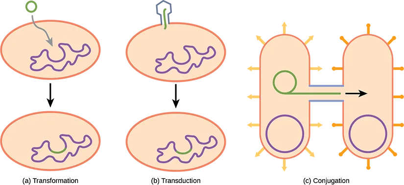
Extended description
Three labeled panels are shown left to right. Panel (a), “Transformation”: a small green circular strand of DNA sits outside an oval cell; a gray arrow shows it entering the cell, and below, a second oval cell shows the same green DNA now merged into the purple, tangled chromosome inside. Panel (b), “Transduction”: an angular blue-and-green shape (a bacteriophage) sits atop a cell injecting a short green strand of DNA; below, a second cell shows that green strand incorporated into the purple chromosome. Panel (c), “Conjugation”: two oval cells, each with orange surface projections and a purple circular chromosome, are joined by a short horizontal bridge; a green loop of DNA is shown partway across the bridge, with a black arrow indicating its direction of transfer from the left cell to the right cell.
Reproduction can be very rapid: a few minutes for some species. This short generation time coupled with mechanisms of genetic recombination and high rates of mutation result in the rapid evolution of prokaryotes, allowing them to respond to environmental changes (such as the introduction of an antibiotic) very quickly.
Evolution Connection. The Evolution of Prokaryotes
How do scientists answer questions about the evolution of prokaryotes? Unlike with animals, artifacts in the fossil record of prokaryotes offer very little information. Fossils of ancient prokaryotes look like tiny bubbles in rock. Some scientists turn to genetics and to the principle of the molecular clock, which holds that the more recently two species have diverged, the more similar their genes (and thus proteins) will be. Conversely, species that diverged long ago will have more genes that are dissimilar.
Scientists at the NASA Astrobiology Institute and at the European Molecular Biology Laboratory collaborated to analyze the molecular evolution of 32 specific proteins common to 72 species of prokaryotes (Battistuzzi, FU, Feijao, A, and Hedges, SB. A genomic timescale of prokaryote evolution: Insights into the origin of methanogenesis, phototrophy, and the colonization of land. BioMed Central: Evolutionary Biology 4 (2004): 44, doi:10.1186/1471-2148-4-44.). The model they derived from their data indicates that three important groups of bacteria—Actinobacteria, Deinococcus, and Cyanobacteria (collectively called Terrabacteria by the authors)—were the first to colonize land. Actinobacteria are a group of very common Gram-positive bacteria that produce branched structures like fungal mycelia, and include species important in decomposition of organic wastes. You will recall that Deinococcus is a genus of bacterium that is highly resistant to ionizing radiation. It has a thick peptidoglycan layer in addition to a second external membrane, so it has features of both Gram-positive and Gram-negative bacteria.
Cyanobacteria are photosynthesizers, and were probably responsible for the production of oxygen on the ancient earth. The timelines of divergence suggest that bacteria (members of the domain Bacteria) diverged from common ancestral species between 2.5 and 3.2 billion years ago, whereas the Archaea diverged earlier: between 3.1 and 4.1 billion years ago. Eukarya later diverged from the archaean line. The work further suggests that stromatolites that formed prior to the advent of cyanobacteria (about 2.6 billion years ago) photosynthesized in an anoxic environment and that because of the modifications of the Terrabacteria for land (resistance to drying and the possession of compounds that protect the organism from excess light), photosynthesis using oxygen may be closely linked to adaptations to survive on land.
Summary
Prokaryotes (domains Archaea and Bacteria) are single-celled organisms that lack a nucleus. They have a single piece of circular DNA in the nucleoid area of the cell. Most prokaryotes have a cell wall that lies outside the boundary of the plasma membrane. Some prokaryotes may have additional structures such as a capsule, flagella, and pili. Bacteria and Archaea differ in the lipid composition of their cell membranes and the characteristics of the cell wall. In archaeal membranes, phytanyl units, rather than fatty acids, are linked to glycerol. Some archaeal membranes are lipid monolayers instead of bilayers.
The cell wall is located outside the cell membrane and prevents osmotic lysis. The chemical composition of cell walls varies between species. Bacterial cell walls contain peptidoglycan. Archaean cell walls do not have peptidoglycan, but they may have pseudopeptidoglycan, polysaccharides, glycoproteins, or protein-based cell walls. Bacteria can be divided into two major groups: Gram positive and Gram negative, based on the Gram stain reaction. Gram-positive organisms have a thick peptidoglycan layer fortified with teichoic acids. Gram-negative organisms have a thin cell wall and an outer envelope containing lipopolysaccharides and lipoproteins.
Prokaryotes can transfer DNA from one cell to another by three mechanisms: transformation (uptake of environmental DNA), transduction (transfer of genomic DNA via viruses), and conjugation (transfer of DNA by direct cell contact).
Key terms
- capsule — external structure that enables a prokaryote to attach to surfaces and protects it from dehydration
- conjugation — process by which prokaryotes move DNA from one individual to another using a pilus
- Gram negative — bacterium whose cell wall contains little peptidoglycan but has an outer membrane
- Gram positive — bacterium that contains mainly peptidoglycan in its cell walls
- peptidoglycan — material composed of polysaccharide chains cross-linked to unusual peptides
- pilus — surface appendage of some prokaryotes used for attachment to surfaces including other prokaryotes
- pseudopeptidoglycan — component of archaea cell walls that is similar to peptidoglycan in morphology but contains different sugars
- S-layer — surface-layer protein present on the outside of cell walls of archaea and bacteria
- teichoic acid — polymer associated with the cell wall of Gram-positive bacteria
- transduction — process by which a bacteriophage moves DNA from one prokaryote to another
- transformation — process by which a prokaryote takes in DNA found in its environment that is shed by other prokaryotes
Practice
Describe the basic structure of a typical prokaryote
The presence of a membrane-enclosed nucleus is a characteristic of ________.
Prokaryotes are defined by the absence of this membrane-bound structure.Which of the following consist of prokaryotic cells?
Fungi, protists, and animals are all eukaryotes.The cell wall is ________.
Re-read the section’s description of what surrounds the plasma membrane in most prokaryotes.An external structure that enables a prokaryote to attach to surfaces and protects it from dehydration is called a ________.
It sits outside the cell wall and also helps resist attack by phagocytic cells.A surface appendage of some prokaryotes used for attachment to surfaces, including other prokaryotes, is called a ________.
Its plural is used earlier in the section for the many short surface projections used for attachment.A protein layer found on the outer surface of both archaeal and bacterial cell walls is called the ________.
Its name is abbreviated with a capital letter and a hyphen.The process by which a prokaryote takes in DNA shed into its environment by other prokaryotes is called ________.
This is the first of the three DNA-exchange mechanisms the section describes.The process by which a bacteriophage moves DNA from one prokaryote to another is called ________.
A virus that infects bacteria carries out this DNA-exchange mechanism.The process by which prokaryotes move DNA from one individual to another using a pilus is called ________.
This mechanism requires direct cell-to-cell contact through a channel.Describe important differences in structure between Archaea and Bacteria
Organisms most likely to be found in extreme environments are ________.
This domain’s members are best known for thriving in extreme conditions.Prokaryotes stain as Gram-positive or Gram-negative because of differences in the cell _______.
Re-read what the Gram stain reaction actually detects.Pseudopeptidoglycan is a characteristic of the walls of ________.
This molecule replaces peptidoglycan in one domain’s cell walls.The lipopolysaccharide layer (LPS) is a characteristic of the wall of ________.
This outer envelope surrounds a thin layer of peptidoglycan.Assign each structural feature from the comparison table to the domain, Bacteria or Archaea, it describes.
Bacteria
Archaea
Mention three differences between bacteria and archaea.
Show model answer
Did your answer mention:
Explain the statement that both types, bacteria and archaea, have the same basic structures, but built from different chemical components.
Show model answer
Did your answer mention:
A scientist isolates a new species of prokaryote. They note that the specimen is a bacillus with a lipid bilayer and cell wall that stains positive for peptidoglycan. Its circular chromosome replicates from a single origin of replication. Is the specimen most likely an Archaea, a Gram-positive bacterium, or a Gram-negative bacterium? How do you know?
Show model answer
Did your answer mention:
Material composed of polysaccharide chains cross-linked to unusual peptides, found in most bacterial cell walls, is called ________.
Its presence or absence is the key difference between bacterial and archaeal cell walls.A bacterium whose cell wall contains little peptidoglycan but is surrounded by an outer membrane is described as ________.
This kind of bacterium’s outer membrane contains lipopolysaccharides.A bacterium whose cell wall is composed mainly of peptidoglycan is described as ________.
This kind of bacterium’s wall is fortified with teichoic acids.A polymer associated with the cell wall of Gram-positive bacteria, sometimes linked to membrane lipids to form a lipid-anchored form, is called ________.
Its lipid-linked form anchors the cell wall to the cell membrane.This section is adapted from Biology 2e, Section 22.2: Structure of Prokaryotes: Bacteria and Archaea by Mary Ann Clark, Jung Choi, Matthew Douglas, and OpenStax, © OpenStax, licensed under CC BY-NC-SA 4.0. Access the original for free at openstax.org. Changes: figures re-encoded as WebP; Figure_22_02_02, Figure_22_02_03-8ae1, Figure_22_02_04f, Figure_B22_02_08b, Figure_22_02_06f, and Figure_22_02_07f re-kinded from the manifest’s file-extension “photo” guess to “diagram” after inspection (a labeled cell diagram, a phylogenetic tree, three infographic tables of bacterial/archaeal phyla with embedded micrographs, and a labeled phospholipid-structure diagram, none of them photographs); a longdesc added for Figure_22_02_02, Figure_22_02_03-8ae1, Figure_22_02_04f, Figure_B22_02_08b, Figure_22_02_06f, Figure_22_02_07f, Figure_22_02_08f, and Figure_B22_02_10 (labels, branching order, table contents, and arrows not carried by the one- or two-line captions); the source alts for Figure_22_02_04f, Figure_B22_02_08b, and Figure_22_02_06f, each well over 600 characters, rewritten to a concise description with the full table walk-through moved into longdesc, and the misspelled organism names in those three vendored table images (“Heliobacter pylori”, “Psuedomonas aeruginosa”, “Vibrio cholera”, “streptomyocin”, “Nanoarchaeotum equitans”, “Rickettsia rickettsia”/“Rickettsii”, “Nitrosomas”, “Crenachaeotes”, “Aneorobic”, and “aneaerobes”) standardized to their correct scientific spelling in the authored alt/longdesc text (the misspellings remain as printed in the images themselves and are reported as suspected source defects); the source’s forward figure references (a link before each figure) reworded as “illustrated below” / “shown below” since figures are not numbered here; the note wrapping the Gram-positive/Gram-negative figure and its question kept as the figure followed by a multiple choice in the body (the Visual Connection Questions section at the end of the module, which repeats the identical item, is not duplicated in Practice); the Evolution Connection feature box kept with its title and its footnote citation, moved to a parenthetical after the sentence it supports; the end-of-section Review Questions and Critical Thinking Questions adapted into the closing interactive Practice block (multiple choice and self-check respectively), and each Critical Thinking model answer’s introductory “Responses will vary. A possible answer is:” framing dropped as assessment-methodology scaffolding rather than section content; rubric checkpoints added to each self-check, decomposing its model answer (the source solution) into check-off clauses with no new claims; the Bacteria-vs-Archaea comparison table (tab-ch22-02-01) transcribed complete as a Markdown table, its spanning title row set as a bold line above it, and also built into one sortbins exercise, drawing one item from each of the table’s eight rows whose Bacteria and Archaea cells differ; and ten key-term recall items added from the glossary (all but pseudopeptidoglycan, which the corresponding Review Question already keys). The prokaryote-shapes long description was corrected to attribute typhus and Rocky Mountain spotted fever to the genus Rickettsia, as the source does, rather than to R. rickettsii alone.