Classifications of Fungi
By the end of this section, you will be able to:
- Identify fungi and place them into the five major phyla according to current classification
- Describe each phylum in terms of major representative species and patterns of reproduction
The kingdom Fungi contains five major phyla that were established according to their mode of sexual reproduction or using molecular data. Unrelated fungi that reproduce without a sexual cycle, were once placed for convenience in a sixth group, the Deuteromycota, called a “form phylum,” because superficially they appeared to be similar, a polyphyletic group (group of organisms that is of mixed evolutionary origin). However, most mycologists have discontinued this practice. Rapid advances in molecular biology and the sequencing of 18S rRNA (ribosomal RNA) continue to show new and different relationships among the various categories of fungi.
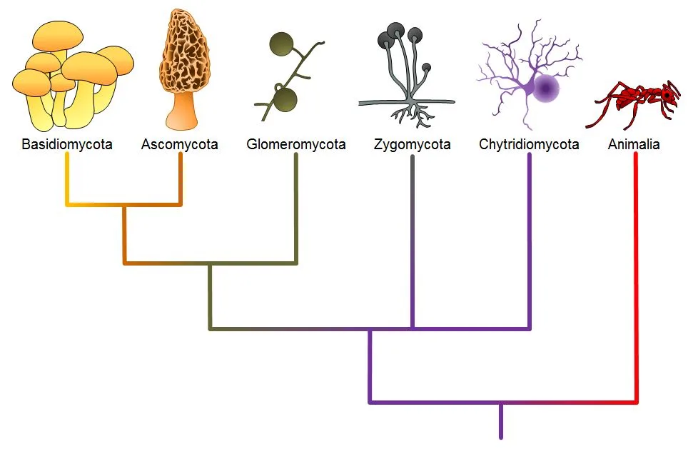
The five true phyla of fungi are the Chytridiomycota (Chytrids), the Zygomycota (conjugated fungi), the Ascomycota (sac fungi), the Basidiomycota (club fungi) and the recently described Phylum Glomeromycota (pictured below).
Extended description
The tree’s root sits at the bottom and its six tip labels — each paired with a small drawn icon — read left to right: Basidiomycota (a cluster of yellow mushrooms), Ascomycota (an orange morel), Glomeromycota (an olive branching structure with round spores), Zygomycota (a gray branching sporangium structure), Chytridiomycota (a purple branching structure), and Animalia (a red ant), each on its own colored vertical line. Basidiomycota’s yellow line and Ascomycota’s orange line join first, closest to the tips, in an orange node. That combined line joins Glomeromycota’s olive line next, in an olive node. That combined line then joins Zygomycota’s gray line, in a purple node, and the result joins Chytridiomycota’s purple line in a second purple node — grouping all five fungal phyla into one lineage. That fungal lineage finally joins Animalia’s red line at the tree’s root, the common ancestor shared by fungi and animals.
Chytridiomycota: The Chytrids
The only class in the Phylum Chytridiomycota is the Chytridiomycetes. The chytrids are the simplest and most primitive Eumycota, or true fungi. The evolutionary record shows that the first recognizable chytrids appeared during the late pre-Cambrian period, more than 500 million years ago. Like all fungi, chytrids have chitin in their cell walls, but one group of chytrids has both cellulose and chitin in the cell wall. Most chytrids are unicellular; however, a few form multicellular organisms and hyphae, which have no septa between cells (coenocytic). The Chytrids are the only fungi that have retained flagella. They produce both gametes and diploid zoospores that swim with the help of a single flagellum. An unusual feature of the chytrids is that both male and female gametes are flagellated.
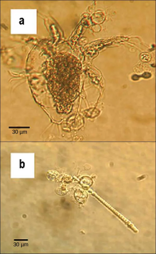
The ecological habitat and cell structure of chytrids have much in common with protists. Chytrids usually live in aquatic environments, although some species live on land. Some species thrive as parasites on plants, insects, or amphibians (pictured below), while others are saprobes. The chytrid species Allomyces is well characterized as an experimental organism. Its reproductive cycle includes both asexual and sexual phases. Allomyces produces diploid or haploid flagellated zoospores in a sporangium.
Zygomycota: The Conjugated Fungi
The zygomycetes are a relatively small group of fungi belonging to the Phylum Zygomycota. They include the familiar bread mold, Rhizopus stolonifer, which rapidly propagates on the surfaces of breads, fruits, and vegetables. Most species are saprobes, living off decaying organic material; a few are parasites, particularly of insects. Zygomycetes play a considerable commercial role. For example, the metabolic products of some species of Rhizopus are intermediates in the synthesis of semi-synthetic steroid hormones.
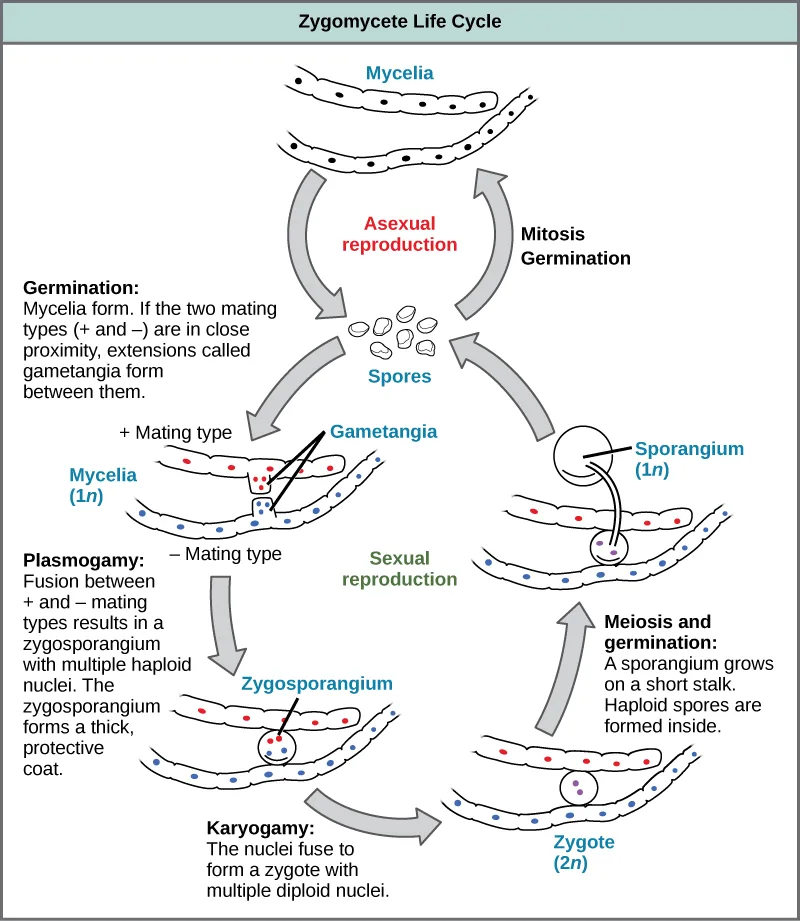
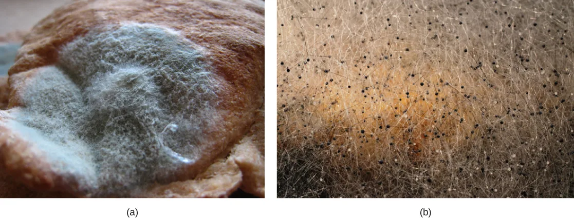
Zygomycetes have a thallus of coenocytic hyphae in which the nuclei are haploid when the organism is in the vegetative stage. The fungi usually reproduce asexually by producing sporangiospores (pictured below). The black tips of bread mold are the swollen sporangia packed with black spores (pictured below). When spores land on a suitable substrate, they germinate and produce a new mycelium. Sexual reproduction starts when environmental conditions become unfavorable. Two opposing mating strains (type + and type –) must be in close proximity for gametangia from the hyphae to be produced and fuse, leading to karyogamy. Each zygospore can contain several diploid nuclei. The developing diploid zygospores have thick coats that protect them from desiccation and other hazards. They may remain dormant until environmental conditions are favorable. When the zygospore germinates, it undergoes meiosis and produces haploid spores, which will, in turn, grow into a new organism. This form of sexual reproduction in fungi is called conjugation (although it differs markedly from conjugation in bacteria and protists), giving rise to the name “conjugated fungi”.
Extended description
The cycle runs counterclockwise from ‘Mycelia’ at the top. Asexual reproduction (the small upper loop): an arrow on the left carries the mycelia down to a cluster of spores, and an arrow on the right, labeled ‘Mitosis, Germination,’ carries spores back up to new mycelia. Sexual reproduction (the large lower loop, running down the left side, along the bottom, and up the right): germination forms mycelia, and if plus and minus mating-type mycelia (labeled ‘+ Mating type’ and ‘– Mating type,’ each cell shown with a red or blue dot) lie close together, extensions called gametangia grow between them. Plasmogamy fuses the two mating types into a zygosporangium containing multiple haploid nuclei and a thick protective coat. Karyogamy then fuses those nuclei into a zygote with multiple diploid (2n) nuclei. Meiosis and germination follow: a sporangium grows on a short stalk, and haploid (1n) spores form inside it, feeding back into the shared spore cluster below the mycelia.
Ascomycota: The Sac Fungi
The majority of known fungi belong to the Phylum Ascomycota, which is characterized by the formation of an ascus (plural, asci), a sac-like structure that contains haploid ascospores. Filamentous ascomycetes produce hyphae divided by perforated septa, allowing streaming of cytoplasm from one cell to another. Conidia and asci, which are used respectively for asexual and sexual reproduction, are usually separated from the vegetative hyphae by blocked (non-perforated) septa. Many ascomycetes are of commercial importance. Some play a beneficial role for humanity, such as the yeasts used in baking, brewing, and wine fermentation, and directly as food delicacies such as truffles and morels. Aspergillus oryzae is used in the fermentation of rice to produce sake. Other ascomycetes parasitize plants and animals, including humans. For example, fungal pneumonia poses a significant threat to AIDS patients who have a compromised immune system. Ascomycetes not only infest and destroy crops directly; they also produce poisonous secondary metabolites that make crops unfit for consumption.
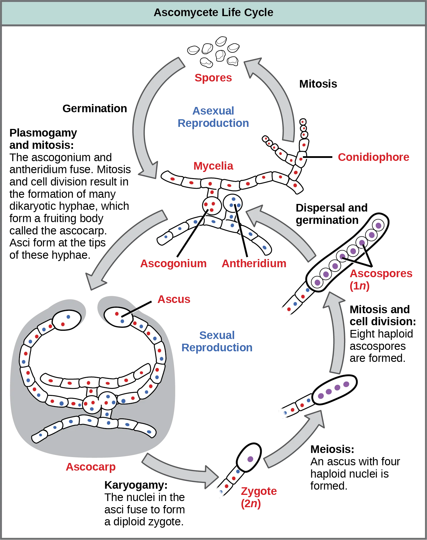
Asexual reproduction is frequent and involves the production of conidiophores that release haploid conidiospores (pictured below). Sexual reproduction starts with the development of special hyphae from either one of two types of mating strains (shown below). The “male” strain produces an antheridium and the “female” strain develops an ascogonium. At fertilization, the antheridium and the ascogonium combine in plasmogamy, without nuclear fusion. Special dikaryotic ascogenous (ascus-producing) hyphae arise from this dikaryon, in which each cell has pairs of nuclei: one from the “male” strain and one from the “female” strain. In each ascus, two haploid nuclei fuse in karyogamy. Thousands of asci fill a fruiting body called the ascocarp. The diploid nucleus in each ascus gives rise to haploid nuclei by meiosis, and spore walls form around each nucleus. The spores in each ascus contain the meiotic products of a single diploid nucleus. The ascospores are then released, germinate, and form hyphae that are disseminated in the environment and start new mycelia (pictured below).
Extended description
The cycle runs counterclockwise from ‘Spores’ at the top. In the small upper loop (Asexual Reproduction): spores form a haploid (1n) mycelium; mitosis produces a conidiophore that buds new spores, which germinate back into mycelia. In the large lower loop (Sexual Reproduction, running down the left side, along the bottom, and up the right): germination and plasmogamy fuse an ascogonium and an antheridium budding from the mycelium; mitosis and cell division form many hyphae, each cell shown with one red and one blue dot, described as dikaryotic, that grow into a fruiting body labeled ‘Ascocarp.’ At the tips of these hyphae, an ascus — drawn with one red and one blue dot — undergoes karyogamy, its two nuclei fusing into a diploid (2n) zygote. The zygote undergoes meiosis, producing an ascus with four haploid (1n) nuclei, drawn as purple dots arranged in a row. Each of the four nuclei then divides once by mitosis, yielding eight ascospores arranged in a row inside the ascus; these are dispersed and germinate, completing the cycle back to mycelia.
Which of the following statements is true?
Trace the diagram above from the Ascus label through Karyogamy, Meiosis, and Mitosis, and note the two colors of dot drawn inside the ascus before karyogamy.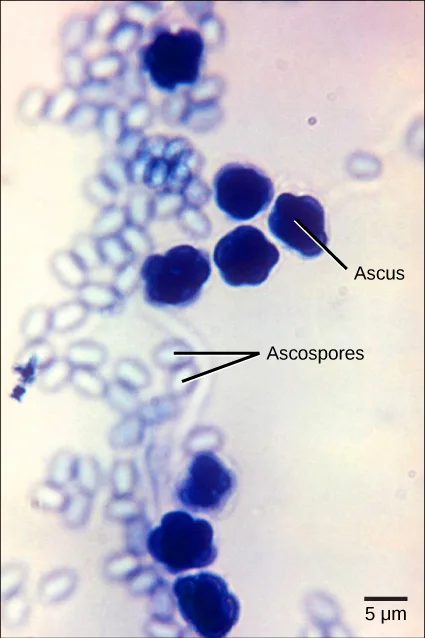
Extended description
Label lines point to two features in the micrograph: ‘Ascus’ labels one of the dark blue-purple, sphere-clustered structures near the middle of the image, and ‘Ascospores’ labels two of the smaller, pale blue oval bodies scattered among the many similar oval cells filling the frame. A scale bar reading 5 µm sits at the lower right.
Basidiomycota: The Club Fungi
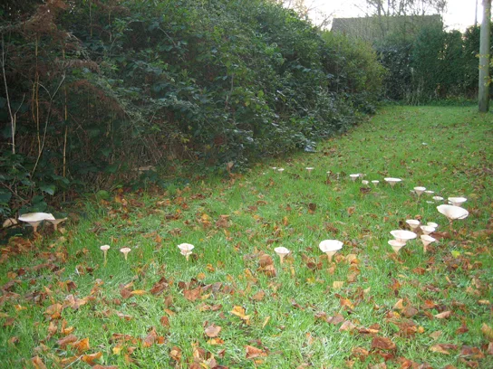
The fungi in the Phylum Basidiomycota are easily recognizable under a light microscope by their club-shaped fruiting bodies called basidia (singular, basidium), which are the swollen terminal cells of hyphae. The basidia, which are the reproductive organs of these fungi, are often contained within the familiar mushroom, commonly seen in fields after rain, on the supermarket shelves, and growing on your lawn (pictured below). These mushroom-producing basidiomycetes are sometimes referred to as “gill fungi” because of the presence of gill-like structures on the underside of the cap. The gills are actually compacted hyphae on which the basidia are borne. This group also includes shelf fungi, which cling to the bark of trees like small shelves. In addition, the basidiomycota include smuts and rusts, which are important plant pathogens. Most edible fungi belong to the Phylum Basidiomycota; however, some basidiomycota are inedible and produce deadly toxins. For example, Cryptococcus neoformans causes severe respiratory illness. The infamous death cap mushroom (Amanita phalloides) is related to the fly agaric seen at the beginning of the previous section.
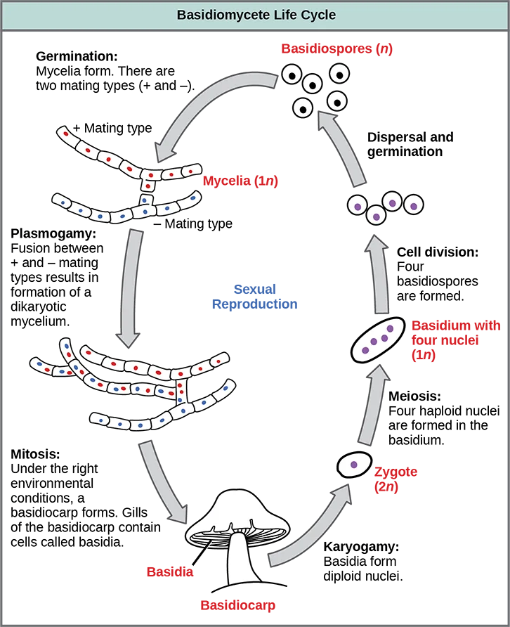
The lifecycle of basidiomycetes includes sexual and asexual reproduction (illustrated below). Most fungi are haploid through most of their life cycles, but the basidiomycetes produce both haploid and dikaryotic mycelia, with the dikaryotic phase being dominant. (Note: The dikaryotic phase is technically not diploid, since the nuclei remain unfused until shortly before spore production.) In the basidiomycetes, sexual spores are more common than asexual spores. The sexual spores form in the club-shaped basidium and are called basidiospores. In the basidium, nuclei of two different mating strains fuse (karyogamy), giving rise to a diploid zygote that then undergoes meiosis. The haploid nuclei migrate into four different chambers appended to the basidium, and then become basidiospores.
Each basidiospore germinates and generates monokaryotic haploid hyphae. The mycelium that results is called a primary mycelium. Mycelia of different mating strains can combine and produce a secondary mycelium that contains haploid nuclei of two different mating strains. This is the dominant dikaryotic stage of the basidiomycete life cycle. Thus, each cell in this mycelium has two haploid nuclei, which will not fuse until formation of the basidium. Eventually, the secondary mycelium generates a basidiocarp, a fruiting body that protrudes from the ground—this is what we think of as a mushroom. The basidiocarp bears the developing basidia on the gills under its cap.
Extended description
The cycle runs counterclockwise from the upper left, down the left side, along the bottom, and up the right. Germination produces plus and minus mating-type mycelia (1n), each cell shown with a red or blue dot. Plasmogamy fuses the two mating types into a dikaryotic mycelium whose cells each carry one red and one blue dot. Under the right conditions, mitosis of this mycelium produces a mushroom-shaped structure labeled ‘Basidiocarp’; club-shaped cells in its gills are labeled ‘Basidia.’ Karyogamy inside each basidium fuses the two nuclei into a diploid (2n) zygote. Meiosis then produces a basidium with four haploid nuclei (1n), drawn as four purple dots. Cell division forms four basidiospores, which are dispersed and germinate, completing the cycle back to new mycelia.
Which of the following statements is true?
Compare the diagram’s own labels for ‘Basidiocarp’ and ‘Basidia’ — which one is drawn as the mushroom shape itself, and which is drawn inside its gills?Asexual Ascomycota and Basidiomycota
Imperfect fungi—those that do not display a sexual phase—were formerly classified in the form phylum Deuteromycota, an invalid taxon no longer used in the present, ever-developing classification of organisms. While Deuteromycota was once a classification taxon, recent molecular analysis has shown that some of the members classified in this group belong to the Ascomycota (pictured below) or the Basidiomycota. Because some members of this group have not yet been appropriately classified, they are less well described in comparison to members of other fungal taxa. Most imperfect fungi live on land, with a few aquatic exceptions. They form visible mycelia with a fuzzy appearance and are commonly known as mold.
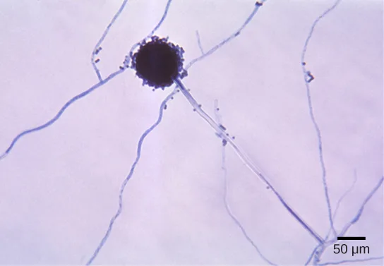
The fungi in this group have a large impact on everyday human life. The food industry relies on them for ripening some cheeses. The blue veins in Roquefort cheese and the white crust on Camembert are the result of fungal growth. The antibiotic penicillin was originally discovered on an overgrown Petri plate, on which a colony of Penicillium fungi had killed the bacterial growth surrounding it. Other fungi in this group cause serious diseases, either directly as parasites (which infect both plants and humans), or as producers of potent toxic compounds, as seen in the aflatoxins released by fungi of the genus Aspergillus.
Glomeromycota
The Glomeromycota is a newly established phylum that comprises about 230 species, all of which are involved in close associations with the roots of trees. Fossil records indicate that trees and their root symbionts share a long evolutionary history. It appears that nearly all members of this family form arbuscular mycorrhizae: the hyphae interact with the root cells forming a mutually beneficial association in which the plants supply the carbon source and energy in the form of carbohydrates to the fungus, and the fungus supplies essential minerals from the soil to the plant. The exception is Geosiphon pyriformis, which hosts the cyanobacterium Nostoc as an endosymbiont.
The glomeromycetes do not reproduce sexually and do not survive without the presence of plant roots. Although they have coenocytic hyphae like the zygomycetes, they do not form zygospores. DNA analysis shows that all glomeromycetes probably descended from a common ancestor, making them a monophyletic lineage.
Summary
Chytridiomycota (chytrids) are considered the most ancestral group of fungi. They are mostly aquatic, and their gametes are the only fungal cells known to have flagella. They reproduce both sexually and asexually; the asexual spores are called zoospores. Zygomycota (conjugated fungi) produce non-septate hyphae with many nuclei. Their hyphae fuse during sexual reproduction to produce a zygospore in a zygosporangium. Ascomycota (sac fungi) form spores in sacs called asci during sexual reproduction. Asexual reproduction is their most common form of reproduction. In the Basidiomycota (club fungi), the sexual phase predominates, producing showy fruiting bodies that contain club-shaped basidia, within which spores form. Most familiar mushrooms belong to this division. Fungi that have no known sexual cycle were originally classified in the “form phylum” Deuteromycota, but many have been classified by comparative molecular analysis with the Ascomycota and Basidiomycota. Glomeromycota form tight associations (called mycorrhizae) with the roots of plants.
Key terms
- Arbuscular mycorrhizae — mycorrhizae commonly involving Glomeromycetes in which the fungal hyphae penetrate the cell walls of the plant root cells (but not the cell membranes)
- ascocarp — fruiting body of ascomycetes
- Ascomycota — (also, sac fungi) phylum of fungi that store spores in a sac called ascus
- basidiocarp — fruiting body that protrudes from the ground and bears the basidia
- Basidiomycota — (also, club fungi) phylum of fungi that produce club-shaped structures (basidia) that contain spores
- basidium — club-shaped fruiting body of basidiomycetes
- Chytridiomycota — (also, chytrids) primitive phylum of fungi that live in water and produce gametes with flagella
- Deuteromycota — former form phylum of fungi that do not have a known sexual reproductive cycle (presently members of two phyla: Ascomycota and Basidiomycota)
- Ectomycorrhizae — mycorrhizae in which the fungal hyphae do not penetrate the root cells of the plant
- Glomeromycota — phylum of fungi that form symbiotic relationships with the roots of trees
- mold — tangle of visible mycelia with a fuzzy appearance
- polyphyletic group — group of organisms that is of mixed evolutionary origin
- Zygomycota — (also, conjugated fungi) phylum of fungi that form a zygote contained in a zygospore
- zygospore — structure with thick cell wall that contains the zygote in zygomycetes
Practice
Identify fungi and place them into the five major phyla according to current classification
The most primitive phylum of fungi is the ________.
This phylum’s members are considered the simplest and most primitive true fungi, with a fossil record dating to the late pre-Cambrian period.Members of which phylum produce a club-shaped structure that contains spores?
This phylum’s fruiting bodies are the mushrooms you see in fields after rain, on supermarket shelves, and on your lawn.Members of which phylum are all involved in close associations with the roots of trees?
Nearly all members of this newly described phylum form arbuscular mycorrhizae with plant roots.The fungi that do not reproduce sexually used to be classified as ________.
This invalid ‘form phylum’ name is no longer used now that molecular analysis has reassigned its members to other phyla.A scientist discovers a new species of fungus that introduces genetic diversity during reproduction by creating a diploid zygote. This new species cannot belong to which modern phylum of fungi?
Re-read which phylum in this section is described as not reproducing sexually at all.The fungal phylum whose spores form inside a specialized structure called an ascus is ________.
Baker’s and brewer’s yeast, along with truffles and morels, belong to this phylum.The fungal phylum that includes the familiar bread mold and produces a zygote inside a thick-walled sporangium-forming structure is called ________.
Rhizopus stolonifer, the fungus that grows on bread and fruit, is this phylum’s best-known example.Describe each phylum in terms of major representative species and patterns of reproduction
What is the advantage for a basidiomycete to produce a showy and fleshy fruiting body?
Show model answer
Did your answer mention:
For each of the four groups of perfect fungi (Chytridiomycota, Zygomycota, Ascomycota, and Basidiomycota), compare the body structure and features, and provide an example.
Show model answer
Did your answer mention:
In zygomycetes, the thick-coated structure that protects the developing zygote from desiccation until conditions favor germination is called a ________.
This structure’s parent phylum’s two mating strains are labeled type + and type – in this section.The club-shaped reproductive cell found on the gills beneath a mushroom’s cap, where spores form, is called a ________.
Its plural form gives the whole phylum, Basidiomycota, its name.In the Basidiomycota (club fungi), the sexual phase predominates, producing showy fruiting bodies that contain club-shaped structures called ________, within which spores form.
This section’s summary sentence about the Basidiomycota names these club-shaped, spore-forming structures directly.This section is adapted from Biology 2e, Section 24.2: Classifications of Fungi by Mary Ann Clark, Jung Choi, Matthew Douglas, and OpenStax, © OpenStax, licensed under CC BY-NC-SA 4.0. Access the original for free at openstax.org. Changes: figures re-encoded as WebP; Figure_B24_02_01 and Figure_B24_02_02 re-kinded from the manifest’s file-extension “photo” guess to “diagram” since both are drawn schematics, not photographs; a longdesc added to five figures whose diagram labels, life-cycle stages, or pointer-line labels are not carried by their one-line captions (Figure_B24_02_01, Figure_B24_02_02, Figure_B24_02_04, Figure_B24_02_07, and Figure_24_02_05); the over-600-character source alts for Figure_B24_02_01, Figure_B24_02_02, Figure_B24_02_04, and Figure_B24_02_07 condensed to short descriptions with their step-by-step walk-throughs moved into a longdesc, and the two Visual-Connection figures’ (Figure_B24_02_04, Figure_B24_02_07) short alts written to describe structure without stating which multiple-choice option is correct; Figure_24_02_03abf’s source alt description of “green mold” replaced with a plain description of the grayish-white fuzz actually visible in the image, matching the source caption’s own “white fuzz”; the Fairy Ring caption’s stray trailing “]” (a source typesetting artifact) dropped; bare in-text figure cross-references rendered as “(pictured below)” / “(shown below)” / “(illustrated below)” since Hugo does not number figures; the two feature boxes wrapping Visual Connection questions rendered as their figure followed by a multiple choice, kept in the body; the end-of-section Review Questions and Critical Thinking Questions adapted into the closing interactive Practice block (multiple choice and self-check respectively); rubric checkpoints added to each self-check, decomposing its model answer (the source solution) into check-off clauses with no new claims; four key-term recall items (Ascomycota, Zygomycota, zygospore, basidium) added from the glossary; one summary-derived select-the-term multiple choice (basidia) added under the second objective, since its two Critical Thinking self-checks alone leave that group under this book’s per-group floor. The mycorrhiza review question’s stem was reworded to this section’s own sentence about the Glomeromycota (“all of which are involved in close associations with the roots of trees”), because the source stem was also true of the Ascomycota and Basidiomycota distractors by the Ecology of Fungi section; options and key unchanged (erratum 418).