Features Used to Classify Animals
By the end of this section, you will be able to:
- Explain the differences in animal body plans that support basic animal classification
- Compare and contrast the embryonic development of protostomes and deuterostomes
Scientists have developed a classification scheme that categorizes all members of the animal kingdom, although there are exceptions to most “rules” governing animal classification (see the phylogenetic tree below). Animals have been traditionally classified according to two characteristics: body plan and developmental pathway. The major feature of the body plan is its symmetry: how the body parts are distributed along the major body axis. Symmetrical animals can be divided into roughly equivalent halves along at least one axis. Developmental characteristics include the number of germ tissue layers formed during development, the origin of the mouth and anus, the presence or absence of an internal body cavity, and other features of embryological development, such as larval types or whether or not periods of growth are interspersed with molting.
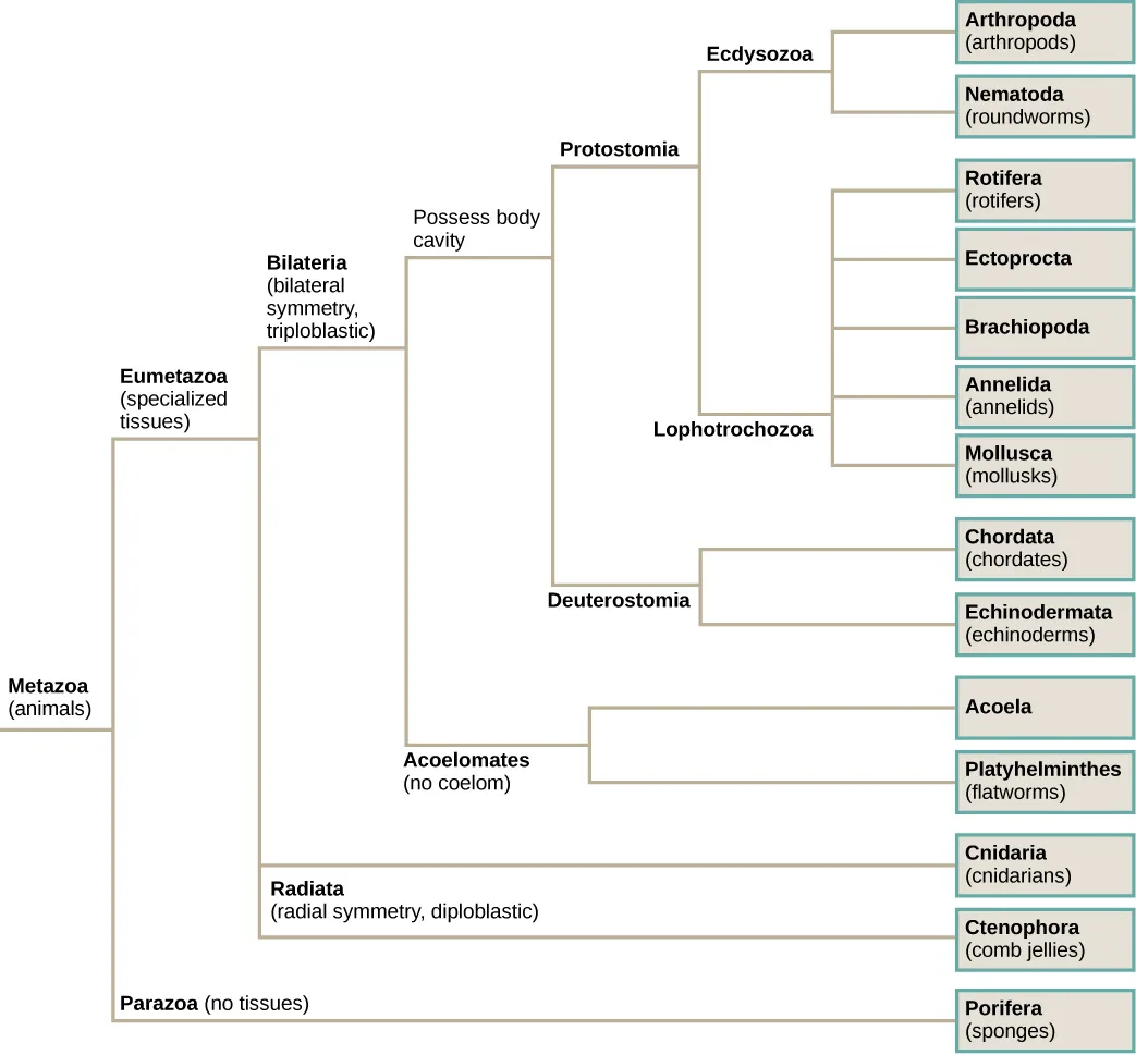
Extended description
Reading the tree from its top branches down: ‘Ecdysozoa’ leads to ‘Arthropoda (arthropods)’ and ‘Nematoda (roundworms).’ Below it, ‘Lophotrochozoa’ leads to ‘Rotifera (rotifers),’ ‘Ectoprocta,’ ‘Brachiopoda,’ ‘Annelida (annelids),’ and ‘Mollusca (mollusks).’ Ecdysozoa and Lophotrochozoa both branch from ‘Protostomia.’ Below that, ‘Deuterostomia’ leads to ‘Chordata (chordates)’ and ‘Echinodermata (echinoderms).’ Protostomia and Deuterostomia both branch from a node labeled ‘Possess body cavity.’ Below that node, ‘Acoelomates (no coelom)’ leads to ‘Acoela’ and ‘Platyhelminthes (flatworms).’ The body-cavity node and Acoelomates both branch from ‘Bilateria (bilateral symmetry, triploblastic).’ Below Bilateria, ‘Radiata (radial symmetry, diploblastic)’ leads to ‘Cnidaria (cnidarians)’ and ‘Ctenophora (comb jellies).’ Bilateria and Radiata both branch from ‘Eumetazoa (specialized tissues),’ which joins at the root, ‘Metazoa (animals),’ with ‘Parazoa (no tissues),’ the bottommost branch, leading to ‘Porifera (sponges).’
Which of the following statements is false?
Trace each group’s branch back to the node that names its symmetry: one of these two groups descends from Bilateria, not from Radiata.Animal Characterization Based on Body Symmetry
At a very basic level of classification, true animals can be largely divided into three groups based on the type of symmetry of their body plan: radially symmetrical, bilaterally symmetrical, and asymmetrical. Asymmetry is seen in two modern clades, the Parazoa (pictured below) and Placozoa. (Although we should note that the ancestral fossils of the Parazoa apparently exhibited bilateral symmetry.) One clade, the Cnidaria, exhibits radial or biradial symmetry: Ctenophores have rotational symmetry. Bilateral symmetry is seen in the largest of the clades, the Bilateria; however the Echinodermata are bilateral as larvae and metamorphose secondarily into radial adults. All types of symmetry are well suited to meet the unique demands of a particular animal’s lifestyle.
Radial symmetry is the arrangement of body parts around a central axis, as is seen in a bicycle wheel or pie. It results in animals having top and bottom surfaces but no left and right sides, nor front or back. If a radially symmetrical animal is divided in any direction along the oral/aboral axis (the side with a mouth is “oral side,” and the side without a mouth is the “aboral side”), the two halves will be mirror images. This form of symmetry marks the body plans of many animals in the phyla Cnidaria, including jellyfish and adult sea anemones. Radial symmetry equips these sea creatures (which may be sedentary or only capable of slow movement or floating) to experience the environment equally from all directions. Bilaterally symmetrical animals, like butterflies, have only a single plane along which the body can be divided into equivalent halves. The Ctenophora, although they look similar to jellyfish, are considered to have rotational symmetry rather than radial or biradial symmetry because division of the body into two halves along the oral/aboral axis divides them into two copies of the same half, with one copy rotated 180°, rather than two mirror images.
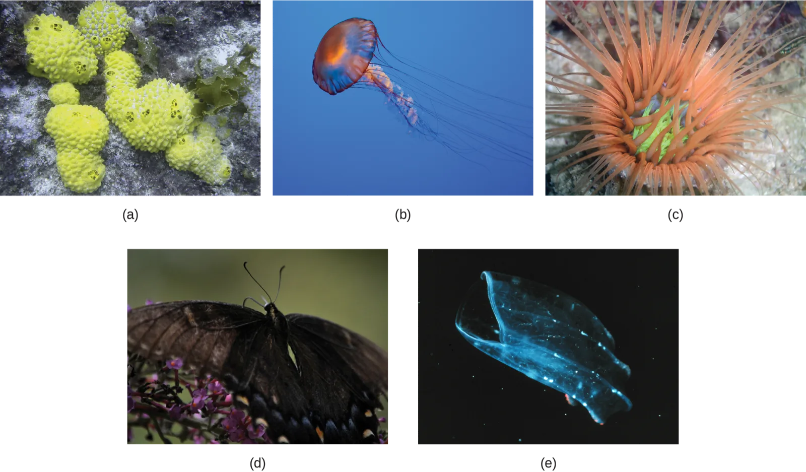
Bilateral symmetry involves the division of the animal through a midsagittal plane, resulting in two superficially mirror images, right and left halves, such as those of a butterfly, crab, or human body. Animals with bilateral symmetry have a “head” and “tail” (anterior vs. posterior), front and back (dorsal vs. ventral), and right and left sides (see the figure below). All Eumetazoa except those with secondary radial symmetry are bilaterally symmetrical. The evolution of bilateral symmetry that allowed for the formation of anterior and posterior (head and tail) ends promoted a phenomenon called cephalization, which refers to the collection of an organized nervous system at the animal’s anterior end. In contrast to radial symmetry, which is best suited for stationary or limited-motion lifestyles, bilateral symmetry allows for streamlined and directional motion. In evolutionary terms, this simple form of symmetry promoted active and controlled directional mobility and increased sophistication of resource-seeking and predator-prey relationships.
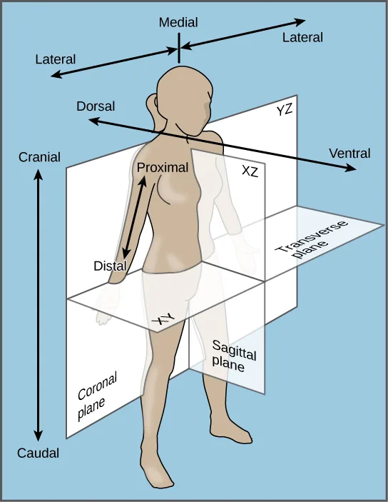
Extended description
At the top, a double-headed arrow crosses the head, labeled ‘Medial’ at its center and ‘Lateral’ at each end. Below it, a longer double-headed arrow runs through the shoulders, labeled ‘Dorsal’ at its back end and ‘Ventral’ at its front end. At the far left, a vertical double-headed arrow spans the whole figure, labeled ‘Cranial’ at the top and ‘Caudal’ at the bottom. On the raised right arm, a short double-headed arrow reads ‘Proximal’ near the shoulder and ‘Distal’ near the hand. Three shaded planes pass through the body: a vertical plane parallel to the body’s front, labeled ‘Coronal plane’ at its lower-left corner and ‘XZ’ near its top, dividing the body into front and back; a second vertical plane at a right angle to the first, labeled ‘Sagittal plane’ at its lower-right corner and ‘YZ’ near its top, dividing the body into left and right; and a horizontal plane at the hips, labeled ‘Transverse plane’ at its right edge, dividing the body into upper and lower.
Animals in the phylum Echinodermata (such as sea stars, sand dollars, and sea urchins) display modified radial symmetry as adults, but as we have noted, their larval stages (such as the bipinnaria) initially exhibit bilateral symmetry until they metamorphose in animals with radial symmetry (this is termed secondary radial symmetry). Echinoderms evolved from bilaterally symmetrical animals; thus, they are classified as bilaterally symmetrical.
Animal Characterization Based on Features of Embryological Development
Most animal species undergo a separation of tissues into germ layers during embryonic development. Recall that these germ layers are formed during gastrulation, and that each germ layer typically gives rise to specific types of embryonic tissues and organs. Animals develop either two or three embryonic germ layers. The animals that display radial, biradial, or rotational symmetry develop two germ layers, an inner layer (endoderm or mesendoderm) and an outer layer (ectoderm). These animals are called diploblasts, and have a nonliving middle layer between the endoderm and ectoderm (although individual cells may be distributed through this middle layer, there is no coherent third layer of tissue). The four clades considered to be diploblastic have different levels of complexity and different developmental pathways, although there is little information about development in Placozoa. More complex animals (usually those with bilateral symmetry) develop three tissue layers: an inner layer (endoderm), an outer layer (ectoderm), and a middle layer (mesoderm). Animals with three tissue layers are called triploblasts.
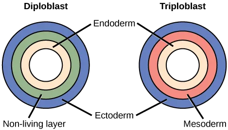
Extended description
The left ring diagram, labeled ‘Diploblast,’ has, from outside to center, an ectoderm ring, a non-living-layer ring, and an endoderm ring surrounding a hollow center. The right ring diagram, labeled ‘Triploblast,’ has, from outside to center, an ectoderm ring, a mesoderm ring in the position the non-living layer occupies at left, and an endoderm ring surrounding a hollow center. A single ‘Endoderm’ label points to the innermost ring of both diagrams, and a single ‘Ectoderm’ label points to the outermost ring of both.
Which of the following statements about diploblasts and triploblasts is false?
Reread the sentence naming which germ layer becomes the central nervous system in the paragraph just after this figure.Each of the three germ layers is programmed to give rise to specific body tissues and organs, although there are variations on these themes. Generally speaking, the endoderm gives rise to the lining of the digestive tract (including the stomach, intestines, liver, and pancreas), as well as to the lining of the trachea, bronchi, and lungs of the respiratory tract, along with a few other structures. The ectoderm develops into the outer epithelial covering of the body surface, the central nervous system, and a few other structures. The mesoderm is the third germ layer; it forms between the endoderm and ectoderm in triploblasts. This germ layer gives rise to all specialized muscle tissues (including the cardiac tissues and muscles of the intestines), connective tissues such as the skeleton and blood cells, and most other visceral organs such as the kidneys and the spleen. Diploblastic animals may have cell types that serve multiple functions, such as epitheliomuscular cells, which serve as a covering as well as contractile cells.
Presence or Absence of a Coelom
Further subdivision of animals with three germ layers (triploblasts) results in the separation of animals that may develop an internal body cavity derived from mesoderm, called a coelom, and those that do not. This epithelial cell-lined coelomic cavity, usually filled with fluid, lies between the visceral organs and the body wall. It houses many organs such as the digestive, urinary, and reproductive systems, the heart and lungs, and also contains the major arteries and veins of the circulatory system. In mammals, the body cavity is divided into the thoracic cavity, which houses the heart and lungs, and the abdominal cavity, which houses the digestive organs. In the thoracic cavity further subdivision produces the pleural cavity, which provides space for the lungs to expand during breathing, and the pericardial cavity, which provides room for movements of the heart. The evolution of the coelom is associated with many functional advantages. For example, the coelom provides cushioning and shock absorption for the major organ systems that it encloses. In addition, organs housed within the coelom can grow and move freely, which promotes optimal organ development and placement. The coelom also provides space for the diffusion of gases and nutrients, as well as body flexibility, promoting improved animal motility.
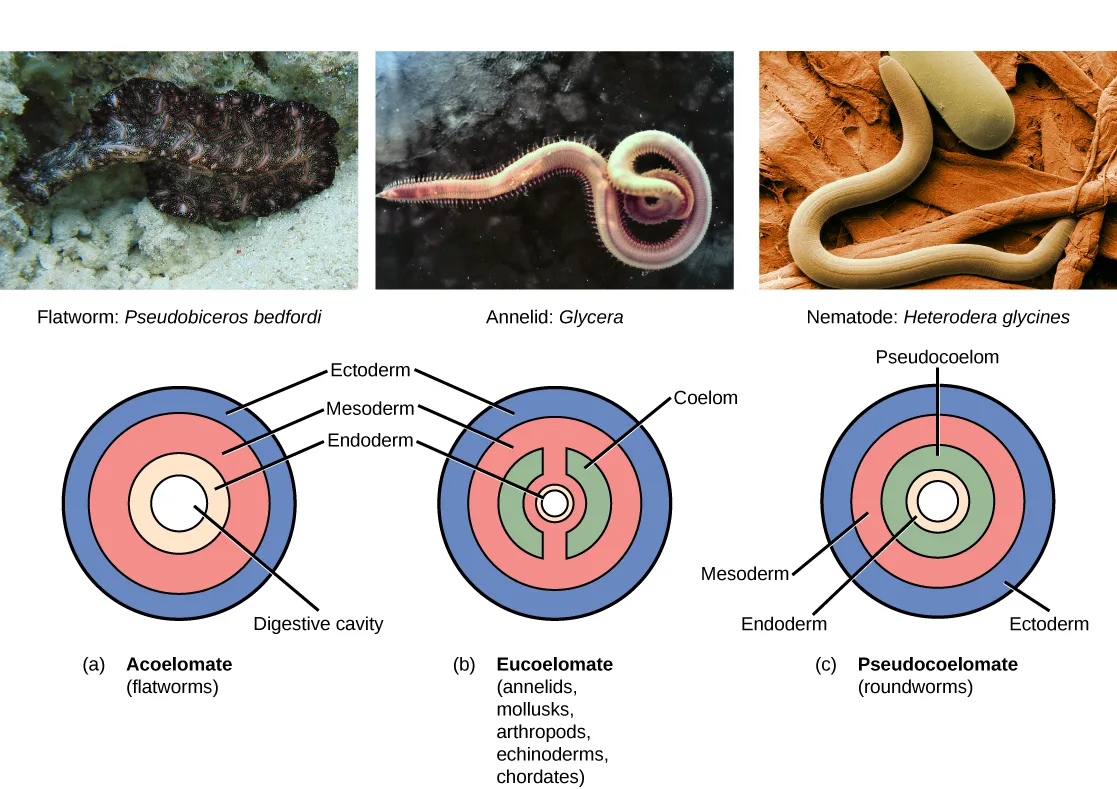
Triploblasts that do not develop a coelom are called acoelomates, and their mesoderm region is completely filled with tissue, although they do still have a gut cavity. Examples of acoelomates include animals in the phylum Platyhelminthes, also known as flatworms. Animals with a true coelom are called eucoelomates (or coelomates). In such cases, a true coelom arises entirely within the mesoderm germ layer and is lined by an epithelial membrane. This membrane also lines the organs within the coelom, connecting and holding them in position while allowing them some freedom of movement. Annelids, mollusks, arthropods, echinoderms, and chordates are all eucoelomates. A third group of triploblasts has a slightly different coelom lined partly by mesoderm and partly by endoderm. Although still functionally a coelom, these are considered “false” coeloms, and so we call these animals pseudocoelomates. The phylum Nematoda (roundworms) is an example of a pseudocoelomate. True coelomates can be further characterized based on other features of their early embryological development.
Extended description
Above the diagrams, three photographs show the phylum representatives: a flatworm (Pseudobiceros bedfordi), an annelid (Glycera), and a nematode (Heterodera glycines). Below them, three ring diagrams. Diagram (a), ‘Acoelomate (flatworms),’ has, from outside to center, an ectoderm ring, a mesoderm ring, an endoderm ring, and a central cavity labeled ‘Digestive cavity.’ Diagram (b), ‘Eucoelomate (annelids, mollusks, arthropods, echinoderms, chordates),’ has the same outer ectoderm ring and a continuous mesoderm ring inside it; two mesoderm spokes cross inward from that ring to a thin inner mesoderm ring, splitting the ring labeled ‘Coelom’ that lies between them into two crescents, and the inner mesoderm ring surrounds an endoderm ring and a small central cavity. Diagram (c), ‘Pseudocoelomate (roundworms),’ has, from outside to center, an ectoderm ring, a mesoderm ring, a ring labeled ‘Pseudocoelom,’ an endoderm ring, and a small central cavity.
Embryonic Development of the Mouth
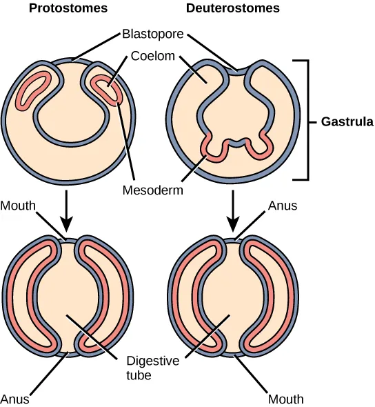
Bilaterally symmetrical, triploblastic eucoelomates can be further divided into two groups based on differences in the origin of the mouth. When the primitive gut forms, the opening that first connects the gut cavity to the outside of the embryo is called the blastopore. Most animals have openings at both ends of the gut: mouth at one end and anus at the other. One of these openings will develop at or near the site of the blastopore. In Protostomes (“mouth first”), the mouth develops at the blastopore. In Deuterostomes (“mouth second”), the mouth develops at the other end of the gut and the anus develops at the site of the blastopore. Protostomes include arthropods, mollusks, and annelids. Deuterostomes include more complex animals such as chordates but also some “simple” animals such as echinoderms. Recent evidence has challenged this simple view of the relationship between the location of the blastopore and the formation of the mouth, however, and the theory remains under debate. Nevertheless, these details of mouth and anus formation reflect general differences in the organization of protostome and deuterostome embryos, which are also expressed in other developmental features.
One of these differences between protostomes and deuterostomes is the method of coelom formation, beginning from the gastrula stage. Since body cavity formation tends to accompany the formation of the mesoderm, the mesoderm of protostomes and deuterostomes forms differently. The coelom of most protostomes is formed through a process called schizocoely. The mesoderm in these organisms is usually the product of specific blastomeres, which migrate into the interior of the embryo and form two clumps of mesodermal tissue. Within each clump, cavities develop and merge to form the hollow opening of the coelom. Deuterostomes differ in that their coelom forms through a process called enterocoely. Here, the mesoderm develops as pouches that are pinched off from the endoderm tissue. These pouches eventually fuse and expand to fill the space between the gut and the body wall, giving rise to the coelom.
Another difference in organization of protostome and deuterostome embryos is expressed during cleavage. Protostomes undergo spiral cleavage, meaning that the cells of one pole of the embryo are rotated, and thus misaligned, with respect to the cells of the opposite pole. This is due to the oblique angle of cleavage relative to the two poles of the embryo. Deuterostomes undergo radial cleavage, where the cleavage axes are either parallel or perpendicular to the polar axis, resulting in the parallel (up-and-down) alignment of the cells between the two poles.
Extended description
Top row, bracketed ‘Gastrula’: the left cutaway sphere, labeled ‘Protostomes,’ shows a ‘Blastopore’ opening at the top leading into a hollow interior, with two crescent-shaped ‘Mesoderm’ masses just inside the opening, one enclosing a small ‘Coelom’ cavity. The right cutaway sphere, labeled ‘Deuterostomes,’ shows the same blastopore opening, but its mesoderm instead forms as two pouches budding inward from the gut lining. An arrow leads down from each gastrula to the bottom row. Bottom left, the mature protostome body shows a continuous ringed ‘Digestive tube’ with ‘Mouth’ labeled at the top opening and ‘Anus’ at the bottom opening. Bottom right, the mature deuterostome body shows the same ringed digestive tube, but ‘Anus’ is labeled at the top opening and ‘Mouth’ at the bottom opening.
A second distinction between the types of cleavage in protostomes and deuterostomes relates to the fate of the resultant blastomeres (cells produced by cleavage). In addition to spiral cleavage, protostomes also undergo determinate cleavage. This means that even at this early stage, the developmental fate of each embryonic cell is already determined. A given cell does not have the ability to develop into any cell type other than its original destination. Removal of a blastomere from an embryo with determinate cleavage can result in missing structures, and embryos that fail to develop. In contrast, deuterostomes undergo indeterminate cleavage, in which cells are not yet fully committed at this early stage to develop into specific cell types. Removal of individual blastomeres from these embryos does not result in the loss of embryonic structures. In fact, twins (clones) can be produced as a result from blastomeres that have been separated from the original mass of blastomere cells. Unlike protostomes, however, if some blastomeres are damaged during embryogenesis, adjacent cells are able to compensate for the missing cells, and the embryo is not damaged. These cells are referred to as undetermined cells. This characteristic of deuterostomes is reflected in the existence of familiar embryonic stem cells, which have the ability to develop into any cell type until their fate is programmed at a later developmental stage.
Evolution Connection. The Evolution of the Coelom. One of the first steps in the classification of animals is to examine the animal’s body. One structure that is used in classification of animals is the body cavity or coelom. The body cavity develops within the mesoderm, so only triploblastic animals can have body cavities. Therefore body cavities are found only within the Bilateria. In other animal clades, the gut is either close to the body wall or separated from it by a jelly-like material. The body cavity is important for two reasons. Fluid within the body cavity protects the organs from shock and compression. In addition, since in triploblastic embryos, most muscle, connective tissue, and blood vessels develop from mesoderm, these tissues developing within the lining of the body cavity can reinforce the gut and body wall, aid in motility, and efficiently circulate nutrients.
To recap what we have discussed above, animals that do not have a coelom are called acoelomates. The major acoelomate group in the Bilateria is the flatworms, including both free-living and parasitic forms such as tapeworms. In these animals, mesenchyme fills the space between the gut and the body wall. Although two layers of muscle are found just under the epidermis, there is no muscle or other mesodermal tissue around the gut. Flatworms rely on passive diffusion for nutrient transport across their body.
In pseudocoelomates, there is a body cavity between the gut and the body wall, but only the body wall has mesodermal tissue. In these animals, the mesoderm forms, but does not develop cavities within it. Major pseudocoelomate phyla are the rotifers and nematodes. Animals that have a true coelom are called eucoelomates; all vertebrates, as well as mollusks, annelids, arthropods, and echinoderms, are eucoelomates. The coelom develops within the mesoderm during embryogenesis. Of the major bilaterian phyla, the mollusks, annelids, and arthropods are schizocoels, in which the mesoderm splits to form the body cavity, while the echinoderms and chordates are enterocoels, in which the mesoderm forms as two or more buds off of the gut. These buds separate from the gut and coalesce to form the body cavity. In the vertebrates, mammals have a subdivided body cavity, with the thoracic cavity separated from the abdominal cavity. The pseudocoelomates may have had eucoelomate ancestors and may have lost their ability to form a complete coelom through genetic mutations. Thus, this step in early embryogenesis—the formation of the coelom—has had a large evolutionary impact on the various species of the animal kingdom.
Summary
Organisms in the animal kingdom are classified based on their body morphology, their developmental pathways, and their genetic affinities. The relationships between the Eumetazoa and more basal clades (Ctenophora, Porifera, and Placozoa) are still being debated. The Eumetazoa (“true animals”) are divided into those with radial versus bilateral symmetry. Generally, the simpler and often nonmotile animals display radial symmetry, which allows them to explore their environment in all directions. Animals with radial symmetry are also generally characterized by the development of two embryological germ layers, the endoderm and ectoderm, whereas animals with bilateral symmetry are generally characterized by the development of a third embryologic germ layer, the mesoderm. Animals with three germ layers, called triploblasts, are further characterized by the presence or absence of an internal body cavity called a coelom. The presence of a coelom affords many advantages, and animals with a coelom may be termed true coelomates or pseudocoelomates, depending the extent to which mesoderm lines the body cavity. Coelomates are further divided into one of two groups called protostomes and deuterostomes, based on a number of developmental characteristics, including differences in zygote cleavage, the method of coelom formation, and the rigidity of the developmental fate of blastomeres.
Key terms
- acoelomate — animal without a body cavity
- bilateral symmetry — type of symmetry in which there is only one plane of symmetry, so the left and right halves of an animal are mirror images
- blastopore — opening into the archenteron that forms during gastrulation
- coelom — lined body cavity
- determinate cleavage — cleavage pattern in which developmental fate of each blastomere is tightly defined
- deuterostome — blastopore develops into the anus, with the second opening developing into the mouth
- diploblast — animal that develops from two germ layers
- enterocoely — mesoderm of deuterostomes develops as pouches that are pinched off from endodermal tissue, cavity contained within the pouches becomes coelom
- eucoelomate — animal with a body cavity completely lined with mesodermal tissue
- indeterminate cleavage — cleavage pattern in which individual blastomeres have the character of “stem cells,” and are not yet predetermined to develop into specific cell types
- protostome — blastopore develops into the mouth of protostomes, with the second opening developing into the anus
- pseudocoelomate — animal with a body cavity located between the mesoderm and endoderm
- radial cleavage — cleavage axes are parallel or perpendicular to the polar axis, resulting in the alignment of cells between the two poles
- radial symmetry — type of symmetry with multiple planes of symmetry, with body parts (rays) arranged around a central disk
- schizocoely — during development of protostomes, a solid mass of mesoderm splits apart and forms the hollow opening of the coelom
- spiral cleavage — cells of one pole of the embryo are rotated or misaligned with respect to the cells of the opposite pole
- triploblast — animal that develops from three germ layers
Practice
Explain the differences in animal body plans that support basic animal classification
Which of the following organisms is most likely to be a diploblast?
A diploblast develops only two germ layers, a pattern that goes with radial, biradial, or rotational symmetry rather than bilateral symmetry.Which of the following is not possible?
A coelom is a body cavity that develops within the mesoderm — and a diploblast, by definition, never forms that third germ layer.Explain some of the advantages brought about through the evolution of bilateral symmetry and coelom formation.
Show model answer
Did your answer mention:
An animal whose body cavity is completely lined with mesodermal tissue — an annelid, mollusk, arthropod, echinoderm, or chordate — is called a(n) ________.
This term names the group with a true coelom, as opposed to the group whose cavity is only partly lined by mesoderm.The lined body cavity that houses the digestive, urinary, and reproductive systems along with the heart, lungs, and major blood vessels is called a(n) ________.
Its subdivisions in mammals include the thoracic and abdominal cavities.Compare and contrast the embryonic development of protostomes and deuterostomes
An animal whose development is marked by radial cleavage and enterocoely is ________.
Match each developmental feature to the group defined by ‘mouth second.’Using the following terms, explain what classifications and groups humans fall into, from the most general to the most specific: symmetry, germ layers, coelom, cleavage, embryological development.
Show model answer
Did your answer mention:
The opening that first connects the primitive gut cavity to the outside of the embryo is called the ________.
In protostomes this site becomes the mouth; in deuterostomes it becomes the anus.An animal in which the mouth develops at the same site where the primitive gut first opened to the outside of the embryo is a(n) ________.
Its name literally means ‘mouth first.’The process by which most protostomes form their coelom, in which a solid mass of mesoderm splits apart to create the cavity, is called ________.
Contrast it with the deuterostome process that pinches mesoderm off from the gut lining instead of splitting a solid mass.The process by which deuterostomes form their coelom, in which mesoderm pinches off from the endoderm as pouches that fuse and expand, is called ________.
Contrast it with the protostome process that splits a solid mass of mesoderm instead of budding pouches from the gut.This section is adapted from Biology 2e, Section 27.2: Features Used to Classify Animals by Mary Ann Clark, Jung Choi, Matthew Douglas, and OpenStax, © OpenStax, licensed under CC BY-NC-SA 4.0. Access the original for free at openstax.org. Changes: figures re-encoded as WebP with a custom alt written from each image rather than the manifest’s guessed text; five figures re-kinded after inspection from the manifest’s file-extension guess — Figure_B27_02_01 (phylogenetic tree), Figure_27_02_03 (labeled body-plane illustration), Figure_27_02_05 (body-cavity ring diagrams with photo insets), and Figure_27_02_06 (protostome/deuterostome development diagram) from “photo” to “diagram,” and Figure_B27_02_02abcd (five photographs of representative animals) from “diagram” to “photo”; a longdesc added to the phylogenetic tree, the labeled body-plane diagram, the diploblast/triploblast ring diagram, the body-cavity ring diagrams, and the protostome/deuterostome development diagram, none of whose labels, rings, or panel arrangement are carried by their one- or two-line captions; both Visual Connection notes rendered as their figure followed by a multiple choice, kept in the body; the Visual Connection question on symmetry keeps the body note’s own wording of option (a) (“Animals that display only radial symmetry during their lifespans are diploblasts”), which differs from the end-of-section Visual Connection Questions list’s wording of the same item (“Animals that display radial symmetry are diploblasts”) without changing the keyed answer; the Link to Learning and Evolution Connection feature boxes rendered as callouts; the end-of-section Review Questions and Critical Thinking Questions adapted into the closing interactive Practice block (multiple choice and self-check respectively); rubric checkpoints added to each self-check, decomposing its model answer (the source solution) into check-off clauses with no new claims; six key-term recall items (eucoelomate, coelom, blastopore, protostome, schizocoely, enterocoely) added from the glossary.