Phylum Cnidaria
By the end of this section, you will be able to:
- Compare structural and organization characteristics of Porifera and Cnidaria
- Describe the progressive development of tissues and their relevance to animal complexity
- Identify the two general body forms found in the Cnidaria
- Describe the identifying features of the major cnidarian classes
Phylum Cnidaria includes animals that exhibit radial or biradial symmetry and are diploblastic, meaning that they develop from two embryonic layers, ectoderm and endoderm. Nearly all (about 99 percent) cnidarians are marine species.
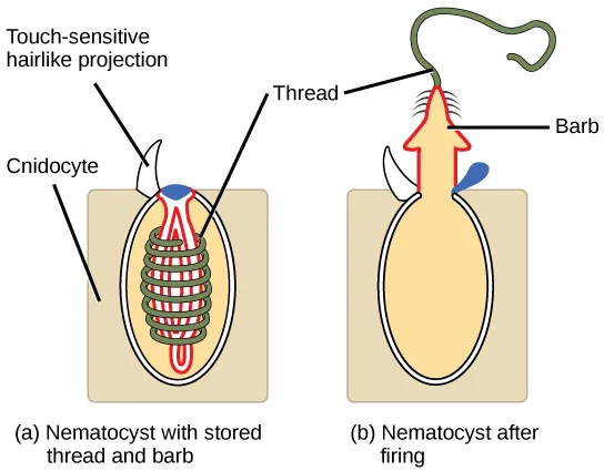
Whereas the defining cell type for the sponges is the choanocyte, the defining cell type for the cnidarians is the cnidocyte, or stinging cell. These cells are located around the mouth and on the tentacles, and serve to capture prey or repel predators. Cnidocytes have large stinging organelles called nematocysts, which usually contain barbs at the base of a long coiled thread. The outer wall of the cell has a hairlike projection called a cnidocil, which is sensitive to tactile stimulation. If the cnidocils are touched, the hollow threads evert with enormous acceleration, approaching 40,000 times that of gravity. The microscopic threads then either entangle the prey or instantly penetrate the flesh of the prey or predator, releasing toxins (including neurotoxins and pore-forming toxins that can lead to cell lysis) into the target, thereby immobilizing it or paralyzing it (see the diagram below).
Extended description
Four labels sit above both panels, each with a line to one of them. ‘Touch-sensitive hairlike projection,’ ‘Cnidocyte,’ and ‘Thread’ all point into panel (a), labeled ‘Nematocyst with stored thread and barb’: a rectangular cnidocyte cell contains a large oval nematocyst, with a hairlike projection reaching from its top to the cell’s outer surface and a coiled thread wound inside it around a small hidden barb. ‘Barb’ points instead into panel (b), labeled ‘Nematocyst after firing’: the same cell now has a lid open at its top, and the barb, trailing the now fully uncoiled thread, projects outward through the opening well beyond the cell’s boundary.
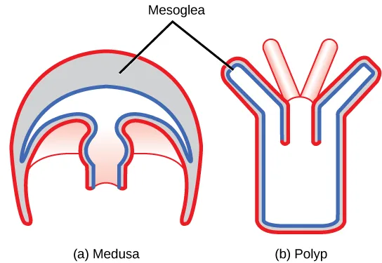
Two distinct body plans are found in Cnidarians: the polyp or tuliplike “stalk” form and the medusa or “bell” form (see the diagram below). An example of the polyp form is found in the genus Hydra, whereas the most typical form of medusa is found in the group called the “sea jellies” (jellyfish). Polyp forms are sessile as adults, with a single opening (the mouth/anus) to the digestive cavity facing up with tentacles surrounding it. Medusa forms are motile, with the mouth and tentacles hanging down from an umbrella-shaped bell.
Extended description
At left, (a) Medusa: an upside-down dome outlined in red on the outside and blue on the inside, with a thick gray band filling most of the dome’s interior above a narrower central body cavity that opens downward. At right, (b) Polyp: an upright, narrower tree-like shape with the same red outer and blue inner outlines, three branch-like tentacles at the top opening into a central body cavity, and only a thin gap between the red and blue outlines running down the stalk. A single label, ‘Mesoglea,’ has two lines: one to the gray band inside the medusa’s dome, the other to the thin gap between the polyp’s outlines, naming both as the same layer at two different thicknesses.
Some cnidarians are dimorphic, that is, they exhibit both body plans during their life cycle. In these species, the polyp serves as the asexual phase, while the medusa serves as the sexual stage and produces gametes. However, both body forms are diploid.
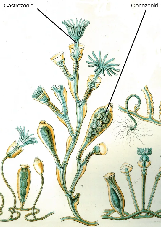
An example of cnidarian dimorphism can be seen in the colonial hydroid Obelia. The sessile asexual colony has two types of polyps, shown below. The first is the gastrozooid, which is adapted for capturing prey and feeding. In Obelia, all polyps are connected through a common digestive cavity called a coenosarc. The other type of polyp is the gonozooid, adapted for the asexual budding and the production of sexual medusae. The reproductive buds from the gonozooid break off and mature into free-swimming medusae, which are either male or female (dioecious). Each medusa has either several testes or several ovaries in which meiosis occurs to produce sperm or egg cells. Interestingly, the gamete-producing cells do not arise within the gonad itself, but migrate into it from the tissues in the gonozooid. This separate origin of gonad and gametes is common throughout the eumetazoa. The gametes are released into the surrounding water, and after fertilization, the zygote develops into a blastula, which soon develops into a ciliated, bilaterally symmetrical planula larva. The planula swims freely for a while, but eventually attaches to a substrate and becomes a single polyp, from which a new colony of polyps is formed by budding.
All cnidarians are diploblastic and thus have two “epithelial” layers in the body that are derived from the endoderm and ectoderm of the embryo. The outer layer (from ectoderm) is called the epidermis and lines the outside of the animal, whereas the inner layer (from endoderm) is called the gastrodermis and lines the digestive cavity. In the planula larva, a layer of ectoderm surrounds a solid mass of endoderm, but as the polyp develops, the digestive or gastrovascular cavity opens within the endoderm. A non-living, jelly-like mesoglea lies between these two epithelial layers. In terms of cellular complexity, cnidarians show the presence of differentiated cell types in each tissue layer, such as nerve cells, contractile epithelial cells, enzyme-secreting cells, and nutrient-absorbing cells, as well as the presence of intercellular connections. However, with a few notable exceptions such as statocysts and rhopalia (see below), the development of organs or organ systems is not advanced in this phylum.
The nervous system is rudimentary, with nerve cells organized in a network scattered across the body. This nerve net may show the presence of groups of cells that form nerve plexi (singular: plexus) or nerve cords. Organization of the nervous system in the motile medusa is more complex than that of the sessile polyp, with a nerve ring around the edge of the medusa bell that controls the action of the tentacles. Cnidarian nerve cells show mixed characteristics of motor and sensory neurons. The predominant signaling molecules in these primitive nervous systems are peptides, which perform both excitatory and inhibitory functions. Despite the simplicity of the nervous system, it is remarkable that it coordinates the complicated movement of the tentacles, the drawing of captured prey to the mouth, the digestion of food, and the expulsion of waste.
The gastrovascular cavity has only one opening that serves as both a mouth and an anus; this arrangement is called an incomplete digestive system. In the gastrovascular cavity, extracellular digestion occurs as food is taken into the gastrovascular cavity, enzymes are secreted into the cavity, and the cells lining the cavity absorb nutrients. However, some intracellular digestion also occurs. The gastrovascular cavity distributes nutrients throughout the body of the animal, with nutrients passing from the digestive cavity across the mesoglea to the epidermal cells. Thus, this cavity serves both digestive and circulatory functions.
Cnidarian cells exchange oxygen and carbon dioxide by diffusion between cells in the epidermis and water in the environment, and between cells in the gastrodermis and water in the gastrovascular cavity. The lack of a circulatory system to move dissolved gases limits the thickness of the body wall and necessitates a non-living mesoglea between the layers. In the cnidarians with a thicker mesoglea, a number of canals help to distribute both nutrients and gases. There is neither an excretory system nor organs, and nitrogenous wastes simply diffuse from the cells into the water outside the animal or into the gastrovascular cavity.
The phylum Cnidaria contains about 10,000 described species divided into two monophyletic clades: the Anthozoa and the Medusozoa. The Anthozoa include the corals, sea fans, sea whips, and the sea anemones. The Medusozoa include several classes of Cnidaria in two clades: The Hydrozoa include sessile forms, some medusoid forms, and swimming colonial forms like the Portuguese man-of-war. The other clade contains various types of jellies including both Scyphozoa and Cubozoa. The Anthozoa contain only sessile polyp forms, while the Medusozoa include species with both polyp and medusa forms in their life cycle.
Class Anthozoa
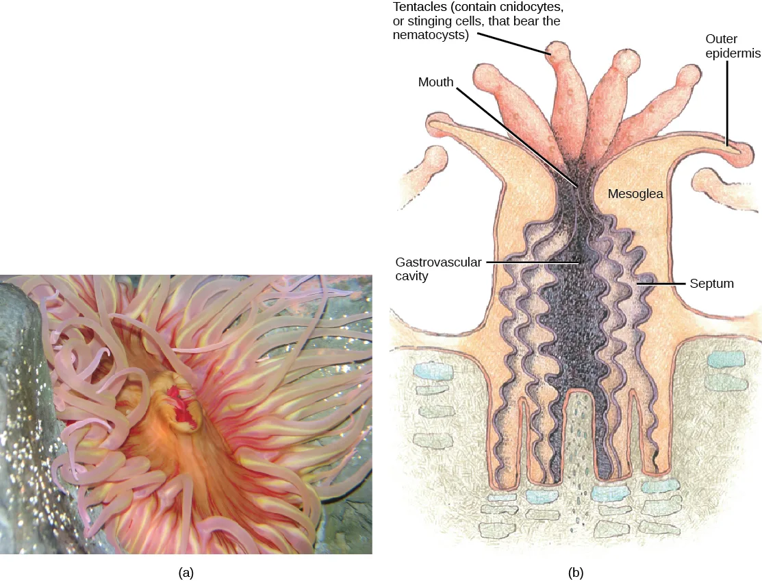
The class Anthozoa (“flower animals”) includes sea anemones (see the photo below), sea pens, and corals, with an estimated number of 6,100 described species. Sea anemones are usually brightly colored and can attain a size of 1.8 to 10 cm in diameter. Individual animals are cylindrical in shape and are attached directly to a substrate.
Extended description
In panel (b), several slender tentacles fan upward from the rim of the anemone’s tube-shaped body, labeled ‘Tentacles (contain cnidocytes, or stinging cells, that bear the nematocysts)’ at upper left. A ‘Mouth’ label points to the opening at the center of the tentacle ring. ‘Outer epidermis’ labels the body’s outer wall at upper right. Below the mouth, a wavy-walled ‘Gastrovascular cavity,’ labeled at lower left, runs down the body’s center, its folded walls forming a ‘Septum,’ labeled at right. ‘Mesoglea,’ labeled at upper right, names the pale layer between the gastrovascular cavity’s wall and the outer epidermis.
The mouth of a sea anemone is surrounded by tentacles that bear cnidocytes. The slit-like mouth opening and flattened pharynx are lined with ectoderm. This structure of the pharynx makes anemones bilaterally symmetrical. A ciliated groove called a siphonoglyph is found on two opposite sides of the pharynx and directs water into it. The pharynx is the muscular part of the digestive system that serves to ingest as well as egest food, and may extend for up to two-thirds the length of the body before opening into the gastrovascular cavity. This cavity is divided into several chambers by longitudinal septa called mesenteries. Each mesentery consists of a fold of gastrodermal tissue with a layer of mesoglea between the sheets of gastrodermis. Mesenteries do not divide the gastrovascular cavity completely, and the smaller cavities coalesce at the pharyngeal opening. The adaptive benefit of the mesenteries appears to be an increase in surface area for absorption of nutrients and gas exchange, as well as additional mechanical support for the body of the anemone.
Sea anemones feed on small fish and shrimp, usually by immobilizing their prey with nematocysts. Some sea anemones establish a mutualistic relationship with hermit crabs when the crab seizes and attaches them to their shell. In this relationship, the anemone gets food particles from prey caught by the crab, and the crab is protected from the predators by the stinging cells of the anemone. Some species of anemone fish, or clownfish, are also able to live with sea anemones because they build up an acquired immunity to the toxins contained within the nematocysts and also secrete a protective mucus that prevents them from being stung.
The structure of coral polyps is similar to that of anemones, although the individual polyps are usually smaller and part of a colony, some of which are massive and the size of small buildings. Coral polyps feed on smaller planktonic organisms, including algae, bacteria, and invertebrate larvae. Some anthozoans have symbiotic associations with dinoflagellate algae called zooxanthellae. The mutually beneficial relationship between zooxanthellae and modern corals—which provides the algae with shelter—gives coral reefs their colors and supplies both organisms with nutrients. This complex mutualistic association began more than 210 million years ago, according to a new study by an international team of scientists. That this symbiotic relationship arose during a time of massive worldwide coral-reef expansion suggests that the interconnection of algae and coral is crucial for the health of coral reefs, which provide habitat for roughly one-fourth of all marine life. Reefs are threatened by a trend in ocean warming that has caused corals to expel their zooxanthellae algae and turn white, a process called coral bleaching.
Anthozoans remain polypoid (note that this term is easily confused with “polyploid”) throughout their lives and can reproduce asexually by budding or fragmentation, or sexually by producing gametes. Male or female gametes produced by a polyp fuse to give rise to a free-swimming planula larva. The larva settles on a suitable substratum and develops into a sessile polyp.
Class Scyphozoa
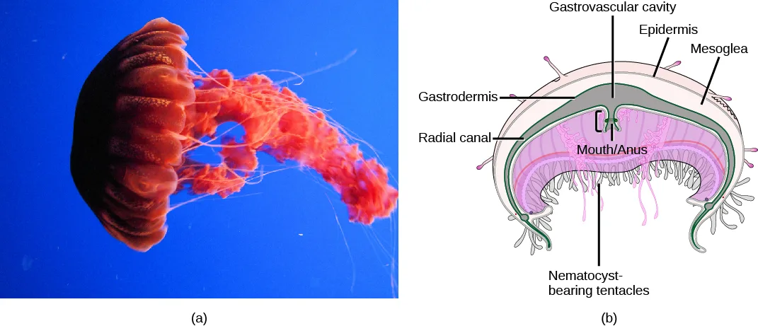
Class Scyphozoa (“cup animals”) includes only marine jellies, with about 200 known species. The medusa is the prominent stage in the life cycle, although there is a polyp stage in the life cycle of most species. Most jellies range from 2 to 40 cm in length but the largest scyphozoan species, Cyanea capillata, can reach a size of two meters in diameter. Scyphozoans display a characteristic bell-like morphology (see the photo below).
Extended description
In panel (b), three labels sit across the top of the dome: ‘Gastrovascular cavity’ at top center, pointing into the dark space inside the dome; ‘Epidermis’ at upper right, pointing to the outer surface; and ‘Mesoglea’ at the far right, pointing to the thick pale layer beneath it. On the left, ‘Gastrodermis’ points to the inner lining and, lower down, ‘Radial canal’ points to a channel running along the dome’s inner edge. A central opening beneath the dome is labeled ‘Mouth/Anus.’ At the bottom, the label ‘Nematocyst-bearing tentacles’ points up to the pink frilly structures hanging beneath the mouth; short tentacles also fringe the bell’s rim.
In the sea jelly, a mouth opening is present on the underside of the animal, surrounded by hollow tentacles bearing nematocysts. Scyphozoans live most of their life cycle as free-swimming, solitary carnivores. The mouth leads to the gastrovascular cavity, which may be sectioned into four interconnected sacs, called diverticuli. In some species, the digestive system may branch further into radial canals. Like the septa in anthozoans, the branched gastrovascular cells serve two functions: to increase the surface area for nutrient absorption and diffusion, and to support the body of the animal.
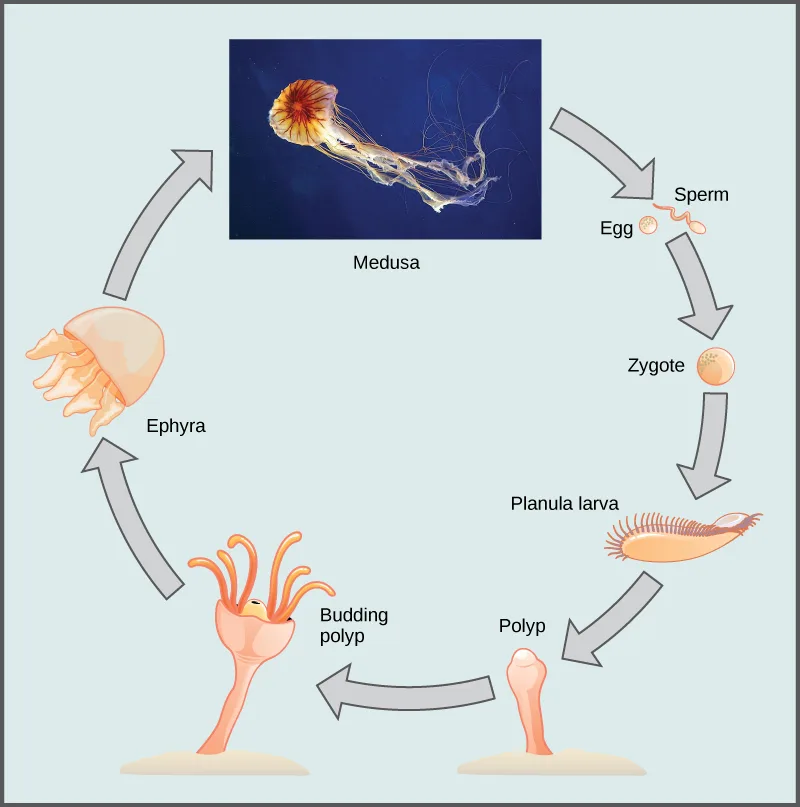
In scyphozoans, nerve cells are organized in a nerve net that extends over the entire body, with a nerve ring around the edge of the bell. Clusters of sensory organs called rhopalia may be present in pockets in the edge of the bell. Jellies have a ring of muscles lining the dome of the body, which provides the contractile force required to swim through water, as well as to draw in food from the water as they swim. Scyphozoans have separate sexes. The gonads are formed from the gastrodermis and gametes are expelled through the mouth. Planula larvae are formed by external fertilization; they settle on a substratum in a polypoid form. These polyps may bud to form additional polyps or begin immediately to produce medusa buds. In a few species, the planula larva may develop directly into the medusa. The life cycle of most scyphozoans, shown below, includes both sexual medusoid and asexual polypoid body forms.
Extended description
Seven stages run clockwise around a circular diagram. At top center, a photographed ‘Medusa’ — a red-and-white jellyfish trailing long tentacles — sits in a navy box. An arrow leads right and down to ‘Sperm’ and ‘Egg’ cells at upper right. A second arrow leads down to a round orange ‘Zygote.’ A third arrow leads down to a spiky, oval ‘Planula larva.’ A fourth arrow leads down and left to a small stalked ‘Polyp’ at bottom center. A fifth arrow leads left to a ‘Budding polyp’ at bottom left, shown sprouting a smaller second polyp from its side. A sixth arrow leads up to a bell-shaped ‘Ephyra’ at left. A seventh arrow leads up and right, closing the loop back at the Medusa.
Class Cubozoa
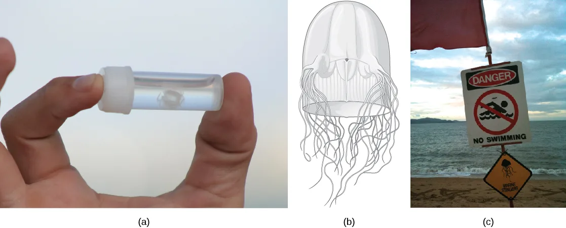
This class includes jellies that have a box-shaped medusa, or a bell that is square in cross-section, and are colloquially known as “box jellyfish.” These species may achieve sizes of 15 to 25 cm, but typically members of the Cubozoa are not as large as those of the Scyphozoa. However, cubozoans display overall morphological and anatomical characteristics that are similar to those of the scyphozoans. A prominent difference between the two classes is the arrangement of tentacles. The cubozoans contain muscular pads called pedalia at the corners of the square bell canopy, with one or more tentacles attached to each pedalium. In some cases, the digestive system may extend into the pedalia. Nematocysts may be arranged in a spiral configuration along the tentacles; this arrangement helps to effectively subdue and capture prey. Cubozoans include the most venomous of all the cnidarians (see the photo below).
These animals are unusual in having image-forming eyes, including a cornea, lens, and retina. Because these structures are made from a number of interactive tissues, they can be called true organs. Eyes are located in four clusters between each pair of pedalia. Each cluster consists of four simple eye spots plus two image-forming eyes oriented in different directions. How images formed by these very complex eyes are processed remains a mystery, since cubozoans have extensive nerve nets but no distinct brain. Nonetheless, the presence of eyes helps the cubozoans to be active and effective hunters of small marine animals like worms, arthropods, and fish.
Cubozoans have separate sexes and fertilization occurs inside the female. Planula larvae may develop inside the female or be released, depending on species. Each planula develops into a polyp. These polyps may bud to form more polyps to create a colony; each polyp then transforms into a single medusa.
Class Hydrozoa
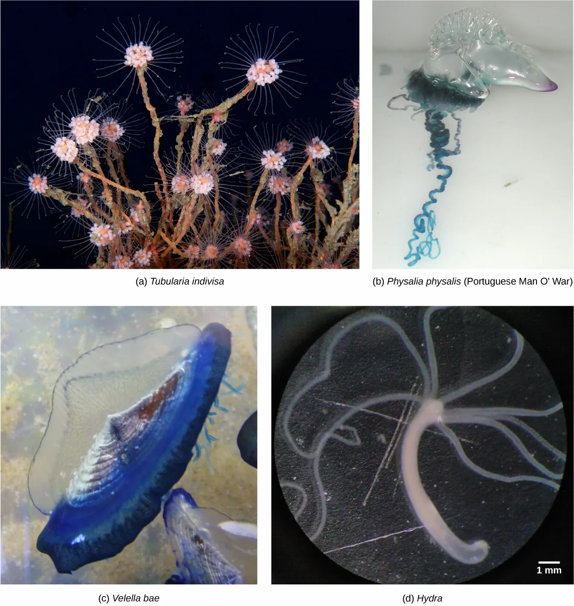
Hydrozoa is a diverse group that includes nearly 3,200 species; most are marine, although some freshwater species are known (see the photo below). Most species exhibit both polypoid and medusoid forms in their lifecycles, although the familiar Hydra has only the polyp form. The medusoid form has a muscular veil or velum below the margin of the bell and for this reason is called a hydromedusa. In contrast, the medusoid form of Scyphozoa lacks a velum and is termed a scyphomedusa.
The polyp form in these animals often shows a cylindrical morphology with a central gastrovascular cavity lined by the gastrodermis. The gastrodermis and epidermis have a simple layer of mesoglea sandwiched between them. A mouth opening, surrounded by tentacles, is present at the oral end of the animal. Many hydrozoans form sessile, branched colonies of specialized polyps that share a common, branching gastrovascular cavity (coenosarc), such as is found in the colonial hydroid Obelia.
Free-floating colonial species called siphonophores contain both medusoid and polypoid individuals that are specialized for feeding, defense, or reproduction. The distinctive rainbow-hued float of the Portuguese man o’ war (Physalia physalis) creates a pneumatophore with which it regulates buoyancy by filling and expelling carbon monoxide gas. At first glance, these complex superorganisms appear to be a single organism; but the reality is that even the tentacles are actually composed of zooids laden with nematocysts. Thus, although it superficially resembles a typical medusozoan jellyfish, P. physalis is a free-floating hydrozoan colony; each specimen is made up of many hundreds of organisms, each specialized for a certain function, including motility and buoyancy, feeding, reproduction and defense. Although they are carnivorous and feed on many soft bodied marine animals, P. physalis lack stomachs and instead have specialized polyps called gastrozooids that they use to digest their prey in the open water.
Physalia has male and female colonies, which release their gametes into the water. The zygote develops into a single individual, which then buds asexually to form a new colony. Siphonophores include the largest known floating cnidarian colonies such as Praya dubia, whose chain of zooids can get up to 50 meters (165 feet) long. Other hydrozoan species are solitary polyps (Hydra) or solitary hydromedusae (Gonionemus). One defining characteristic shared by the hydrozoans is that their gonads are derived from epidermal tissue, whereas in all other cnidarians they are derived from gastrodermal tissue.
Summary
Cnidarians represent a more complex level of organization than Porifera. They possess outer and inner tissue layers that sandwich a noncellular mesoglea between them. Cnidarians possess a well-formed digestive system and carry out extracellular digestion in a digestive cavity that extends through much of the animal. The mouth is surrounded by tentacles that contain large numbers of cnidocytes—specialized cells bearing nematocysts used for stinging and capturing prey as well as discouraging predators. Cnidarians have separate sexes and many have a lifecycle that involves two distinct morphological forms—medusoid and polypoid—at various stages in their life cycles. In species with both forms, the medusa is the sexual, gamete-producing stage and the polyp is the asexual stage. Cnidarian species include individual or colonial polypoid forms, floating colonies, or large individual medusa forms (sea jellies).
Key terms
- Cnidaria — phylum of animals that are diploblastic and have radial symmetry.
- cnidocyte — specialized stinging cell found in Cnidaria.
- epidermis — outer layer (from ectoderm) that lines the outside of the animal.
- extracellular digestion — food is taken into the gastrovascular cavity, enzymes are secreted into the cavity, and the cells lining the cavity absorb nutrients.
- gastrodermis — inner layer (from endoderm) that lines the digestive cavity.
- gastrovascular cavity — opening that serves as both a mouth and an anus, which is termed an incomplete digestive system.
- medusa — free-floating cnidarian body plan with mouth on underside and tentacles hanging down from a bell.
- mesoglea — non-living, gel-like matrix present between ectoderm and endoderm in cnidarians.
- nematocyst — harpoon-like organelle within cnidocyte with pointed projectile and poison to stun and entangle prey.
- polyp — stalk-like sessile life form of a cnidarians with mouth and tentacles facing upward, usually sessile but may be able to glide along surface.
- polymorphic — possessing multiple body plans within the lifecycle of a group of organisms.
- siphon — tubular structure that serves as an inlet for water into the mantle cavity.
Practice
Compare structural and organization characteristics of Porifera and Cnidaria
Cnidocytes are found in _____.
This stinging cell type is what defines the phylum, replacing the sponge’s choanocyte.Compare the structural differences between Porifera and Cnidaria.
Show model answer
Did your answer mention:
Explain the function of nematocysts in cnidarians.
Show model answer
Did your answer mention:
The specialized stinging cell found in Cnidaria is called a ________.
This cell type replaces the sponge’s choanocyte as the phylum’s defining feature, and houses the harpoon-like nematocyst.Describe the progressive development of tissues and their relevance to animal complexity
The outer layer of a cnidarian, derived from ectoderm, that lines the outside of the animal is called the ________.
Its inner counterpart, derived from endoderm, lines the digestive cavity instead.The inner layer of a cnidarian, derived from endoderm, that lines the digestive cavity is called the ________.
Its outer counterpart, derived from ectoderm, lines the outside of the animal instead.The non-living, gel-like matrix present between the ectoderm and endoderm in cnidarians is called the ________.
It is thicker in the dome of the bell-shaped body form than in the stalk-shaped one.Digestion in which food is taken into a cavity, enzymes are secreted into it, and the lining cells absorb the released nutrients is called ________.
This is how the single-opening gastrovascular cavity processes most food, though some digestion also happens inside individual cells.Identify the two general body forms found in the Cnidaria
The sessile, stalk-like cnidarian body form, with mouth and tentacles facing upward, is called the ________.
This is the asexual phase in species exhibiting both body forms, and the only form Hydra ever takes.The free-floating, bell-shaped cnidarian body form, with mouth and tentacles hanging downward, is called the ________.
This motile form is the sexual, gamete-producing stage in species that also have a stalk-shaped stage.An organism or group of organisms possessing multiple body plans within its life cycle is described as ________.
Obelia’s alternation between an asexual budding stage and a free-swimming, gamete-producing stage is one example.Which of the following is one of the two general cnidarian body forms, sessile as an adult, with a single mouth/anus opening facing upward and tentacles surrounding it?
It stays fixed in place, unlike the free-swimming bell shape; the other two options are life-cycle stages rather than one of the two general body forms.Describe the identifying features of the major cnidarian classes
Cubozoans are ________.
This class’s polyps eventually transform into a single medusa, so members pass through more than one body plan over a lifetime.While collecting specimens, a marine biologist finds a sessile Cnidarian. The medusas that bud from it swim by contracting a ring of muscle in their bells. To which class does this specimen belong?
This class’s jellies have a ring of muscles lining the dome of the body that provides the contractile force needed for swimming.Compare the differences in sexual reproduction between Porifera and Cubozoans. How does the difference in fertilization provide an evolutionary advantage to the Cubozoans?
Show model answer
Did your answer mention:
Which cnidarian clade includes only sessile polyp forms, never a medusa stage?
This clade’s members — anemones, sea pens, and corals — remain polypoid throughout their lives.This section is adapted from Biology 2e, Section 28.2: Phylum Cnidaria by Mary Ann Clark, Jung Choi, Matthew Douglas, and OpenStax, © OpenStax, licensed under CC BY-NC-SA 4.0. Access the original for free at openstax.org. Changes: figures re-encoded as WebP; four figures re-kinded after inspection — Figure_28_02_01, Figure_28_02_02, Figure_28_02_03, and Figure_28_02_06 — from the manifest’s file-extension “photo” guess to “diagram” (line drawings, not captured photographs), while Figure_28_02_04, Figure_28_02_05ab, Figure_28_02_07, and Figure_28_02_08abcd keep the manifest’s “photo” guess (each is a single vendored image whose photographed panel(s) dominate, even where a labeled schematic panel sits alongside); a longdesc added to the nematocyst-firing diagram (Figure_28_02_01), the body-plan diagram (Figure_28_02_02), the labeled sea-anemone and sea-jelly cross-sections (panel (b) of Figure_28_02_04 and Figure_28_02_05ab), and the scyphozoan life-cycle diagram (Figure_28_02_06, walking its seven clockwise stages) — none of whose captions carry their label or arrow content; two Link to Learning notes rendered as callouts, replacing their “click here”/bare “video” link text with a description of each destination; the end-of-section Review Questions and Critical Thinking Questions adapted into the closing interactive Practice block (multiple choice and self-check respectively); rubric checkpoints added to each self-check, decomposing its model answer (the source solution) into check-off clauses with no new claims; eight key-term recall items (cnidocyte, epidermis, gastrodermis, mesoglea, extracellular digestion, polyp, medusa, and polymorphic, each once) added from the glossary — the glossary’s “siphon” entry (a mantle-cavity water inlet) is kept in Key terms for completeness but has no counterpart anywhere in this module’s text and was not used for a recall item, since the section teaches no such structure — reported as a source defect; two locally written multiple-choice items added (distinguishing the polyp from the medusa and other life-cycle stages, and identifying Anthozoa as the only polyp-only clade), both built strictly from the section’s own sentences, to round out the “body forms” and “cnidarian classes” objective groups.