Reproductive Development and Structure
By the end of this section, you will be able to:
- Describe the two stages of a plant’s lifecycle
- Compare and contrast male and female gametophytes and explain how they form in angiosperms
- Describe the reproductive structures of a plant
- Describe the components of a complete flower
- Describe the development of microsporangium and megasporangium in gymnosperms
Sexual reproduction takes place with slight variations in different groups of plants. Plants have two distinct stages in their lifecycle: the gametophyte stage and the sporophyte stage. The haploid gametophyte produces the male and female gametes by mitosis in distinct multicellular structures. Fusion of the male and female gametes forms the diploid zygote, which develops into the sporophyte. After reaching maturity, the diploid sporophyte produces spores by meiosis, which in turn divide by mitosis to produce the haploid gametophyte. The new gametophyte produces gametes, and the cycle continues. This is the alternation of generations, and is typical of plant reproduction, shown below.
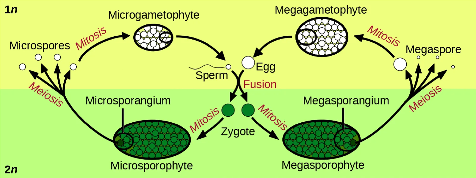
Extended description
Reading left to right, top (1n, haploid) to bottom (2n, diploid): the diploid Microsporophyte (dark green oval, lower left) sends a Meiosis-labeled arrow up into the 1n band to a cluster of Microspores (small circles); a Mitosis arrow leads right to an oval Microgametophyte packed with small circles. From the microgametophyte, a squiggly line marked Sperm curves down to a point marked Fusion. Mirroring this on the right, the diploid Megasporophyte (dark green oval, lower right) sends a Meiosis arrow up to a cluster of Megaspore cells; a Mitosis arrow leads left to a larger oval Megagametophyte, from which a circle marked Egg curves down to the same Fusion point. Sperm and egg fuse to form two green dots labeled Zygote, sitting on the boundary between the 1n and 2n bands. Two Mitosis arrows lead from the zygote back down into the Microsporophyte (left) and the Megasporophyte (right), closing the cycle.
The life cycle of higher plants is dominated by the sporophyte stage, with the gametophyte borne on the sporophyte. In ferns, the gametophyte is free-living and very distinct in structure from the diploid sporophyte. In bryophytes, such as mosses, the haploid gametophyte is more developed than the sporophyte.
During the vegetative phase of growth, plants increase in size and produce a shoot system and a root system. As they enter the reproductive phase, some of the branches start to bear flowers. Many flowers are borne singly, whereas some are borne in clusters. The flower is borne on a stalk known as a receptacle. Flower shape, color, and size are unique to each species, and are often used by taxonomists to classify plants.
Sexual Reproduction in Angiosperms
The lifecycle of angiosperms follows the alternation of generations explained previously. The haploid gametophyte alternates with the diploid sporophyte during the sexual reproduction process of angiosperms. Flowers contain the plant’s reproductive structures.
Flower Structure
A typical flower has four main parts—or whorls—known as the calyx, corolla, androecium, and gynoecium, shown below. The outermost whorl of the flower has green, leafy structures known as sepals. The sepals, collectively called the calyx, help to protect the unopened bud. The second whorl is comprised of petals—usually, brightly colored—collectively called the corolla. The number of sepals and petals varies depending on whether the plant is a monocot or dicot. In monocots, petals usually number three or multiples of three; in dicots, the number of petals is four or five, or multiples of four and five. Together, the calyx and corolla are known as the perianth. The third whorl contains the male reproductive structures and is known as the androecium. The androecium has stamens with anthers that contain the microsporangia. The innermost group of structures in the flower is the gynoecium, or the female reproductive component(s). The carpel is the individual unit of the gynoecium and has a stigma, style, and ovary. A flower may have one or multiple carpels.
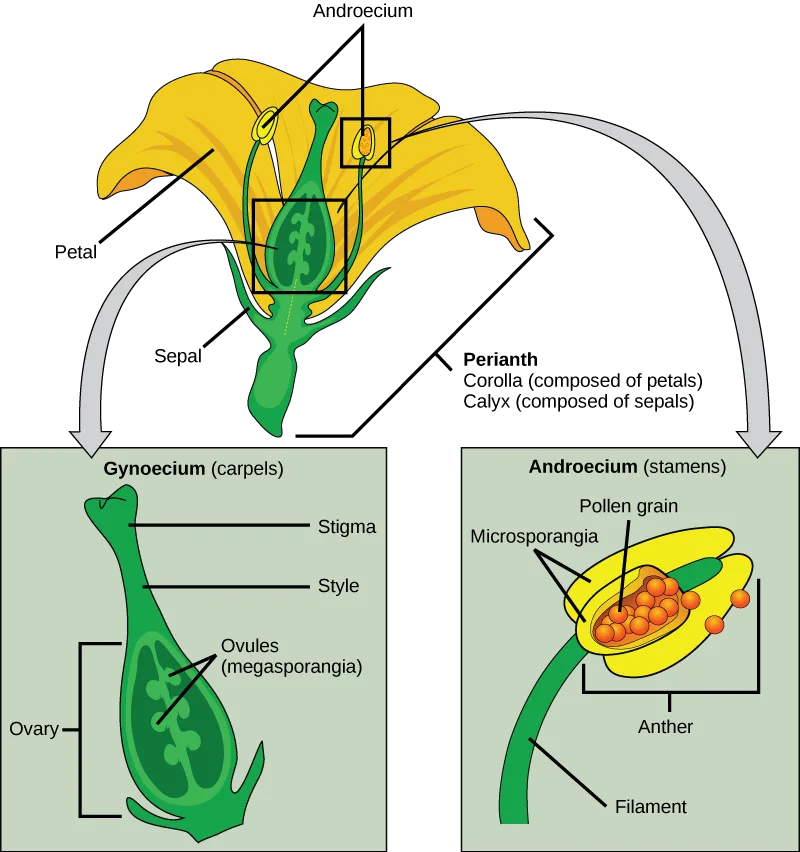
Extended description
At the top, a whole yellow flower is drawn with a central green carpel and one stamen, each outlined by a black box. Lines from the flower point to Petal (the yellow lobe) and Sepal (the green base); a bracket labeled Perianth spans Corolla (composed of petals) and Calyx (composed of sepals); two lines from the label Androecium point to two of the stamens, one of them boxed. A gray arrow leads down-left from the boxed carpel to the bottom-left panel, Gynoecium (carpels): a vase-shaped green drawing labeled, top to bottom, Stigma (the tip), Style (the narrow neck), and Ovules (megasporangia) (oval shapes inside), with a bracket labeling the whole lower bulb Ovary. A second gray arrow leads down-right from the boxed stamen to the bottom-right panel, Androecium (stamens): a green filament topped by a yellow anther, labeled Filament (the stalk), Anther (a bracket around the yellow lobe), Microsporangia (two lines to the anther’s internal chambers), and Pollen grain (a line to the orange dots spilling from the anther).
If the anther is missing, what type of reproductive structure will the flower be unable to produce? What term is used to describe an incomplete flower lacking the androecium? What term describes an incomplete flower lacking a gynoecium?
Show model answer
Did your answer mention:
If all four whorls (the calyx, corolla, androecium, and gynoecium) are present, the flower is described as complete. If any of the four parts is missing, the flower is known as incomplete. Flowers that contain both an androecium and a gynoecium are called perfect, androgynous or hermaphrodites. There are two types of incomplete flowers: staminate flowers contain only an androecium, and carpellate flowers have only a gynoecium, shown below.
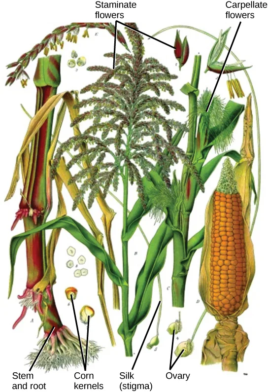
If both male and female flowers are borne on the same plant, the species is called monoecious (meaning “one home”): examples are corn and pea. Species with male and female flowers borne on separate plants are termed dioecious, or “two homes,” examples of which are C. papaya and Cannabis. The ovary, which may contain one or multiple ovules, may be placed above other flower parts, which is referred to as superior; or, it may be placed below the other flower parts, referred to as inferior, shown below.
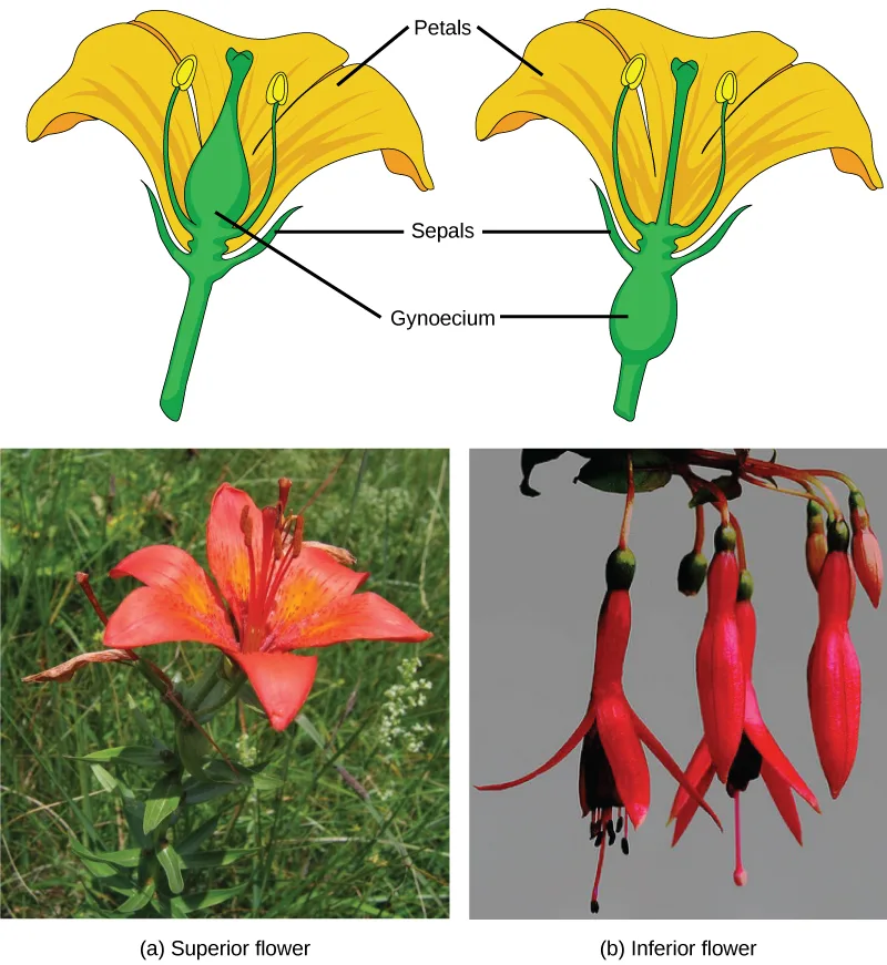
Extended description
Two labeled line drawings of a generic flower sit above two photographs. In the left drawing, the green gynoecium bulges above where the petals attach, matching (a) below it; in the right drawing, the gynoecium bulges below the petals, matching (b). Both drawings carry the same three labels — Petals (the yellow lobes), Sepals (the green base), and Gynoecium (the swollen green structure) — pointing to the corresponding part in each. Below them, photo (a) shows an orange-red lily in a grassy field, its petals spread wide with the gynoecium visible rising from the flower’s center; photo (b) shows three fuchsia flowers hanging point-down from a stem, their swollen ovary near the stem and the narrow petals trailing below.
Male Gametophyte (The Pollen Grain)
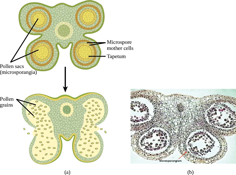
The male gametophyte develops and reaches maturity in an immature anther. In a plant’s male reproductive organs, development of pollen takes place in a structure known as the microsporangium, shown below. The microsporangia, which are usually bilobed, are pollen sacs in which the microspores develop into pollen grains. These are found in the anther, which is at the end of the stamen—the long filament that supports the anther.
Extended description
Panel (a), top: an immature anther in cross section is a four-lobed, clover-shaped cluster of green tissue. Each of the four lobes holds a yellow center ringed by an orange band. A line from the label Pollen sacs (microsporangia) points to two of the four lobes (the left pair); lines from Microspore mother cells and Tapetum point to the yellow center and orange ring of a third lobe (lower right). A small pale circle of tissue sits at the very center of the cross, between the four lobes, unlabeled. A black arrow points down to panel (a), bottom: the mature anther, where each pair of adjacent lobes has fused and split open, leaving two large yellow chambers full of small oval pollen grains — two lines from Pollen grains point into the left chamber — with narrow gaps visible at the top and bottom center where the wall has split. Panel (b), to the right: a micrograph of a similar four-lobed cross section, each lobe packed with dark-stained grains, with an arrow pointing to one lobe labeled Microsporangium.
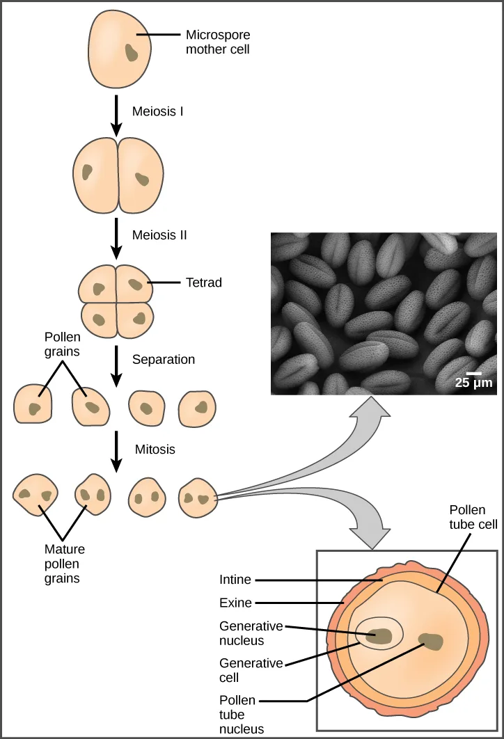
Within the microsporangium, each of the microspore mother cells divides by meiosis to give rise to four microspores, each of which will ultimately form a pollen grain, shown below. An inner layer of cells, known as the tapetum, provides nutrition to the developing microspores and contributes key components to the pollen wall. Mature pollen grains contain two cells: a generative cell and a pollen tube cell. The generative cell is contained within the larger pollen tube cell. Upon germination, the tube cell forms the pollen tube through which the generative cell migrates to enter the ovary. During its transit inside the pollen tube, the generative cell divides to form two male gametes (sperm cells). Upon maturity, the microsporangia burst, releasing the pollen grains from the anther.
Extended description
A vertical sequence of five stages, each connected to the next by a downward black arrow. (1) Microspore mother cell: one oval cell with a single nucleus. Arrow labeled Meiosis I leads to (2) two joined cells, each with its own nucleus. Arrow labeled Meiosis II leads to (3) a four-celled Tetrad in a 2×2 cluster. Arrow labeled Separation leads to (4) four individual single-nucleus cells in a row; a bracket labeled Pollen grains points to the leftmost two. Arrow labeled Mitosis leads to (5) four cells in a row, each now containing two nuclei; a bracket labeled Mature pollen grains again points to the leftmost two. From the rightmost of these four two-nucleus cells, two curved gray arrows branch off: the upper one points to an inset scanning electron micrograph showing dozens of oval pollen grains with a 25 µm scale bar; the lower one points to an enlarged cross section of a single pollen grain, labeled from outside in — Pollen tube cell (the outer boundary), Intine and Exine (the two wall layers), and inside, Generative nucleus, Generative cell, and Pollen tube nucleus.
Each pollen grain has two coverings: the exine (thicker, outer layer) and the intine, shown above. The exine contains sporopollenin, a complex waterproofing substance supplied by the tapetal cells. Sporopollenin allows the pollen to survive under unfavorable conditions and to be carried by wind, water, or biological agents without undergoing damage.
Female Gametophyte (The Embryo Sac)
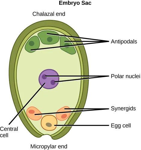
While the details may vary between species, the development of the female gametophyte occurs in a two-phase process called megasporogenesis. In the first phase, a single cell in the diploid megasporangium—an area of tissue in the ovules—undergoes meiosis to produce four megaspores, only one of which survives. During the second phase, megagametogenesis, the surviving haploid megaspore undergoes mitosis to produce an eight-nucleate, seven-cell female gametophyte, also known as the megagametophyte or embryo sac. Two of the nuclei—the polar nuclei—move to the equator and fuse, forming a single, diploid central cell. This central cell later fuses with a sperm to form the triploid endosperm. Three nuclei position themselves on the end of the embryo sac opposite the micropyle and develop into the antipodal cells, which later degenerate. The nucleus closest to the micropyle becomes the female gamete, or egg cell, and the two adjacent nuclei develop into synergid cells, shown below. The synergids help guide the pollen tube for successful fertilization, after which they disintegrate. Once fertilization is complete, the resulting diploid zygote develops into the embryo, and the fertilized ovule forms the other tissues of the seed.
A double-layered integument protects the megasporangium and, later, the embryo sac. The integument will develop into the seed coat after fertilization and protect the entire seed. The ovule wall will become part of the fruit. The integuments, while protecting the megasporangium, do not enclose it completely, but leave an opening called the micropyle. The micropyle allows the pollen tube to enter the female gametophyte for fertilization.
Extended description
Reading from the chalazal end (top) to the micropylar end (bottom): at the top, three green Antipodal cells cluster together, with three lines converging on the label Antipodals. Below them, a large purple Central cell fills the middle of the sac; two small dots inside it are labeled Polar nuclei, and a separate line from the label Central cell (at lower left) points to the same purple cell. Near the bottom, two pink Synergid cells sit side by side next to one yellow Egg cell; a line points to each synergid and another to the egg cell.
An embryo sac is missing the synergids. What specific impact would you expect this to have on fertilization?
Recall what the synergids’ chemical signals actually accomplish once a pollen tube is already growing, and note that the egg cell’s nucleus is separate from the two synergid nuclei.Sexual Reproduction in Gymnosperms
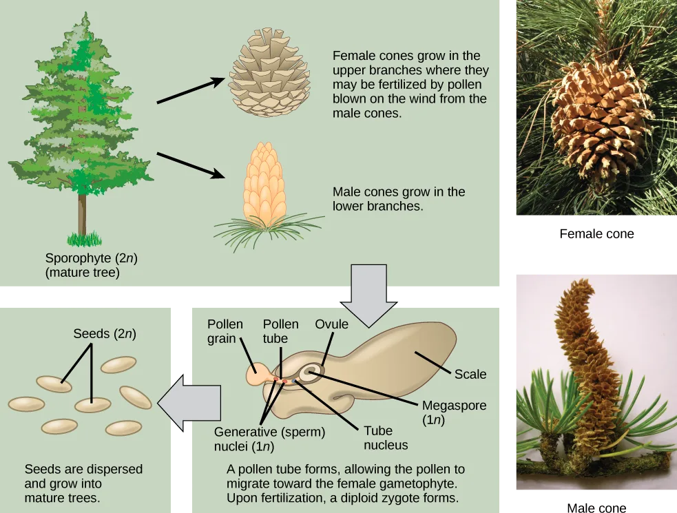
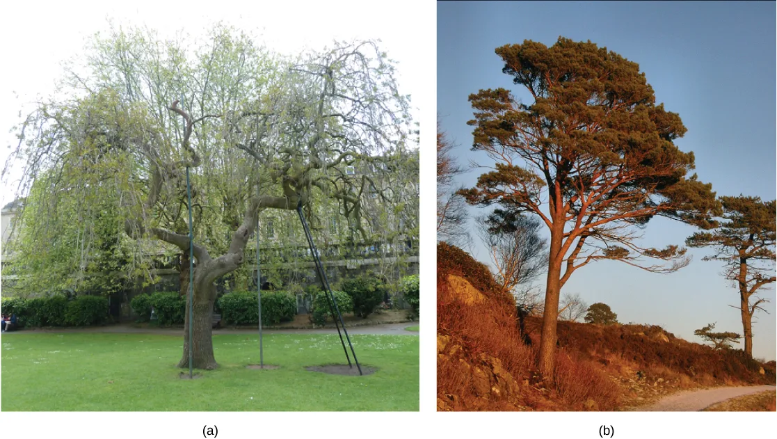
As with angiosperms, the lifecycle of a gymnosperm is also characterized by alternation of generations. In conifers such as pines, the green leafy part of the plant is the sporophyte, and the cones contain the male and female gametophytes, shown below. The female cones are larger than the male cones and are positioned towards the top of the tree; the small, male cones are located in the lower region of the tree. Because the pollen is shed and blown by the wind, this arrangement makes it difficult for a gymnosperm to self-pollinate.
Extended description
Three pale-green panels are linked by gray block arrows, with two photographs alongside. The top panel, spanning the width, is labeled Sporophyte (2n) (mature tree) and shows a conifer tree; two black arrows lead right from it, one to a drawing of a closed brown cone captioned ‘Female cones grow in the upper branches where they may be fertilized by pollen blown on the wind from the male cones,’ the other to a drawing of a slender tan cone captioned ‘Male cones grow in the lower branches.’ A gray block arrow points down from the top panel to the bottom-middle panel, a cutaway drawing of a cone scale labeled Pollen grain, Pollen tube, and Ovule at the top, Scale at the right, and Megaspore (1n), Generative (sperm) nuclei (1n), and Tube nucleus at the bottom, captioned ‘A pollen tube forms, allowing the pollen to migrate toward the female gametophyte. Upon fertilization, a diploid zygote forms.’ A second gray block arrow points left from that panel to the bottom-left panel, labeled Seeds (2n), showing seven oval seed shapes, captioned ‘Seeds are dispersed and grow into mature trees.’ To the right of the diagram, two separate photographs show an actual Female cone (large, brown, and closed) above an actual Male cone (slender, upright, and shedding pollen).
Male Gametophyte
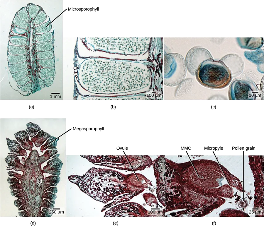
A male cone has a central axis on which bracts, a type of modified leaf, are attached. The bracts are known as microsporophylls, shown below, and are the sites where microspores will develop. The microspores develop inside the microsporangium. Within the microsporangium, cells known as microsporocytes divide by meiosis to produce four haploid microspores. Further mitosis of the microspore produces two nuclei: the generative nucleus, and the tube nucleus. Upon maturity, the male gametophyte (pollen) is released from the male cones and is carried by the wind to land on the female cone.
Female Gametophyte
The female cone also has a central axis on which bracts known as megasporophylls, shown below, are present. In the female cone, megaspore mother cells are present in the megasporangium. The megaspore mother cell divides by meiosis to produce four haploid megaspores. One of the megaspores divides to form the multicellular female gametophyte, while the others divide to form the rest of the structure. The female gametophyte is contained within a structure called the archegonium.
Reproductive Process
Upon landing on the female cone, the tube cell of the pollen forms the pollen tube, through which the generative cell migrates towards the female gametophyte through the micropyle. It takes approximately one year for the pollen tube to grow and migrate towards the female gametophyte. The male gametophyte containing the generative cell splits into two sperm nuclei, one of which fuses with the egg, while the other degenerates. After fertilization of the egg, the diploid zygote is formed, which divides by mitosis to form the embryo. The scales of the cones are closed during development of the seed. The seed is covered by a seed coat, which is derived from the female sporophyte. Seed development takes another one to two years. Once the seed is ready to be dispersed, the bracts of the female cones open to allow the dispersal of seed; no fruit formation takes place because gymnosperm seeds have no covering.
Angiosperms versus Gymnosperms
Gymnosperm reproduction differs from that of angiosperms in several ways, shown below. In angiosperms, the female gametophyte exists in an enclosed structure—the ovule—which is within the ovary; in gymnosperms, the female gametophyte is present on exposed bracts of the female cone. Double fertilization is a key event in the lifecycle of angiosperms, but is completely absent in gymnosperms. The male and female gametophyte structures are present on separate male and female cones in gymnosperms, whereas in angiosperms, they are a part of the flower. Lastly, wind plays an important role in pollination in gymnosperms because pollen is blown by the wind to land on the female cones. Although many angiosperms are also wind-pollinated, animal pollination is more common.
Summary
The flower contains the reproductive structures of a plant. All complete flowers contain four whorls: the calyx, corolla, androecium, and gynoecium. The stamens are made up of anthers, in which pollen grains are produced, and a supportive strand called the filament. The pollen contains two cells— a generative cell and a tube cell—and is covered by two layers called the intine and the exine. The carpels, which are the female reproductive structures, consist of the stigma, style, and ovary. The female gametophyte is formed from mitotic divisions of the megaspore, forming an eight-nuclei ovule sac. This is covered by a layer known as the integument. The integument contains an opening called the micropyle, through which the pollen tube enters the embryo sac.
The diploid sporophyte of angiosperms and gymnosperms is the conspicuous and long-lived stage of the life cycle. The sporophytes differentiate specialized reproductive structures called sporangia, which are dedicated to the production of spores. The microsporangium contains microspore mother cells, which divide by meiosis to produce haploid microspores. The microspores develop into male gametophytes that are released as pollen. The megasporangium contains megaspore mother cells, which divide by meiosis to produce haploid megaspores. A megaspore develops into a female gametophyte containing a haploid egg. A new diploid sporophyte is formed when a male gamete from a pollen grain enters the ovule sac and fertilizes this egg.
Key terms
- androecium — sum of all the stamens in a flower.
- antipodals — the three cells away from the micropyle.
- exine — outermost covering of pollen.
- gametophyte — multicellular stage of the plant that gives rise to haploid gametes or spores.
- gynoecium — the sum of all the carpels in a flower.
- intine — inner lining of the pollen.
- megagametogenesis — second phase of female gametophyte development, during which the surviving haploid megaspore undergoes mitosis to produce an eight-nucleate, seven-cell female gametophyte, also known as the megagametophyte or embryo sac.
- megasporangium — tissue found in the ovary that gives rise to the female gamete or egg.
- megasporogenesis — first phase of female gametophyte development, during which a single cell in the diploid megasporangium undergoes meiosis to produce four megaspores, only one of which survives.
- megasporophyll — bract (a type of modified leaf) on the central axis of a female cone.
- micropyle — opening on the ovule sac through which the pollen tube can gain entry.
- microsporangium — tissue that gives rise to the microspores or the pollen grain.
- microsporophyll — bract (a type of modified leaf) on the central axis of a male cone.
- perianth — (also, petal or sepal) part of the flower consisting of the calyx and/or corolla; forms the outer envelope of the flower.
- polar nuclei — found in the ovule sac; fusion with one sperm cell forms the endosperm.
- sporophyte — multicellular diploid stage in plants that is formed after the fusion of male and female gametes.
- synergid — type of cell found in the ovule sac that secretes chemicals to guide the pollen tube towards the egg.
Practice
Describe the two stages of a plant’s lifecycle
Describe the two-stage lifecycle of plants: the gametophyte stage and the sporophyte stage.
Show model answer
Did your answer mention:
The multicellular stage of a plant that gives rise to haploid gametes or spores is called the ________.
This stage is haploid and produces gametes by mitosis, not meiosis.The multicellular diploid stage in plants that forms after the fusion of male and female gametes is called the ________.
This stage is diploid and eventually produces spores by meiosis.Compare and contrast male and female gametophytes and explain how they form in angiosperms
In a plant’s male reproductive organs, development of pollen takes place in a structure known as the ________.
This tissue lies inside the anther, where the microspore mother cells actually divide by meiosis — not the anther itself, which houses it.The type of cell in the embryo sac that secretes chemicals to guide the pollen tube toward the egg is called a ________.
There are two of these cells, positioned right beside the egg cell at the micropylar end of the embryo sac.The two nuclei in the embryo sac that fuse together and then fuse with a sperm cell to form the endosperm are called the ________.
These two nuclei move to the equator of the embryo sac and fuse into a single diploid central cell before fertilization.Describe the reproductive structures of a plant
Describe the reproductive organs inside a flower.
Show model answer
Did your answer mention:
The pollen lands on which part of the flower?
This is the flat structure at the top of the carpel, reached before the pollen tube grows down through the narrow neck beneath it.The sum of all the stamens in a flower is called the ________.
This whorl holds the anthers with their microsporangia — the flower’s male reproductive part, not its female part.The sum of all the carpels in a flower is called the ________.
This is the flower’s innermost whorl and its female reproductive part, made up of one or more carpels with a stigma, style, and ovary.Describe the components of a complete flower
The stamen consists of a long stalk called the filament that supports the ________.
This structure sits atop the filament and holds the microsporangia, or pollen sacs — it is not part of the gynoecium at all.The ________ are collectively called the calyx.
These are the outermost, usually green, leafy structures that protect the unopened bud.Describe the four main parts, or whorls, of a flower.
Show model answer
Did your answer mention:
Discuss the differences between a complete flower and an incomplete flower.
Show model answer
Did your answer mention:
The part of the flower made up of the calyx and corolla together, forming its outer envelope, is called the ________.
This term combines the flower’s two outermost whorls — the leafy protective whorl and the often colorful whorl inside it.Describe the development of microsporangium and megasporangium in gymnosperms
The tissue that gives rise to the microspores, or pollen grain, is called the ________.
In a male gymnosperm cone, this tissue lies within the microsporophylls, where microsporocytes divide by meiosis into four haploid microspores.The tissue that gives rise to the female gamete, or egg, is called the ________.
In a female gymnosperm cone, this tissue lies within the megasporophylls, where a megaspore mother cell divides by meiosis into four haploid megaspores.A bract on the central axis of a male gymnosperm cone, where microspores develop, is called a ________.
A male cone bears roughly 20 of these bracts, each producing hundreds of pollen grains.A bract on the central axis of a female gymnosperm cone that carries the megaspore mother cells is called a ________.
A female cone bears roughly 15 of these bracts, each containing an ovule with the megaspore mother cell inside.This section is adapted from Biology 2e, Section 32.1: Reproductive Development and Structure by Mary Ann Clark, Jung Choi, Matthew Douglas, and OpenStax, © OpenStax, licensed under CC BY-NC-SA 4.0. Access the original for free at openstax.org. Changes: figures re-encoded as WebP with a custom alt written from each image rather than reusing the source’s own alt text; four figures re-kinded after inspection from the manifest’s file-extension guess of “photo” to “diagram” — Figure_32_01_03 (a hand-colored botanical illustration of a corn plant, not a photograph), Figure_32_01_04ab (a labeled line-drawing comparison of ovary position, paired with two photographs), and Figure_32_01_06f (a hand-drawn pollen-development flow chart with one inset micrograph) — and Figure_37_01_10 from “diagram” to “photo” (a six-panel series of stained microscope images, not a hand-drawn diagram); a longdesc added to the two life-cycle diagrams (Figure_37_01_01, Figure_32_01_08), the two Visual Connection diagrams (Figure_32_01_02, Figure_32_01_07), the pollen-development diagrams (Figure_37_01_02, Figure_32_01_06f), and the superior/inferior ovary-position schematic (Figure_32_01_04ab), each counted and walked in reading order; both notes wrapping a Visual Connection rendered as their figure followed by a self-check or multiple choice, kept in the body, and not repeated in Practice; both Link to Learning notes kept as callouts with the module’s own openstax.org/l/ redirect URLs; the end-of-section Review Questions and Critical Thinking Questions adapted into the closing interactive Practice block; rubric checkpoints added to each self-check, decomposing its model answer (the source solution) into check-off clauses with no new claims; eleven key-term recall items (gametophyte, sporophyte, synergid, polar nuclei, androecium, gynoecium, perianth, microsporangium, megasporangium, microsporophyll, megasporophyll) added from the glossary; a typo in the opening paragraph, “the male and females gametes,” corrected to “the male and female gametes” — reported as a source defect; the glossary definition of “microsporophyll,” which reads “central axis of a male cone on which bracts … are attached,” corrected to “bract (a type of modified leaf) on the central axis of a male cone,” because the section’s own sentence “The bracts are known as microsporophylls” identifies the bracts themselves, not the axis, as the microsporophylls — reported as a source defect; the glossary definition of “megasporophyll” corrected from “central axis of a female gametophyte” to “central axis of a female cone,” because the section’s own sentence places the bracts “on a central axis” belonging to “the female cone,” not the female gametophyte (the haploid generation the cone’s megasporophylls give rise to) — reported as a source defect.