Animal Primary Tissues
By the end of this section, you will be able to:
- Describe epithelial tissues
- Discuss the different types of connective tissues in animals
- Describe three types of muscle tissues
- Describe nervous tissue
The tissues of multicellular, complex animals are four primary types: epithelial, connective, muscle, and nervous. Recall that tissues are groups of similar cells (cells carrying out related functions). These tissues combine to form organs—like the skin or kidney—that have specific, specialized functions within the body. Organs are organized into organ systems to perform functions; examples include the circulatory system, which consists of the heart and blood vessels, and the digestive system, consisting of several organs, including the stomach, intestines, liver, and pancreas. Organ systems come together to create an entire organism.
Epithelial Tissues
Epithelial tissues cover the outside of organs and structures in the body and line the lumens of organs in a single layer or multiple layers of cells. The types of epithelia are classified by the shapes of cells present and the number of layers of cells. Epithelia composed of a single layer of cells is called simple epithelia; epithelial tissue composed of multiple layers is called stratified epithelia. The table below summarizes the different types of epithelial tissues.
| Cell shape | Description | Location |
|---|---|---|
| squamous | flat, irregular round shape | simple: lung alveoli, capillaries; stratified: skin, mouth, vagina |
| cuboidal | cube shaped, central nucleus | glands, renal tubules |
| columnar | tall, narrow, nucleus toward base; tall, narrow, nucleus along cell | simple: digestive tract; pseudostratified: respiratory tract |
| transitional | round, simple but appear stratified | urinary bladder |
Squamous Epithelia
Squamous epithelial cells are generally round, flat, and have a small, centrally located nucleus. The cell outline is slightly irregular, and cells fit together to form a covering or lining. When the cells are arranged in a single layer (simple epithelia), they facilitate diffusion in tissues, such as the areas of gas exchange in the lungs and the exchange of nutrients and waste at blood capillaries.
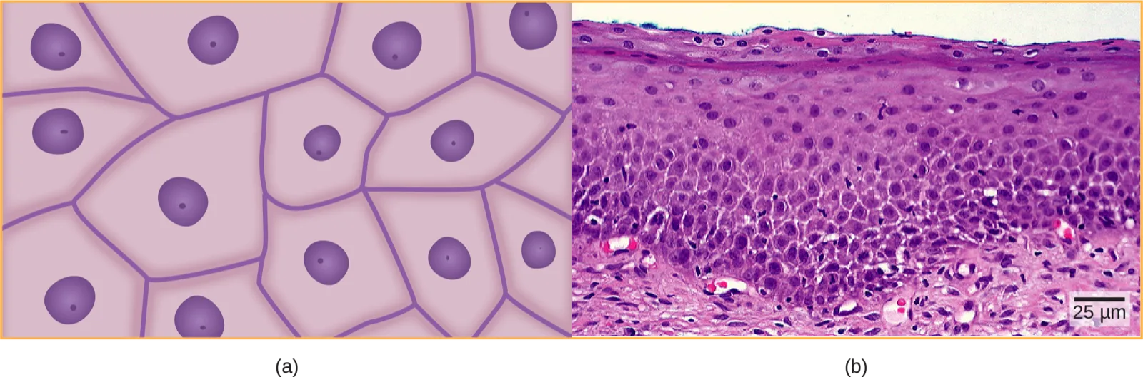
Figure (a) above illustrates a layer of squamous cells with their membranes joined together to form an epithelium. Image (b) illustrates squamous epithelial cells arranged in stratified layers, where protection is needed on the body from outside abrasion and damage. This is called a stratified squamous epithelium and occurs in the skin and in tissues lining the mouth and vagina.
Cuboidal Epithelia
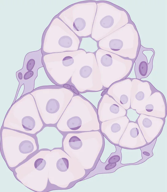
Cuboidal epithelial cells, shown below, are cube-shaped with a single, central nucleus. They are most commonly found in a single layer representing a simple epithelia in glandular tissues throughout the body where they prepare and secrete glandular material. They are also found in the walls of tubules and in the ducts of the kidney and liver.
Columnar Epithelia
Columnar epithelial cells are taller than they are wide: they resemble a stack of columns in an epithelial layer, and are most commonly found in a single-layer arrangement. The nuclei of columnar epithelial cells in the digestive tract appear to be lined up at the base of the cells, as illustrated below. These cells absorb material from the lumen of the digestive tract and prepare it for entry into the body through the circulatory and lymphatic systems.
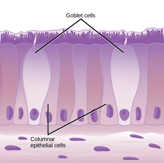
Extended description
Two guide lines labeled ‘Goblet cells’ at the top point down to two pale, swollen, flask-shaped cells that reach up to the ciliated surface, wider than their neighbors. A second label, ‘Columnar epithelial cells,’ has two guide lines pointing to the tall, narrow cells flanking the goblet cells, each with an oval purple nucleus set near its base and short cilia fringing its top edge. Beneath the row of columnar and goblet cells lies a scattered layer of small, rounded purple cells forming the basal tissue.
Columnar epithelial cells lining the respiratory tract appear to be stratified. However, each cell is attached to the base membrane of the tissue and, therefore, they are simple tissues. The nuclei are arranged at different levels in the layer of cells, making it appear as though there is more than one layer, as seen below. This is called pseudostratified, columnar epithelia. This cellular covering has cilia at the apical, or free, surface of the cells. The cilia enhance the movement of mucus and trapped particles out of the respiratory tract, helping to protect the system from invasive microorganisms and harmful material that has been breathed into the body. Goblet cells are interspersed in some tissues (such as the lining of the trachea). The goblet cells contain mucus that traps irritants, which in the case of the trachea keep these irritants from getting into the lungs.
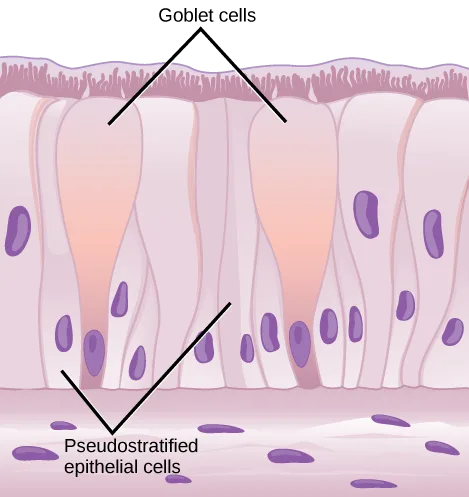
Extended description
Two guide lines from the label ‘Goblet cells’ at the top point to two pale, swollen, flask-shaped cells that widen toward the ciliated surface. A second label, ‘Pseudostratified epithelial cells,’ has two guide lines pointing to the surrounding tall and short cells: the tall cells’ nuclei sit high, near the middle of the layer, while shorter cells wedged between them have nuclei set lower, close to the base — the staggered nuclear heights are what make the single layer look stratified. Cilia fringe the free surface at top. Beneath the row lies a scattered layer of small, rounded purple cells forming the basal tissue.
Transitional Epithelia
Transitional or uroepithelial cells appear only in the urinary system, primarily in the bladder and ureter. These cells are arranged in a stratified layer, but they have the capability of appearing to pile up on top of each other in a relaxed, empty bladder, as illustrated below. As the urinary bladder fills, the epithelial layer unfolds and expands to hold the volume of urine introduced into it. As the bladder fills, it expands and the lining becomes thinner. In other words, the tissue transitions from thick to thin.
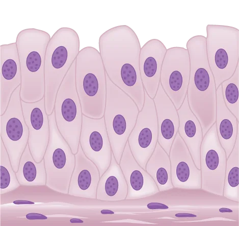
Which of the following statements about types of epithelial cells is false?
Check which epithelial type the table above actually lists for the lung’s alveoli.Connective Tissues
Connective tissues are made up of a matrix consisting of living cells and a nonliving substance, called the ground substance. The ground substance is made of an organic substance (usually a protein) and an inorganic substance (usually a mineral or water). The principal cell of connective tissues is the fibroblast. This cell makes the fibers found in nearly all of the connective tissues. Fibroblasts are motile, able to carry out mitosis, and can synthesize whichever connective tissue is needed. Macrophages, lymphocytes, and, occasionally, leukocytes can be found in some of the tissues. Some tissues have specialized cells that are not found in the others. Connective tissues are made up of living cells and a nonliving matrix of organic substances (usually proteins) and inorganic substances (usually a mineral or water).
The organic portion or protein fibers found in connective tissues are either collagen, elastic, or reticular fibers. Collagen fibers provide strength to the tissue, preventing it from being torn or separated from the surrounding tissues. Elastic fibers are made of the protein elastin; this fiber can stretch to one and one half of its length and return to its original size and shape. Elastic fibers provide flexibility to the tissues. Reticular fibers are the third type of protein fiber found in connective tissues. This fiber consists of thin strands of collagen that form a network of fibers to support the tissue and other organs to which it is connected. The table below summarizes the various types of connective tissues, the types of cells and fibers they are made of, and sample locations of the tissues.
| Tissue | Cells | Fibers | Location |
|---|---|---|---|
| loose/areolar | fibroblasts, macrophages, some lymphocytes, some neutrophils | few: collagen, elastic, reticular | around blood vessels; anchors epithelia |
| dense, fibrous connective tissue | fibroblasts, macrophages | mostly collagen | irregular: skin; regular: tendons, ligaments |
| cartilage | chondrocytes, chondroblasts | hyaline: few: collagen; fibrocartilage: large amount of collagen | shark skeleton, fetal bones, human ears, intervertebral discs |
| bone | osteoblasts, osteocytes, osteoclasts | some: collagen, elastic | vertebrate skeletons |
| adipose | adipocytes | few | adipose (fat) |
| blood | red blood cells, white blood cells | none | blood |
Loose/Areolar Connective Tissue
Loose connective tissue, also called areolar connective tissue, has a sampling of all of the components of a connective tissue. As illustrated below, loose connective tissue has some fibroblasts; macrophages are present as well. Collagen fibers are relatively wide and stain a light pink, while elastic fibers are thin and stain dark blue to black. The material in the connective tissue gives it a loose consistency similar to a cotton ball that has been pulled apart. Loose connective tissue is found around every blood vessel and helps to keep the vessel in place. The tissue is also found around and between most body organs. In summary, areolar tissue is tough, yet flexible, and comprises membranes.
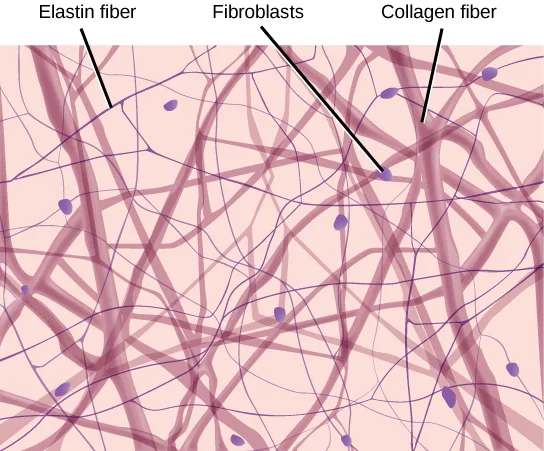
Extended description
Three guide lines run from labels at the top down into the fiber mesh. ‘Elastin fiber,’ at left, points to one of the thin, wavy strands crossing the field. ‘Fibroblasts,’ in the middle, points to one of the small, oval, purple-staining cells scattered loosely among the fibers. ‘Collagen fiber,’ at right, points to one of the wider, more heavily shaded bands running through the mesh. Fibers of both widths cross and loop through the field in no consistent direction, with open space visible between them.
Fibrous Connective Tissue
Fibrous connective tissues contain large amounts of collagen fibers and few cells or matrix material. The fibers can be arranged irregularly or regularly with the strands lined up in parallel. Irregularly arranged fibrous connective tissues are found in areas of the body where stress occurs from all directions, such as the dermis of the skin. Regular fibrous connective tissue, shown below, is found in tendons (which connect muscles to bones) and ligaments (which connect bones to bones).
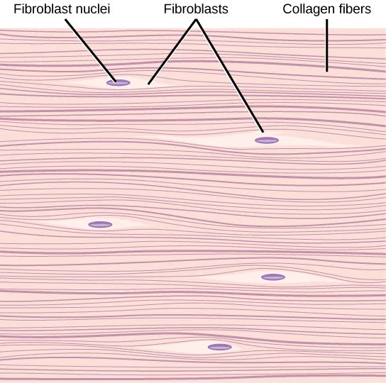
Extended description
Three guide lines point from labels at the top into the parallel fiber bundles. ‘Fibroblast nuclei,’ at left, points to a small, oval, purple-staining nucleus lying flush between fiber strands, with no visible cell outline around it. ‘Fibroblasts,’ in the middle, points to a longer, spindle-shaped cell with a visible nucleus, oriented in the same left-to-right direction as the fibers. ‘Collagen fibers,’ at right, points to one of the wavy pink bands running the width of the image. The whole field is made up of these parallel bands with the same small nuclei repeated at intervals between them.
Cartilage
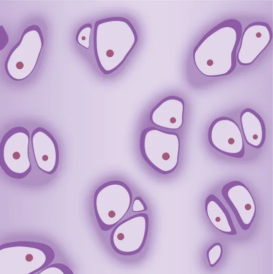
Cartilage is a connective tissue with a large amount of the matrix of variable composition. The cells, called chondrocytes, make the matrix and fibers of the tissue. Chondrocytes are found in spaces within the tissue called lacunae.
A cartilage with few collagen and elastic fibers is hyaline cartilage, illustrated below. The lacunae are randomly scattered throughout the tissue and the matrix takes on a milky or scrubbed appearance with routine histological stains. Sharks have cartilaginous skeletons, as does nearly the entire human skeleton during a specific pre-birth developmental stage. A remnant of this cartilage persists in the outer portion of the human nose. Hyaline cartilage is also found at the ends of long bones, reducing friction and cushioning the articulations of these bones.
Elastic cartilage has a large amount of elastic fibers, giving it tremendous flexibility. The ears of most vertebrate animals contain this cartilage as do portions of the larynx, or voice box. Fibrocartilage contains a large amount of collagen fibers, giving the tissue tremendous strength. Fibrocartilage comprises the intervertebral discs in vertebrate animals. Hyaline cartilage found in movable joints such as the knee and shoulder becomes damaged as a result of age or trauma. Damaged hyaline cartilage is replaced by fibrocartilage and results in the joints becoming “stiff.”
Bone
Bone, or osseous tissue, is a connective tissue that has a large amount of two different types of matrix material. The organic matrix is similar to the matrix material found in other connective tissues, including some amount of collagen and elastic fibers. This gives strength and flexibility to the tissue. The inorganic matrix consists of mineral salts—mostly calcium salts—that give the tissue hardness. Without adequate organic material in the matrix, the tissue breaks; without adequate inorganic material in the matrix, the tissue bends.
There are three types of cells in bone: osteoblasts, osteocytes, and osteoclasts. Osteoblasts are active in making bone for growth and remodeling. Osteoblasts deposit bone material into the matrix and, after the matrix surrounds them, they continue to live, but in a reduced metabolic state as osteocytes. Osteocytes are found in lacunae of the bone. Osteoclasts are active in breaking down bone for bone remodeling, and they provide access to calcium stored in tissues. Osteoclasts are usually found on the surface of the tissue.
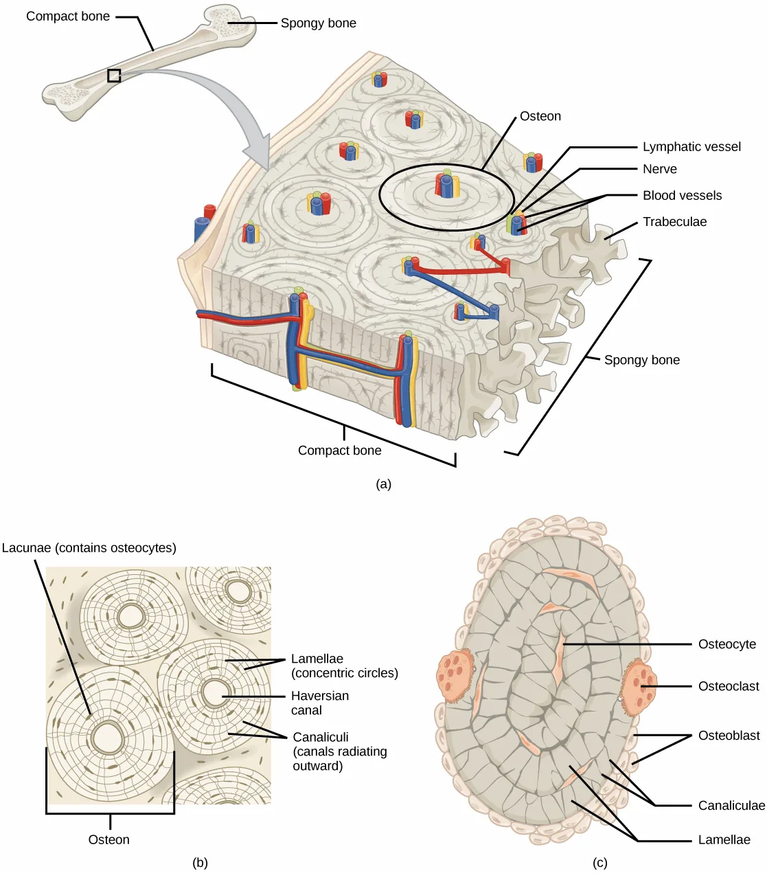
Bone can be divided into two types: compact and spongy. Compact bone is found in the shaft (or diaphysis) of a long bone and the surface of the flat bones, while spongy bone is found in the end (or epiphysis) of a long bone. Compact bone is organized into subunits called osteons, as illustrated below. A blood vessel and a nerve are found in the center of the structure within the Haversian canal, with radiating circles of lacunae around it known as lamellae. The wavy lines seen between the lacunae are microchannels called canaliculi; they connect the lacunae to aid diffusion between the cells. Spongy bone is made of tiny plates called trabeculae; these plates serve as struts to give the spongy bone strength. Over time, these plates can break causing the bone to become less resilient. Bone tissue forms the internal skeleton of vertebrate animals, providing structure to the animal and points of attachment for tendons.
Extended description
Panel (a), upper left: a small whole long bone is labeled ‘Compact bone’ at its shaft and ‘Spongy bone’ at its widened end; an arrow leads from a boxed region of the shaft down to a large 3D block of bone tissue. On the block, a circled region is labeled ‘Osteon’; three lines from ‘Lymphatic vessel,’ ‘Nerve,’ and ‘Blood vessels’ point to a bundle of colored tubes running through the block’s central channel. ‘Trabeculae’ labels the web-like mesh at the block’s wider end. Brackets at the bottom and right of the block are labeled ‘Compact bone’ and ‘Spongy bone,’ naming the block’s dense outer region and porous inner region. Panel (b), lower left: a flat, square cross section shows several circular osteons packed together, each made of concentric rings. ‘Lacunae (contains osteocytes)’ points to small dark flecks scattered between the rings; ‘Lamellae (concentric circles)’ points to the rings themselves; ‘Haversian canal’ points to the open circle at the center of one osteon; ‘Canaliculi (canals radiating outward)’ points to the fine lines radiating from the rings toward the lacunae. A bracket beneath one osteon is labeled ‘Osteon.’ Panel (c), lower right: a single osteon is drawn as a cutaway oval. ‘Osteocyte’ points to a cell within the rings; ‘Osteoclast’ points to one of two larger, multi-nucleated cells at the oval’s left and right edges; ‘Osteoblast’ points to small cells lining the oval’s outer edge; ‘Canaliculi’ and ‘Lamellae’ point to the fine radiating lines and concentric rings inside the oval.
Adipose Tissue
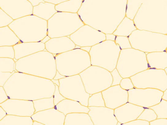
Adipose tissue, or fat tissue, is considered a connective tissue even though it does not have fibroblasts or a real matrix. Adipose tissue is made up of cells called adipocytes that collect and store fat in the form of triglycerides, for energy metabolism. Adipose tissues additionally serve as insulation to help maintain body temperatures, allowing animals to be endothermic, and they function as cushioning against damage to body organs. Under a microscope, adipose tissue cells appear empty due to the extraction of fat during the processing of the material for viewing, as seen below. The thin lines in the image are the cell membranes, and the nuclei are the small, black dots at the edges of the cells.
Blood
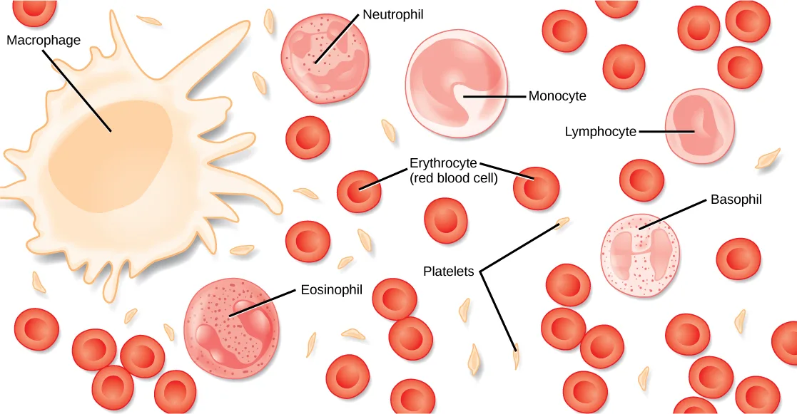
Blood is considered a connective tissue because it has a matrix, as shown below. The living cell types are red blood cells (RBC), also called erythrocytes, and white blood cells (WBC), also called leukocytes. The fluid portion of whole blood, its matrix, is commonly called plasma.
Extended description
Reading roughly left to right across the field: at far left, ‘Macrophage’ labels a very large, irregular cell with finger-like projections and a large oval nucleus. Above and right of it, ‘Neutrophil’ labels a round cell with a lobed, multi-part nucleus and fine granules. ‘Monocyte,’ at upper middle, labels a large round cell with a kidney-bean-shaped nucleus. ‘Lymphocyte,’ at upper right, labels a round cell with a large, smooth, off-center nucleus that fills most of the cell. In the lower middle, ‘Eosinophil’ labels a round, coarsely granular cell with a two-lobed nucleus. At right, ‘Basophil’ labels a round cell packed with dark granules around an H-shaped nucleus. ‘Erythrocyte (red blood cell)’ labels one of the many small, disc-shaped red cells with a lighter central dimple that fill the rest of the field. ‘Platelets’ labels several small, tan, irregular fragments scattered among the red cells.
The cell found in greatest abundance in blood is the erythrocyte. Erythrocytes are counted in millions in a blood sample: the average number of red blood cells in primates is 4.7 to 5.5 million cells per microliter. Erythrocytes are consistently the same size in a species, but vary in size between species. For example, the average diameter of a primate red blood cell is 7.5 µm, a dog is close at 7.0 µm, but a cat’s RBC diameter is 5.9 µm. Sheep erythrocytes are even smaller at 4.6 µm. Mammalian erythrocytes lose their nuclei and mitochondria when they are released from the bone marrow where they are made. Fish, amphibian, and avian red blood cells maintain their nuclei and mitochondria throughout the cell’s life. The principal job of an erythrocyte is to carry and deliver oxygen to the tissues.
Leukocytes are the predominant white blood cells found in the peripheral blood. Leukocytes are counted in the thousands in the blood with measurements expressed as ranges: primate counts range from 4,800 to 10,800 cells per µl, dogs from 5,600 to 19,200 cells per µl, cats from 8,000 to 25,000 cells per µl, cattle from 4,000 to 12,000 cells per µl, and pigs from 11,000 to 22,000 cells per µl.
Lymphocytes function primarily in the immune response to foreign antigens or material. Different types of lymphocytes make antibodies tailored to the foreign antigens and control the production of those antibodies. Neutrophils are phagocytic cells and they participate in one of the early lines of defense against microbial invaders, aiding in the removal of bacteria that have entered the body. Another leukocyte that is found in the peripheral blood is the monocyte. Monocytes give rise to phagocytic macrophages that clean up dead and damaged cells in the body, whether they are foreign or from the host animal. Two additional leukocytes in the blood are eosinophils and basophils—both help to facilitate the inflammatory response.
The slightly granular material among the cells is a cytoplasmic fragment of a cell in the bone marrow. This is called a platelet or thrombocyte. Platelets participate in the stages leading up to coagulation of the blood to stop bleeding through damaged blood vessels. Blood has a number of functions, but primarily it transports material through the body to bring nutrients to cells and remove waste material from them.
Muscle Tissues
There are three types of muscle in animal bodies: smooth, skeletal, and cardiac. They differ by the presence or absence of striations or bands, the number and location of nuclei, whether they are voluntarily or involuntarily controlled, and their location within the body. The table below summarizes these differences.
| Type of Muscle | Striations | Nuclei | Control | Location |
|---|---|---|---|---|
| smooth | no | single, in center | involuntary | visceral organs |
| skeletal | yes | many, at periphery | voluntary | skeletal muscles |
| cardiac | yes | single, in center | involuntary | heart |
Smooth Muscle
Smooth muscle does not have striations in its cells. It has a single, centrally located nucleus, as shown below. Constriction of smooth muscle occurs under involuntary, autonomic nervous control and in response to local conditions in the tissues. Smooth muscle tissue is also called non-striated as it lacks the banded appearance of skeletal and cardiac muscle. The walls of blood vessels, the tubes of the digestive system, and the tubes of the reproductive systems are composed of mostly smooth muscle.

Extended description
Three tissue samples are shown side by side, each captioned with its own title above it. Left, ‘Smooth muscle cells’: elongated spindle-shaped cells run roughly parallel, each with a single dark oval nucleus near its middle and no visible banding. Middle, ‘Skeletal muscle cells’: long, straight fibers cross the panel, each ruled with fine, evenly spaced light cross-stripes (striations) and studded along its edges with several small, dark nuclei. Right, ‘Cardiac muscle cells’: shorter fibers with the same fine cross-striations branch and rejoin, each fiber holding one nucleus near its center; a guide line labeled ‘Intercalated disc’ points to one of several bright vertical lines where the end of one fiber meets the next.
Skeletal Muscle
Skeletal muscle has striations across its cells caused by the arrangement of the contractile proteins actin and myosin. These muscle cells are relatively long and have multiple nuclei along the edge of the cell. Skeletal muscle is under voluntary, somatic nervous system control and is found in the muscles that move bones. The figure above illustrates the histology of skeletal muscle.
Cardiac Muscle
Cardiac muscle, shown above, is found only in the heart. Like skeletal muscle, it has cross striations in its cells, but cardiac muscle has a single, centrally located nucleus. Cardiac muscle is not under voluntary control but can be influenced by the autonomic nervous system to speed up or slow down. An added feature to cardiac muscle cells is a line than extends along the end of the cell as it abuts the next cardiac cell in the row. This line is called an intercalated disc: it assists in passing electrical impulse efficiently from one cell to the next and maintains the strong connection between neighboring cardiac cells.
Nervous Tissues
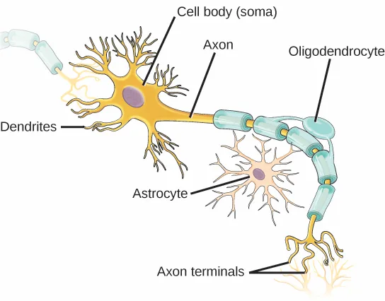
Nervous tissues are made of cells specialized to receive and transmit electrical impulses from specific areas of the body and to send them to specific locations in the body. The main cell of the nervous system is the neuron, illustrated below. The large structure with a central nucleus is the cell body of the neuron. Projections from the cell body are either dendrites specialized in receiving input or a single axon specialized in transmitting impulses. Some glial cells are also shown. Astrocytes regulate the chemical environment of the nerve cell, and oligodendrocytes insulate the axon so the electrical nerve impulse is transferred more efficiently. Other glial cells that are not shown support the nutritional and waste requirements of the neuron. Some of the glial cells are phagocytic and remove debris or damaged cells from the tissue. A nerve consists of neurons and glial cells.
Extended description
At the upper left, a branching yellow dendrite continues in from off-frame. ‘Cell body (soma)’ labels the large, star-shaped yellow cell with a round purple nucleus at the image’s left-center; many thin, branching dendrites radiate from its edges. ‘Dendrites’ labels this same branching fringe. From the opposite side of the cell body, ‘Axon’ labels a long, narrow yellow projection that runs right and down across the image, wrapped at intervals in short teal segments. ‘Oligodendrocyte’ labels one of these teal segments partway along the axon. Below the axon’s midpoint, ‘Astrocyte’ labels a smaller, star-shaped tan cell with its own short branching projections, sitting beside but not touching the axon. At the lower right, the axon ends in a spray of thin, branching yellow filaments labeled ‘Axon terminals.’
Summary
The basic building blocks of complex animals are four primary tissues. These are combined to form organs, which have a specific, specialized function within the body, such as the skin or kidney. Organs are organized together to perform common functions in the form of systems. The four primary tissues are epithelia, connective tissues, muscle tissues, and nervous tissues.
Key terms
- canaliculus — microchannel that connects the lacunae and aids diffusion between cells.
- cartilage — type of connective tissue with a large amount of ground substance matrix, cells called chondrocytes, and some amount of fibers.
- chondrocyte — cell found in cartilage.
- columnar epithelia — epithelia made of cells taller than they are wide, specialized in absorption.
- connective tissue — type of tissue made of cells, ground substance matrix, and fibers.
- cuboidal epithelia — epithelia made of cube-shaped cells, specialized in glandular functions.
- epithelial tissue — tissue that either lines or covers organs or other tissues.
- fibrous connective tissue — type of connective tissue with a high concentration of fibers.
- lacuna — space in cartilage and bone that contains living cells.
- loose (areolar) connective tissue — type of connective tissue with small amounts of cells, matrix, and fibers; found around blood vessels.
- matrix — component of connective tissue made of both living and nonliving (ground substances) cells.
- osteon — subunit of compact bone.
- pseudostratified — layer of epithelia that appears multilayered, but is a simple covering.
- simple epithelia — single layer of epithelial cells.
- squamous epithelia — type of epithelia made of flat cells, specialized in aiding diffusion or preventing abrasion.
- stratified epithelia — multiple layers of epithelial cells.
- trabecula — tiny plate that makes up spongy bone and gives it strength.
- transitional epithelia — epithelia that can transition for appearing multilayered to simple; also called uroepithelial.
Practice
Describe epithelial tissues
Which type of epithelial cell is best adapted to aid diffusion?
Look back at the epithelial-tissue table for which cell shape lines the lung’s air sacs and the walls of capillaries — thin cells make for a short diffusion path.Which type of epithelial cell is found in glands?
This shape prepares and secretes material in glandular tissue and lines kidney and liver tubules.Which type of epithelial cell is found in the urinary bladder?
This epithelium changes thickness as an organ that stores fluid fills and empties.A layer of epithelia that appears multilayered, but is actually a simple covering, is called ________.
This tissue lines the respiratory tract, where nuclei sit at staggered heights within a single layer of cells.How can squamous epithelia both facilitate diffusion and prevent damage from abrasion?
Show model answer
Did your answer mention:
Discuss the different types of connective tissues in animals
Which type of connective tissue has the most fibers?
This tissue type is found in tendons and ligaments, where cells and matrix are sparse compared to the fiber content.Which type of connective tissue has a mineralized different matrix?
Only one of these connective tissues gets its hardness from calcium salts deposited in its matrix.The cell found in bone that breaks it down is called an ________.
This cell type provides access to calcium stored in bone tissue by breaking the tissue down, and is usually found on the tissue’s surface.The cell found in bone that makes the bone is called an ________.
This cell type deposits new bone material during growth and remodeling, then continues living at a reduced metabolic rate once the matrix surrounds it.Plasma is the ________.
This is the fluid, non-cellular portion of whole blood.A space in cartilage and bone that contains living cells is called a ________.
Osteocytes and chondrocytes both sit inside these small cavities within their tissue’s matrix.What are the similarities between cartilage and bone?
Show model answer
Did your answer mention:
Describe three types of muscle tissues
The type of muscle cell under voluntary control is the ________.
This muscle type has multiple nuclei at the cell periphery and moves the bones under conscious control.Why are intercalated discs essential to the function of cardiac muscle?
Think about what has to happen electrically for the whole heart to contract together rather than one cell at a time.The line that extends along the end of a cardiac muscle cell, joining it to the next cell in the row and helping pass an electrical impulse between them, is called the ________.
Cardiac cells are shorter than skeletal muscle fibers, and each one connects end to end to its neighbor at this membrane structure.When a person leads a sedentary life his skeletal muscles atrophy, but his smooth muscles do not. Why?
Show model answer
Did your answer mention:
Describe nervous tissue
The part of a neuron that contains the nucleus is the
Picture the neuron’s parts: the nucleus sits inside the largest, most central structure, not in any of its thread-like projections.The glial cells that regulate the chemical environment of the nerve cell are the ________.
Their star-shaped projections sit alongside the axon but do not wrap around it, unlike their neighboring glial cell.The glial cells that insulate a neuron’s axon so the electrical nerve impulse is transferred more efficiently are the ________.
Their segments wrap directly around the axon, forming a sheath, unlike the star-shaped glial cells that only sit nearby.Multiple sclerosis is a debilitating autoimmune disease that results in the loss of the insulation around neuron axons. What cell type is the immune system attacking, and how does this disrupt the transfer of messages by the nervous system?
Show model answer
Did your answer mention:
This section is adapted from Biology 2e, Section 33.2: Animal Primary Tissues by Mary Ann Clark, Jung Choi, Matthew Douglas, and OpenStax, © OpenStax, licensed under CC BY-NC-SA 4.0. Access the original for free at openstax.org. Changes: figures re-encoded as WebP with a custom alt written from each image rather than reusing the source’s own alt text; eleven figures re-kinded after inspection — Figure_33_02_02, Figure_33_02_03, Figure_33_02_06, Figure_33_02_07, Figure_33_02_08, Figure_33_02_09, Figure_33_02_10, Figure_33_02_11, Figure_33_02_12abc, and Figure_33_02_13 from the manifest’s file-extension “photo” guess to “diagram” (all ten are drawn, flat-color textbook illustrations, not photographs), and Figure_B33_02_01ab from “diagram” to “photo” (its panel (b) is a genuine photomicrograph of human cervix tissue, described alongside the drawn panel (a) in one alt); a longdesc added to the eight figures whose printed labels are not carried by their one-line captions (Figure_33_02_03, Figure_33_02_04, Figure_33_02_06, Figure_33_02_07, Figure_33_02_09, Figure_33_02_11, Figure_33_02_12abc, Figure_33_02_13); Figure 33.15(c)’s own printed label “Canaliculae” (its sibling panel (b) prints the correct “Canaliculi”) corrected to “Canaliculi” in the longdesc — reported as a source defect, the artwork itself left as printed; the Visual Connection Questions exercise’s option “Pseudostratisfied columnar epithilia occur in a single layer, but the arrangement of nuclei makes it appear that more than one layer is present” (also printed the same way inside the body’s Visual Connection note) corrected to “Pseudostratified columnar epithelia occur in a single layer, but the arrangement of nuclei makes it appear that more than one layer is present” — reported as a source defect; the note wrapping the Transitional Epithelia Visual Connection rendered as its figure followed by a multiple choice (the exercise-set copy, identical in wording to the note’s own copy), kept in the body; the end-of-chapter Review Questions and Critical Thinking Questions for this section adapted into the closing interactive Practice block (multiple choice and self-check respectively); rubric checkpoints added to each self-check, decomposing its model answer (the source solution) into check-off clauses with no new claims; two key-term recall items (pseudostratified, lacuna) added from the glossary, plus three local recall items (intercalated disc, astrocyte, oligodendrocyte) built strictly from the section’s own sentences, since the glossary defines no muscle- or nervous-tissue vocabulary; the Review Question “Which type of epithelial cell is best adapted to aid diffusion?” keeps its printed options but is re-keyed from the source’s “columnar” to “squamous”, because the section’s own text (“they facilitate diffusion in tissues, such as the areas of gas exchange in the lungs”) and its glossary (“squamous epithelia… specialized in aiding diffusion or preventing abrasion”) both support squamous, not columnar — reported as a source defect. A semicolon was inserted in the connective-tissue table’s cartilage fibers cell (“hyaline: few: collagen; fibrocartilage: large amount of collagen”), which the source prints run together.