Digestive Systems
By the end of this section, you will be able to:
- Explain the processes of digestion and absorption
- Compare and contrast different types of digestive systems
- Explain the specialized functions of the organs involved in processing food in the body
- Describe the ways in which organs work together to digest food and absorb nutrients
Animals obtain their nutrition from the consumption of other organisms. Depending on their diet, animals can be classified into the following categories: plant eaters (herbivores), meat eaters (carnivores), and those that eat both plants and animals (omnivores). The nutrients and macromolecules present in food are not immediately accessible to the cells. There are a number of processes that modify food within the animal body in order to make the nutrients and organic molecules accessible for cellular function. As animals evolved in complexity of form and function, their digestive systems have also evolved to accommodate their various dietary needs.
Herbivores, Omnivores, and Carnivores
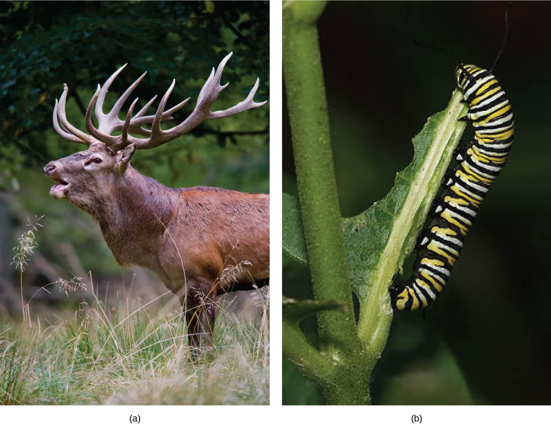
Herbivores are animals whose primary food source is plant-based. Examples of herbivores include vertebrates like deer, koalas, and some bird species, as well as invertebrates such as crickets and caterpillars, shown below. These animals have evolved digestive systems capable of handling large amounts of plant material. Herbivores can be further classified into frugivores (fruit-eaters), granivores (seed eaters), nectivores (nectar feeders), and folivores (leaf eaters).
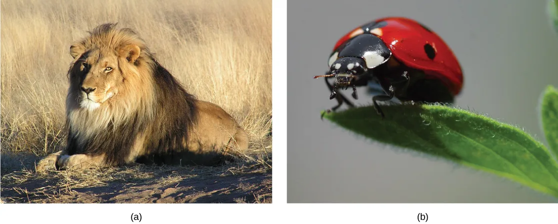
Carnivores are animals that eat other animals. The word carnivore is derived from Latin and literally means “meat eater.” Wild cats such as lions and tigers are examples of vertebrate carnivores, as are snakes and sharks, while invertebrate carnivores include sea stars, spiders, and ladybugs, shown below. Obligate carnivores are those that rely entirely on animal flesh to obtain their nutrients; examples of obligate carnivores are members of the cat family, such as lions and cheetahs. Facultative carnivores are those that also eat non-animal food in addition to animal food. Note that there is no clear line that differentiates facultative carnivores from omnivores; dogs would be considered facultative carnivores.
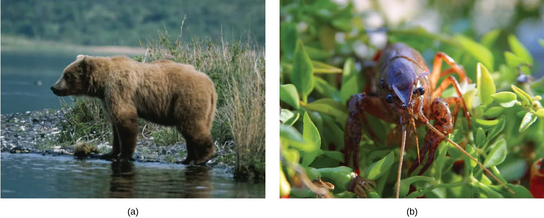
Omnivores are animals that eat both plant- and animal-derived food. In Latin, omnivore means to eat everything. Humans, bears, and chickens are examples of vertebrate omnivores; invertebrate omnivores include cockroaches and crayfish, shown below.
Invertebrate Digestive Systems
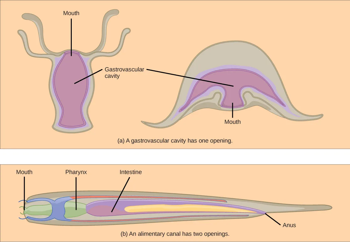
Animals have evolved different types of digestive systems to aid in the digestion of the different foods they consume. The simplest example is that of a gastrovascular cavity and is found in organisms with only one opening for digestion. Platyhelminthes (flatworms), Ctenophora (comb jellies), and Cnidaria (coral, jelly fish, and sea anemones) use this type of digestion. Gastrovascular cavities, shown below, are typically a blind tube or cavity with only one opening, the “mouth”, which also serves as an “anus”. Ingested material enters the mouth and passes through a hollow, tubular cavity. Cells within the cavity secrete digestive enzymes that break down the food. The food particles are engulfed by the cells lining the gastrovascular cavity.
The alimentary canal, also shown below, is a more advanced system: it consists of one tube with a mouth at one end and an anus at the other. Earthworms are an example of an animal with an alimentary canal. Once the food is ingested through the mouth, it passes through the esophagus and is stored in an organ called the crop; then it passes into the gizzard where it is churned and digested. From the gizzard, the food passes through the intestine, the nutrients are absorbed, and the waste is eliminated as feces, called castings, through the anus.
Extended description
The figure has two panels. (a) On the left, a vase-shaped hydra stands upright: a leader line labeled ‘Mouth’ points to the opening at the top center, ringed by tentacles, and a second leader line labeled ‘Gastrovascular cavity’ points to the shaded interior chamber that fills most of the body. Beside it, a bell-shaped jellyfish medusa lies on its side with tentacles trailing from the rim of the bell; a leader line labeled ‘Mouth’ points to the opening at the center underside of the bell, which opens into the same shaded gastrovascular cavity — no separate cavity label is drawn for the jellyfish since it shares the panel’s single cavity shading. (b) On the right, a long, tube-shaped nematode is drawn front to back with four leader lines in this order: ‘Mouth’ at the wide front end, ‘Pharynx’ just behind it, ‘Intestine’ along the long shaded middle section, and ‘Anus’ near the tapered tail end.
Vertebrate Digestive Systems
Vertebrates have evolved more complex digestive systems to adapt to their dietary needs. Some animals have a single stomach, while others have multi-chambered stomachs. Birds have developed a digestive system adapted to eating unmasticated food.
Monogastric: Single-chambered Stomach
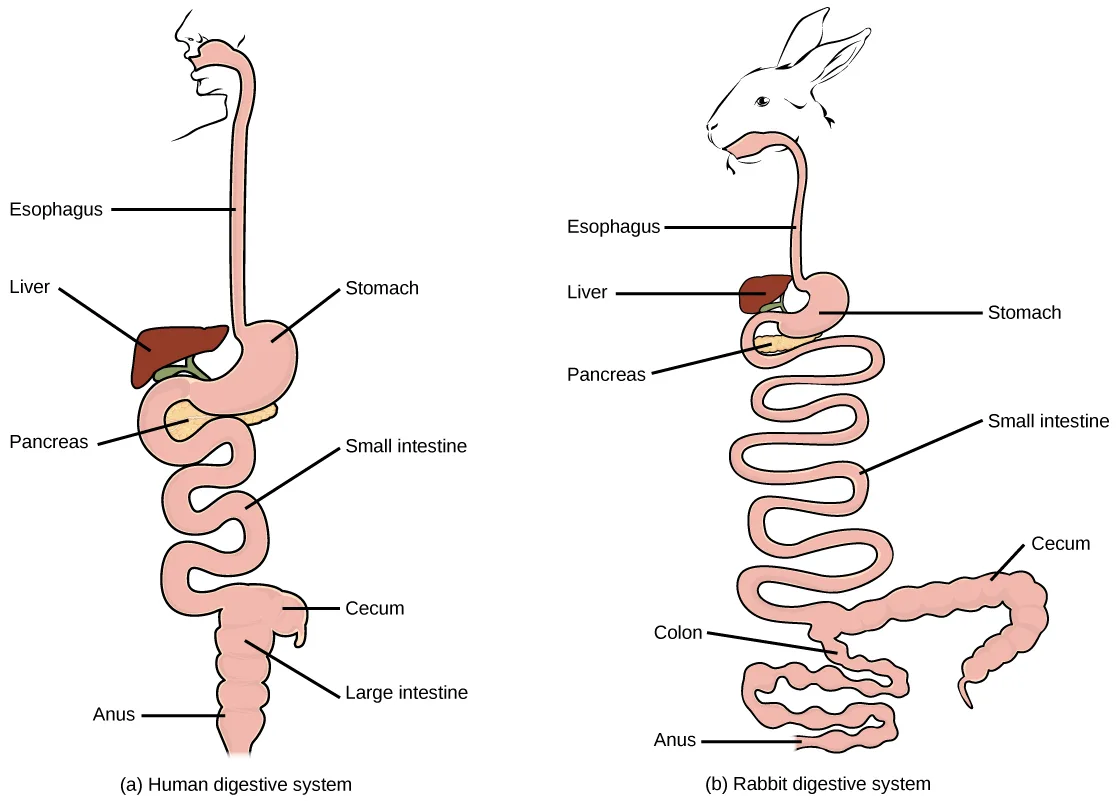
As the word monogastric suggests, this type of digestive system consists of one (“mono”) stomach chamber (“gastric”). Humans and many animals have a monogastric digestive system, shown below. The process of digestion begins with the mouth and the intake of food. The teeth play an important role in masticating (chewing) or physically breaking down food into smaller particles. The enzymes present in saliva also begin to chemically break down food. The esophagus is a long tube that connects the mouth to the stomach. Using peristalsis, or wave-like smooth muscle contractions, the muscles of the esophagus push the food towards the stomach. In order to speed up the actions of enzymes in the stomach, the stomach is an extremely acidic environment, with a pH between 1.5 and 2.5. The gastric juices, which include enzymes in the stomach, act on the food particles and continue the process of digestion. Further breakdown of food takes place in the small intestine where enzymes produced by the liver, the small intestine, and the pancreas continue the process of digestion. The nutrients are absorbed into the bloodstream across the epithelial cells lining the walls of the small intestines. The waste material travels on to the large intestine where water is absorbed and the drier waste material is compacted into feces; it is stored until it is excreted through the rectum.
Extended description
Two side-view drawings, each showing a tube-like digestive tract with leader-line labels from the mouth down to the anus. (a) Human digestive system, eight labels top to bottom: Esophagus (the straight tube from the mouth), Liver (a dark red wedge sitting against the stomach), Stomach (the kidney-shaped pouch below the esophagus), Pancreas (a pale organ tucked beneath the stomach), Small intestine (a tightly coiled tube below the stomach), Cecum (a small pouch where the small intestine meets the large intestine), Large intestine (the wider tube framing the coils), and Anus (the tract’s opening at the bottom). (b) Rabbit digestive system, the same eight organs in the same top-to-bottom order — Esophagus, Liver, Stomach, Pancreas, Small intestine, Cecum, Colon (labeled in place of ’large intestine’), and Anus — but drawn with a much longer, more tightly wound small intestine and a noticeably larger, coiled cecum before the colon.
Avian
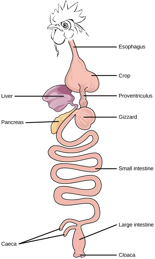
Birds face special challenges when it comes to obtaining nutrition from food. They do not have teeth and so their digestive system, shown below, must be able to process un-masticated food. Birds have evolved a variety of beak types that reflect the vast variety in their diet, ranging from seeds and insects to fruits and nuts. Because most birds fly, their metabolic rates are high in order to efficiently process food and keep their body weight low. The stomach of birds has two chambers: the proventriculus, where gastric juices are produced to digest the food before it enters the stomach, and the gizzard, where the food is stored, soaked, and mechanically ground. The undigested material forms food pellets that are sometimes regurgitated. Most of the chemical digestion and absorption happens in the intestine and the waste is excreted through the cloaca.
Extended description
A rooster’s head and neck lead down into a labeled tube with ten labels top to bottom: Esophagus (the tube from the beak), Crop (an upside-down heart-shaped pouch bulging from the esophagus), Liver (a purple organ beside the crop, on the left), Proventriculus (a narrow tube below the crop), Pancreas (a pale yellow organ beside the gizzard, on the left), Gizzard (a round chamber below the proventriculus), Small intestine (a long, tightly coiled tube below the gizzard), and, at the bottom, Large intestine (the short final segment) with Caeca (two thin blind tubes branching off where the small and large intestines meet) and Cloaca (the tract’s single opening at the very bottom).
Evolution Connection. Avian Adaptations
Birds have a highly efficient, simplified digestive system. Recent fossil evidence has shown that the evolutionary divergence of birds from other land animals was characterized by streamlining and simplifying the digestive system. Unlike many other animals, birds do not have teeth to chew their food. In place of lips, they have sharp pointy beaks. The horny beak, lack of jaws, and the smaller tongue of the birds can be traced back to their dinosaur ancestors. The emergence of these changes seems to coincide with the inclusion of seeds in the bird diet. Seed-eating birds have beaks that are shaped for grabbing seeds and the two-compartment stomach allows for delegation of tasks. Since birds need to remain light in order to fly, their metabolic rates are very high, which means they digest their food very quickly and need to eat often. Contrast this with the ruminants, where the digestion of plant matter takes a very long time.
Ruminants
Ruminants are mainly herbivores like cows, sheep, and goats, whose entire diet consists of eating large amounts of roughage or fiber. They have evolved digestive systems that help them digest vast amounts of cellulose. An interesting feature of the ruminants’ mouth is that they do not have upper incisor teeth. They use their lower teeth, tongue and lips to tear and chew their food. From the mouth, the food travels to the esophagus and on to the stomach.
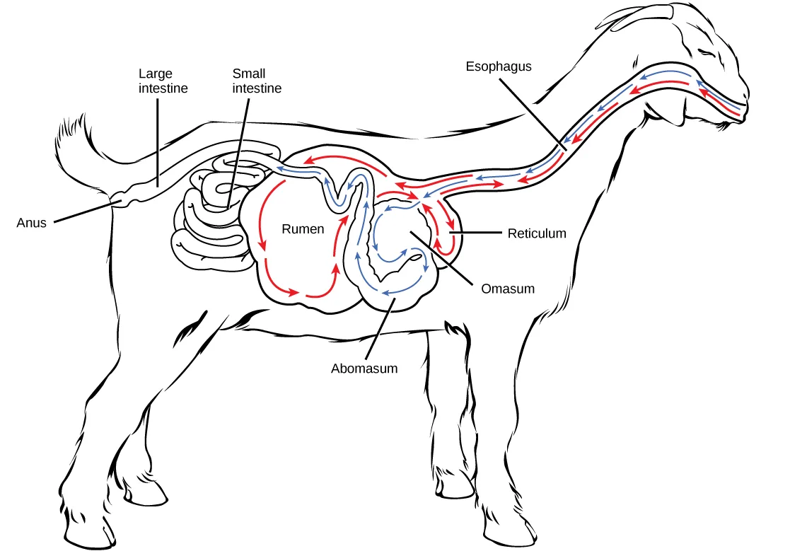
To help digest the large amount of plant material, the stomach of the ruminants is a multi-chambered organ, shown below. The four compartments of the stomach are called the rumen, reticulum, omasum, and abomasum. These chambers contain many microbes that breakdown cellulose and ferment ingested food. The abomasum is the “true” stomach and is the equivalent of the monogastric stomach chamber where gastric juices are secreted. The four-compartment gastric chamber provides larger space and the microbial support necessary to digest plant material in ruminants. The fermentation process produces large amounts of gas in the stomach chamber, which must be eliminated. As in other animals, the small intestine plays an important role in nutrient absorption, and the large intestine helps in the elimination of waste.
Extended description
A goat stands in profile, head and neck at the right. Two parallel tracks of small arrows, one red and one blue, run the length of the esophagus between the mouth and the stomach chambers, both pointing down the neck toward the body; near the base of the neck a single red arrow points back up the esophagus toward the mouth. The esophagus leads into the Rumen, the largest chamber (labeled, center-left), where red arrows trace a closed loop around its inner wall — arriving from the Reticulum side, circling along the top and around to the bottom, and returning to the Reticulum side rather than exiting anywhere else. The Reticulum (labeled, a smaller rounded pouch just below and to the right of the rumen) carries both red and blue arrows, connecting the rumen’s red loop up to the esophagus and passing blue arrows onward. From the reticulum, blue arrows spiral inward through the Omasum (labeled, a coiled pouch beside the reticulum) and continue down into the Abomasum (labeled, the pouch at the bottom of the chamber cluster), which the caption identifies as the ’true’ stomach. Three further leader lines, unconnected to the arrow tracks, label the Small intestine and Large intestine (coiled tubes at the goat’s rear flank) and the Anus (where the large intestine ends, near the tail).
Pseudo-ruminants
Some animals, such as camels and alpacas, are pseudo-ruminants. They eat a lot of plant material and roughage. Digesting plant material is not easy because plant cell walls contain the polymeric sugar molecule cellulose. The digestive enzymes of these animals cannot breakdown cellulose, but microorganisms present in the digestive system can. Therefore, the digestive system must be able to handle large amounts of roughage and break down the cellulose. Pseudo-ruminants have a three-chamber stomach in the digestive system. However, their cecum—a pouched organ at the beginning of the large intestine containing many microorganisms that are necessary for the digestion of plant materials—is large and is the site where the roughage is fermented and digested. These animals do not have a rumen but have an omasum, abomasum, and reticulum.
Parts of the Digestive System
The vertebrate digestive system is designed to facilitate the transformation of food matter into the nutrient components that sustain organisms.
Oral Cavity
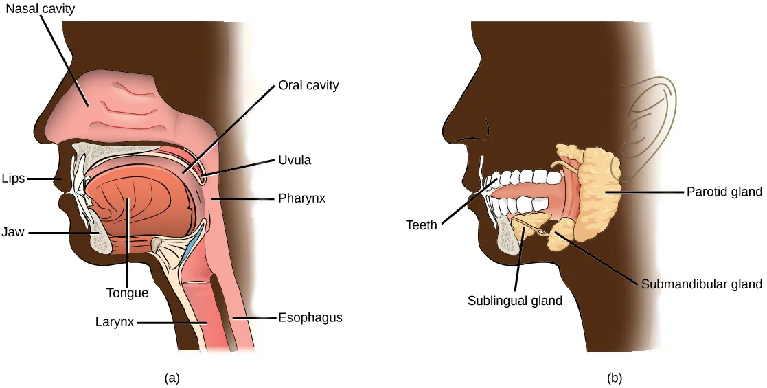
The oral cavity, or mouth, is the point of entry of food into the digestive system, illustrated below. The food consumed is broken into smaller particles by mastication, the chewing action of the teeth. All mammals have teeth and can chew their food.
The extensive chemical process of digestion begins in the mouth. As food is being chewed, saliva, produced by the salivary glands, mixes with the food. Saliva is a watery substance produced in the mouths of many animals. There are three major glands that secrete saliva—the parotid, the submandibular, and the sublingual. Saliva contains mucus that moistens food and buffers the pH of the food. Saliva also contains immunoglobulins and lysozymes, which have antibacterial action to reduce tooth decay by inhibiting growth of some bacteria. Saliva also contains an enzyme called salivary amylase that begins the process of converting starches in the food into a disaccharide called maltose. Another enzyme called lipase is produced by the cells in the tongue. Lipases are a class of enzymes that can breakdown triglycerides. The lingual lipase begins the breakdown of fat components in the food. The chewing and wetting action provided by the teeth and saliva prepare the food into a mass called the bolus for swallowing. The tongue helps in swallowing—moving the bolus from the mouth into the pharynx. The pharynx opens to two passageways: the trachea, which leads to the lungs, and the esophagus, which leads to the stomach. The trachea has an opening called the glottis, which is covered by a cartilaginous flap called the epiglottis. When swallowing, the epiglottis closes the glottis and food passes into the esophagus and not the trachea. This arrangement allows food to be kept out of the trachea.
Extended description
Two cutaway side-view drawings of a human head in profile. (a) Nine leader lines: Nasal cavity (the pink chamber above the mouth), Oral cavity (the space inside the open mouth), Uvula (the small flap hanging at the back of the mouth), Pharynx (the passage behind the uvula), Lips and Jaw (the mouth’s outer structures), Tongue (the muscle filling the lower mouth), Larynx and Esophagus (the two tubes at the base of the throat, larynx toward the front, esophagus toward the back). (b) Four leader lines: Teeth (the row inside the open mouth), Parotid gland (a large gland behind the jaw, toward the ear), Submandibular gland (a smaller gland beneath the jaw), and Sublingual gland (a gland further forward, beneath the tongue).
Esophagus
The esophagus is a tubular organ that connects the mouth to the stomach. The chewed and softened food passes through the esophagus after being swallowed. The smooth muscles of the esophagus undergo a series of wave like movements called peristalsis that push the food toward the stomach, shown below. The peristalsis wave is unidirectional—it moves food from the mouth to the stomach, and reverse movement is not possible. The peristaltic movement of the esophagus is an involuntary reflex; it takes place in response to the act of swallowing.
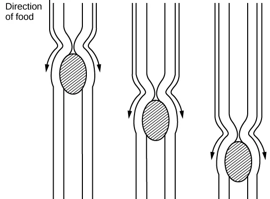
Extended description
Three near-identical drawings sit side by side, each a vertical tube formed by two parallel walls. In each one a hatched oval (the food bolus) sits inside the tube, with the walls pinched inward directly behind it and two small arrows pointing down and outward from that pinch point, showing the constriction pressing the bolus onward. A ‘Direction of food’ label with a downward arrow sits beside the leftmost drawing. Reading left to right, the pinch point sits progressively lower in the tube in each successive drawing, showing the same wave of constriction traveling farther down the esophagus and pushing the bolus ahead of it.
A ring-like muscle called a sphincter forms valves in the digestive system. The gastro-esophageal sphincter is located at the stomach end of the esophagus. In response to swallowing and the pressure exerted by the bolus of food, this sphincter opens, and the bolus enters the stomach. When there is no swallowing action, this sphincter is shut and prevents the contents of the stomach from traveling up the esophagus. Many animals have a true sphincter; however, in humans, there is no true sphincter, but the esophagus remains closed when there is no swallowing action. Acid reflux or “heartburn” occurs when the acidic digestive juices escape into the esophagus.
Stomach
A large part of digestion occurs in the stomach, shown below. The stomach is a saclike organ that secretes gastric digestive juices. The pH in the stomach is between 1.5 and 2.5. This highly acidic environment is required for the chemical breakdown of food and the extraction of nutrients. When empty, the stomach is a rather small organ; however, it can expand to up to 20 times its resting size when filled with food. This characteristic is particularly useful for animals that need to eat when food is available.
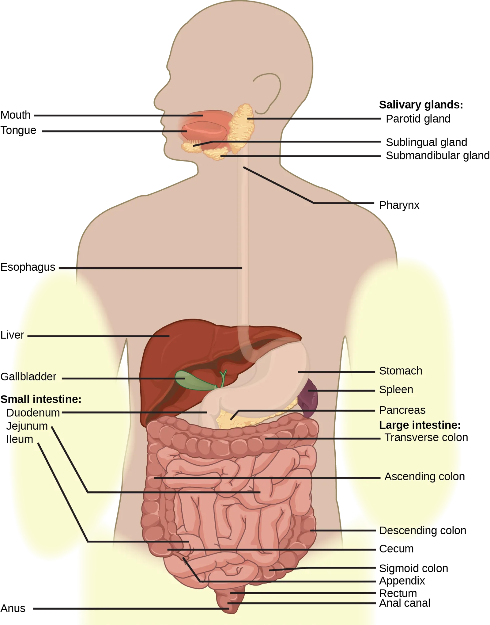
Extended description
A body outline faces forward, labeled from top to bottom with leader lines alternating left and right. At the top: Mouth and Tongue (left) beside Salivary glands — Parotid gland, Sublingual gland, and Submandibular gland (right); below that, Pharynx (right). Down the neck: Esophagus (left). In the torso, left side: Liver, Gallbladder, then Small intestine — Duodenum, Jejunum, Ileum. Torso, right side: Stomach, Spleen, Pancreas, then Large intestine — Transverse colon, Ascending colon, Descending colon, Cecum, Sigmoid colon, Appendix, Rectum, Anal canal. At the bottom left: Anus. In all, 24 labeled structures trace the tract from mouth to anus.
Which of the following statements about the digestive system is false?
Follow the tract in the figure above from the stomach onward — which two organs come right after each other, and in what order?The stomach is also the major site for protein digestion in animals other than ruminants. Protein digestion is mediated by an enzyme called pepsin in the stomach chamber. Pepsin is secreted by the chief cells in the stomach in an inactive form called pepsinogen. Pepsin breaks peptide bonds and cleaves proteins into smaller polypeptides; it also helps activate more pepsinogen, starting a positive feedback mechanism that generates more pepsin. Another cell type—parietal cells—secrete hydrogen and chloride ions, which combine in the lumen to form hydrochloric acid, the primary acidic component of the stomach juices. Hydrochloric acid helps to convert the inactive pepsinogen to pepsin. The highly acidic environment also kills many microorganisms in the food and, combined with the action of the enzyme pepsin, results in the hydrolysis of protein in the food. Chemical digestion is facilitated by the churning action of the stomach. Contraction and relaxation of smooth muscles mixes the stomach contents about every 20 minutes. The partially digested food and gastric juice mixture is called chyme. Chyme passes from the stomach to the small intestine. Further protein digestion takes place in the small intestine. Gastric emptying occurs within two to six hours after a meal. Only a small amount of chyme is released into the small intestine at a time. The movement of chyme from the stomach into the small intestine is regulated by the pyloric sphincter.
When digesting protein and some fats, the stomach lining must be protected from getting digested by pepsin. There are two points to consider when describing how the stomach lining is protected. First, as previously mentioned, the enzyme pepsin is synthesized in the inactive form. This protects the chief cells, because pepsinogen does not have the same enzyme functionality of pepsin. Second, the stomach has a thick mucus lining that protects the underlying tissue from the action of the digestive juices. When this mucus lining is ruptured, ulcers can form in the stomach. Ulcers are open wounds in or on an organ caused by bacteria (Helicobacter pylori) when the mucus lining is ruptured and fails to reform.
Small Intestine
Chyme moves from the stomach to the small intestine. The small intestine is the organ where the digestion of protein, fats, and carbohydrates is completed. The small intestine is a long tube-like organ with a highly folded surface containing finger-like projections called the villi. The apical surface of each villus has many microscopic projections called microvilli. These structures, illustrated below, are lined with epithelial cells on the luminal side and allow for the nutrients to be absorbed from the digested food and absorbed into the bloodstream on the other side. The villi and microvilli, with their many folds, increase the surface area of the intestine and increase absorption efficiency of the nutrients. Absorbed nutrients in the blood are carried into the hepatic portal vein, which leads to the liver. There, the liver regulates the distribution of nutrients to the rest of the body and removes toxic substances, including drugs, alcohol, and some pathogens.
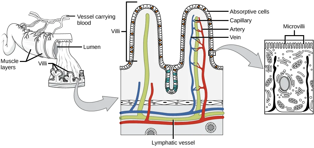
Extended description
Three panels connected left to right by two gray arrows. Left panel: a block of intestinal tissue labeled with four leader lines — Vessel carrying blood (branching vessels at the top), Lumen (the hollow opening at the cut face), Muscle layers (the outer wall of the block), and Villi (the small fingerlike projections on the inner surface). A gray arrow leads to the middle panel: an enlarged cross-section of two upside-down-U-shaped villi, each covered along its outer edge by a row of Absorptive cells (labeled at upper right); inside each villus run a red vessel labeled Capillary, a second red vessel labeled Artery, and a blue vessel labeled Vein, all bracketed together under the label Villi at the top; a green vessel at the base of the panel is labeled Lymphatic vessel. A second gray arrow leads to the right panel: a close-up cross-section of one absorptive cell, its bristly upper border labeled Microvilli, its interior showing scattered organelles and a central nucleus.
Which of the following statements about the small intestine is false?
In the figure above, the blood and lymphatic vessels run up inside the villi themselves — check which structure, one level smaller, is a single cell’s own surface projections instead.The human small intestine is over 6m long and is divided into three parts: the duodenum, the jejunum, and the ileum. The “C-shaped,” fixed part of the small intestine is called the duodenum. The duodenum is separated from the stomach by the pyloric sphincter which opens to allow chyme to move from the stomach to the duodenum. In the duodenum, chyme is mixed with pancreatic juices in an alkaline solution rich in bicarbonate that neutralizes the acidity of chyme and acts as a buffer. Pancreatic juices also contain several digestive enzymes. Digestive juices from the pancreas, liver, and gallbladder, as well as from gland cells of the intestinal wall itself, enter the duodenum. Bile is produced in the liver and stored and concentrated in the gallbladder. Bile contains bile salts which emulsify lipids while the pancreas produces enzymes that catabolize starches, disaccharides, proteins, and fats. These digestive juices break down the food particles in the chyme into glucose, triglycerides, and amino acids. The bulk of chemical digestion of food takes place in the duodenum. Absorption of fatty acids also takes place in the duodenum.
The second part of the small intestine is called the jejunum. Here, hydrolysis of nutrients is continued while most of the carbohydrates and amino acids are absorbed through the intestinal lining. Some chemical digestion and the bulk of nutrient absorption occurs in the jejunum.
The ileum is the last part of the small intestine and here the bile salts and vitamins are absorbed into the bloodstream. The undigested food is sent to the colon from the ileum via peristaltic movements of the muscle. The ileum ends and the large intestine begins at the ileocecal valve. The vermiform, “worm-like,” appendix is located at the ileocecal valve. The appendix of humans secretes no enzymes and has only a minor role in immunity. (Source note: the source says “an insignificant role”; this book’s Adaptive Immune Response section lists the appendix among the mucosal immune induction sites.)
Large Intestine
The large intestine, illustrated below, reabsorbs the water from the undigested food material and processes the waste material. The human large intestine is much smaller in length compared to the small intestine but larger in diameter. It has three parts: the cecum, the colon, and the rectum. The cecum joins the ileum to the colon and is the receiving pouch for the waste matter. The colon is home to many bacteria or “intestinal flora” that aid in the digestive processes. The colon can be divided into four regions, the ascending colon, the transverse colon, the descending colon, and the sigmoid colon. The main functions of the colon are to extract the water and mineral salts from undigested food, and to store waste material. Carnivorous mammals have a shorter large intestine compared to herbivorous mammals due to their diet.
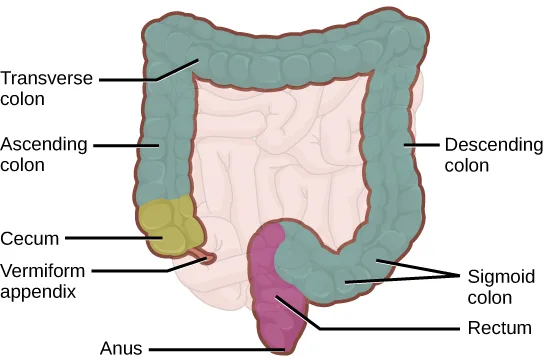
Extended description
The large intestine is drawn as a squared-off loop with eight leader-line labels. On the left, from bottom to top: Vermiform appendix (a thin projection hanging off the cecum), Cecum (a small rounded pouch above it), and Ascending colon (the teal segment rising from the cecum). Across the top: Transverse colon (the teal segment running left to right). On the right, from top to bottom: Descending colon (the teal segment descending), then Sigmoid colon (the curving pink segment at the bottom), Rectum (the pink segment below it), and Anus (the tract’s opening at the very bottom, labeled at lower left).
Rectum and Anus
The rectum is the terminal end of the large intestine. The primary role of the rectum is to store the feces until defecation. The feces are propelled using peristaltic movements during elimination. The anus is an opening at the far-end of the digestive tract and is the exit point for the waste material. Two sphincters between the rectum and anus control elimination: the inner sphincter is involuntary and the outer sphincter is voluntary.
Accessory Organs
The organs discussed above are the organs of the digestive tract through which food passes. Accessory organs are organs that add secretions (enzymes) that catabolize food into nutrients. Accessory organs include salivary glands, the liver, the pancreas, and the gallbladder. The liver, pancreas, and gallbladder are regulated by hormones in response to the food consumed.
The liver is the largest internal organ in humans and it plays a very important role in digestion of fats and detoxifying blood. The liver produces bile, a digestive juice that is required for the breakdown of fatty components of the food in the duodenum. The liver also processes the vitamins and fats and synthesizes many plasma proteins.
The pancreas is another important gland that secretes digestive juices. The chyme produced from the stomach is highly acidic in nature; the pancreatic juices contain high levels of bicarbonate, an alkali that neutralizes the acidic chyme. Additionally, the pancreatic juices contain a large variety of enzymes that are required for the digestion of protein and carbohydrates.
The gallbladder is a small organ that aids the liver by storing bile and concentrating bile salts. When chyme containing fatty acids enters the duodenum, the bile is secreted from the gallbladder into the duodenum.
Summary
Different animals have evolved different types of digestive systems specialized to meet their dietary needs. Humans and many other animals have monogastric digestive systems with a single-chambered stomach. Birds have evolved a digestive system that includes a gizzard where the food is crushed into smaller pieces. This compensates for their inability to masticate. Ruminants that consume large amounts of plant material have a multi-chambered stomach that digests roughage. Pseudo-ruminants have similar digestive processes as ruminants but do not have the four-compartment stomach. Processing food involves ingestion (eating), digestion (mechanical and enzymatic breakdown of large molecules), absorption (cellular uptake of nutrients), and elimination (removal of undigested waste as feces).
Many organs work together to digest food and absorb nutrients. The mouth is the point of ingestion and the location where both mechanical and chemical breakdown of food begins. Saliva contains an enzyme called amylase that breaks down carbohydrates. The food bolus travels through the esophagus by peristaltic movements to the stomach. The stomach has an extremely acidic environment. An enzyme called pepsin digests protein in the stomach. Further digestion and absorption take place in the small intestine. The large intestine reabsorbs water from the undigested food and stores waste until elimination.
Key terms
- alimentary canal — tubular digestive system with a mouth and anus.
- anus — exit point for waste material.
- bile — digestive juice produced by the liver; important for digestion of lipids.
- bolus — mass of food resulting from chewing action and wetting by saliva.
- carnivore — animal that consumes animal flesh.
- chyme — mixture of partially digested food and stomach juices.
- duodenum — first part of the small intestine where a large part of digestion of carbohydrates and fats occurs.
- esophagus — tubular organ that connects the mouth to the stomach.
- gallbladder — organ that stores and concentrates bile.
- gastrovascular cavity — digestive system consisting of a single opening.
- gizzard — muscular organ that grinds food.
- herbivore — animal that consumes a strictly plant diet.
- ileum — last part of the small intestine; connects the small intestine to the large intestine; important for absorption of B-12.
- jejunum — second part of the small intestine.
- large intestine — digestive system organ that reabsorbs water from undigested material and processes waste matter.
- lipase — enzyme that chemically breaks down lipids.
- liver — organ that produces bile for digestion and processes vitamins and lipids.
- monogastric — digestive system that consists of a single-chambered stomach.
- omnivore — animal that consumes both plants and animals.
- pancreas — gland that secretes digestive juices.
- pepsin — enzyme found in the stomach whose main role is protein digestion.
- pepsinogen — inactive form of pepsin.
- peristalsis — wave-like movements of muscle tissue.
- proventriculus — glandular part of a bird’s stomach.
- rectum — area of the body where feces is stored until elimination.
- roughage — component of food that is low in energy and high in fiber.
- ruminant — animal with a stomach divided into four compartments.
- salivary amylase — enzyme found in saliva, which converts carbohydrates to maltose.
- small intestine — organ where digestion of protein, fats, and carbohydrates is completed.
- sphincter — band of muscle that controls movement of materials throughout the digestive tract.
- stomach — saclike organ containing acidic digestive juices.
- villi — folds on the inner surface of the small intestine whose role is to increase absorption area.
Practice
Explain the processes of digestion and absorption
The acidic nature of chyme is neutralized by ________.
Look at what the pancreatic juices are rich in when they mix with chyme in the duodenum — an alkaline salt, not a strong industrial base or an acid.Explain how the villi and microvilli aid in absorption.
Show model answer
Did your answer mention:
The mixture of partially digested food and stomach juices is called ________.
It forms in a saclike organ whose contents have a pH between 1.5 and 2.5, before moving on to the small intestine.Compare and contrast different types of digestive systems
Which of the following is a pseudo-ruminant?
The section names two example animals as pseudo-ruminants, ones with a three-chambered stomach and a large fermenting cecum; rule out the true ruminant, the monogastric omnivore, and the bird.Which of the following statements is untrue?
The section explains why birds need to eat often and digest quickly — check whether that reason is about covering distance in flight or about something else flight requires.A scientist dissects a new species of animal. If the animal’s digestive system has a single stomach with an extended small intestine, to which animal could the dissected specimen be closely related?
Look at which animal’s monogastric system the section describes as having an enlarged small intestine and cecum to digest more plant material — it is neither the carnivore nor the two animals without a true single stomach chamber.How does the polygastric digestive system aid in digesting roughage?
Show model answer
Did your answer mention:
An animal with a stomach divided into four compartments is called a ________.
Cows, sheep, and goats are the section’s examples of this animal type, whose entire diet is roughage.Explain the specialized functions of the organs involved in processing food in the body
The digestive juices from the liver are delivered to the ________.
Bile from the liver, along with pancreatic secretions, enters the first, C-shaped part of the small intestine — not the stomach or the colon.What is the role of the accessory organs in digestion?
Show model answer
Did your answer mention:
The organ that stores and concentrates bile is called the ________.
This organ sits between the liver and the duodenum, releasing its stored contents when fatty acids enter the duodenum.Describe the ways in which organs work together to digest food and absorb nutrients
How do birds digest their food in the absence of teeth?
Show model answer
Did your answer mention:
Name two components of the digestive system that perform mechanical digestion. Describe how mechanical digestion contributes to acquiring nutrients from food.
Show model answer
Did your answer mention:
Saliva contains an enzyme called ________ that breaks down carbohydrates.
The section names this same carbohydrate-digesting enzyme earlier as part of what the salivary glands secrete to begin converting starches into maltose.This section is adapted from Biology 2e, Section 34.1: Digestive Systems by Mary Ann Clark, Jung Choi, Matthew Douglas, and OpenStax, © OpenStax, licensed under CC BY-NC-SA 4.0. Access the original for free at openstax.org. Changes: figures re-encoded as WebP with a custom alt written from each image rather than reusing the source’s own alt text; seven figures re-kinded after inspection from the manifest’s file-extension “photo” guess to “diagram” — Figure_34_01_04ab, Figure_34_01_05ab, Figure_34_01_06, Figure_34_01_07, Figure_34_01_08ab, Figure_34_01_09, and Figure_34_01_12 (drawn line illustrations, not captured photographs, despite Figure_34_01_09’s own source alt beginning “Photo shows”), while Figure_B34_01_10 and Figure_34_01_11f already carried the manifest’s correct “diagram” guess; a longdesc added to the nine labeled diagrams (Figure_34_01_04ab, Figure_34_01_05ab, Figure_34_01_06, Figure_34_01_07, Figure_34_01_08ab, Figure_34_01_09, Figure_B34_01_10, Figure_34_01_11f, Figure_34_01_12) whose leader-line labels, panels, or arrow tracks are not carried by their one- or two-line captions; the two Visual Connections (the stomach’s digestive-system overview and the small intestine’s villi cross-section) rendered as their figure followed by a multiple choice, kept in the body where the module prints them; the end-of-section Review Questions and Critical Thinking Questions adapted into the closing interactive Practice block (multiple choice and self-check respectively); rubric checkpoints added to each self-check, decomposing its model answer (the source solution) into check-off clauses with no new claims; three key-term recall items (chyme, ruminant, gallbladder) added from the glossary; one cloze recall item (amylase, accepting the section’s own compound form “salivary amylase”) added from the Section Summary to round out the “organs work together” objective. the Review Question “Which of the following is a pseudo-ruminant?” has its keyed option “horse” replaced by “camel” (the three distractors kept as printed), because the section names only camels and alpacas as pseudo-ruminants and defines them by a three-chambered stomach (omasum, abomasum, reticulum) with cecal fermentation, a description a single-stomached horse does not meet — reported as a source defect. One claim is softened with a visible Source note: the appendix has a minor, not “insignificant,” role in immunity, since this book’s immunology chapter names it an induction site (erratum 440).