Digestive System Processes
By the end of this section, you will be able to:
- Describe the process of digestion
- Detail the steps involved in digestion and absorption
- Define elimination
- Explain the role of both the small and large intestines in absorption
Obtaining nutrition and energy from food is a multistep process. For true animals, the first step is ingestion, the act of taking in food. This is followed by digestion, absorption, and elimination. In the following sections, each of these steps will be discussed in detail.
Ingestion
The large molecules found in intact food cannot pass through the cell membranes. Food needs to be broken into smaller particles so that animals can harness the nutrients and organic molecules. The first step in this process is ingestion. Ingestion is the process of taking in food through the mouth. In vertebrates, the teeth, saliva, and tongue play important roles in mastication (preparing the food into bolus). While the food is being mechanically broken down, the enzymes in saliva begin to chemically process the food as well. The combined action of these processes modifies the food from large particles to a soft mass that can be swallowed and can travel the length of the esophagus.
Digestion and Absorption
Digestion is the mechanical and chemical breakdown of food into small organic fragments. It is important to break down macromolecules into smaller fragments that are of suitable size for absorption across the digestive epithelium. Large, complex molecules of proteins, polysaccharides, and lipids must be reduced to simpler particles such as simple sugar before they can be absorbed by the digestive epithelial cells. Different organs play specific roles in the digestive process. The animal diet needs carbohydrates, protein, and fat, as well as vitamins and inorganic components for nutritional balance. How each of these components is digested is discussed in the following sections.
Carbohydrates
The digestion of carbohydrates begins in the mouth. The salivary enzyme amylase begins the breakdown of food starches into maltose, a disaccharide. As the bolus of food travels through the esophagus to the stomach, no significant digestion of carbohydrates takes place. The esophagus produces no digestive enzymes but does produce mucous for lubrication. The acidic environment in the stomach stops the action of the amylase enzyme.
The next step of carbohydrate digestion takes place in the duodenum. Recall that the chyme from the stomach enters the duodenum and mixes with the digestive secretion from the pancreas, liver, and gallbladder. Pancreatic juices also contain amylase, which continues the breakdown of starch and glycogen into maltose, a disaccharide. The disaccharides are broken down into monosaccharides by enzymes called maltases, sucrases, and lactases, which are also present in the brush border of the small intestinal wall. Maltase breaks down maltose into glucose. Other disaccharides, such as sucrose and lactose are broken down by sucrase and lactase, respectively. Sucrase breaks down sucrose (or “table sugar”) into glucose and fructose, and lactase breaks down lactose (or “milk sugar”) into glucose and galactose. The monosaccharides (glucose) thus produced are absorbed and then can be used in metabolic pathways to harness energy. The monosaccharides are transported across the intestinal epithelium into the bloodstream to be transported to the different cells in the body. The steps in carbohydrate digestion are summarized in the figure and table below.
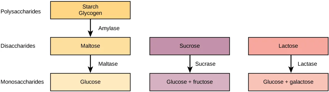
Extended description
Three columns run top to bottom through three tiers labeled at the left margin — Polysaccharides, Disaccharides, Monosaccharides. Left column: a box reading ‘Starch Glycogen’ at the Polysaccharides tier; an arrow labeled ‘Amylase’ points down to a box reading ‘Maltose’ at the Disaccharides tier; a second arrow labeled ‘Maltase’ points down to a box reading ‘Glucose’ at the Monosaccharides tier. Middle column, starting at the Disaccharides tier: a box reading ‘Sucrose’; an arrow labeled ‘Sucrase’ points down to a box reading ‘Glucose + fructose’ at the Monosaccharides tier. Right column, also starting at the Disaccharides tier: a box reading ‘Lactose’; an arrow labeled ‘Lactase’ points down to a box reading ‘Glucose + galactose’ at the Monosaccharides tier. In all, seven boxes and four downward arrows.
| Enzyme | Produced By | Site of Action | Substrate Acting On | End Products |
|---|---|---|---|---|
| Salivary amylase | Salivary glands | Mouth | Polysaccharides (Starch) | Disaccharides (maltose), oligosaccharides |
| Pancreatic amylase | Pancreas | Small intestine | Polysaccharides (starch) | Disaccharides (maltose), monosaccharides |
| Oligosaccharidases | Lining of the intestine; brush border membrane | Small intestine | Disaccharides | Monosaccharides (e.g., glucose, fructose, galactose) |
Protein
A large part of protein digestion takes place in the stomach. The enzyme pepsin plays an important role in the digestion of proteins by breaking down the intact protein to peptides, which are short chains of four to nine amino acids. In the duodenum, other enzymes—trypsin, elastase, and chymotrypsin—act on the peptides reducing them to smaller peptides. Trypsin, elastase, carboxypeptidase, and chymotrypsin are produced by the pancreas and released into the duodenum where they act on the chyme. Further breakdown of peptides to single amino acids is aided by enzymes called peptidases (those that break down peptides). Specifically, carboxypeptidase, dipeptidase, and aminopeptidase play important roles in reducing the peptides to free amino acids. The amino acids are absorbed into the bloodstream through the small intestines. The steps in protein digestion are summarized in the figure and table below.
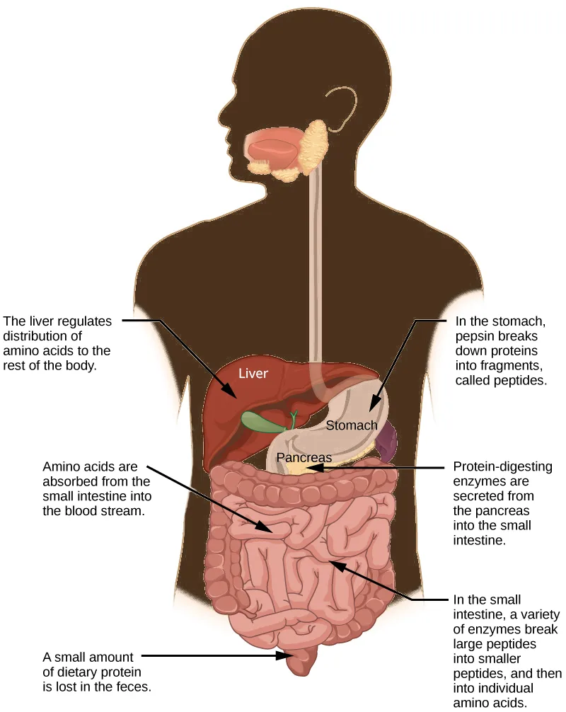
Extended description
A brown silhouette of a human head and upper torso, seen from behind with the head turned to show the profile of the mouth, contains the digestive organs drawn in color inside it. Directly labeled on the illustration are the liver (a large maroon organ at upper left of the abdomen), the stomach (a pink pouch beside it, fed by the esophagus descending from the mouth), and the pancreas (a small cream-colored gland below the stomach); the small intestine fills the lower abdomen as coiled pink loops, ending at a short pouch near the bottom. Five captioned arrows point from the surrounding text to these structures. On the left, top to bottom: ‘The liver regulates distribution of amino acids to the rest of the body,’ pointing to the liver; ‘Amino acids are absorbed from the small intestine into the blood stream,’ pointing into the coiled small intestine; and ‘A small amount of dietary protein is lost in the feces,’ pointing to the pouch at the bottom of the intestine. On the right, top to bottom: ‘In the stomach, pepsin breaks down proteins into fragments, called peptides,’ pointing to the stomach; and ‘Protein-digesting enzymes are secreted from the pancreas into the small intestine,’ pointing from the pancreas into the coiled intestine.
| Enzyme | Produced By | Site of Action | Substrate Acting On | End Products |
|---|---|---|---|---|
| Pepsin | Stomach chief cells | Stomach | Proteins | Peptides |
| Trypsin, Elastase, Chymotrypsin | Pancreas | Small intestine | Peptides | Smaller Peptides |
| Carboxypeptidase | Pancreas | Small intestine | Peptides | Amino acids and peptides |
| Aminopeptidase, Dipeptidase | Lining of intestine | Small intestine | Peptides | Amino acids |
Lipids
Lipid digestion begins in the stomach with the aid of lingual lipase and gastric lipase. However, the bulk of lipid digestion occurs in the small intestine due to pancreatic lipase. When chyme enters the duodenum, the hormonal responses trigger the release of bile, which is produced in the liver and stored in the gallbladder. Bile aids in the digestion of lipids, primarily triglycerides by emulsification. Emulsification is a process in which large lipid globules are broken down into several small lipid globules. These small globules are more widely distributed in the chyme rather than forming large aggregates. Lipids are hydrophobic substances: in the presence of water, they will aggregate to form globules to minimize exposure to water. Bile contains bile salts, which are amphipathic, meaning they contain hydrophobic and hydrophilic parts. Thus, the bile salts hydrophilic side can interface with water on one side and the hydrophobic side interfaces with lipids on the other. By doing so, bile salts emulsify large lipid globules into small lipid globules.
Why is emulsification important for digestion of lipids? Pancreatic juices contain enzymes called lipases (enzymes that breakdown lipids). If the lipid in the chyme aggregates into large globules, very little surface area of the lipids is available for the lipases to act on, leaving lipid digestion incomplete. By forming an emulsion, bile salts increase the available surface area of the lipids manyfold. The pancreatic lipases can then act on the lipids more efficiently and digest them, as detailed in the figure below. Lipases break down the lipids into fatty acids and glycerides. These molecules can pass through the plasma membrane of the cell and enter the epithelial cells of the intestinal lining. The bile salts surround long-chain fatty acids and monoglycerides forming tiny spheres called micelles. The micelles move into the brush border of the small intestine absorptive cells where the long-chain fatty acids and monoglycerides diffuse out of the micelles into the absorptive cells leaving the micelles behind in the chyme. The long-chain fatty acids and monoglycerides recombine in the absorptive cells to form triglycerides, which aggregate into globules and become coated with proteins. These large spheres are called chylomicrons. Chylomicrons contain triglycerides, cholesterol, and other lipids and have proteins on their surface. The surface is also composed of the hydrophilic phosphate “heads” of phospholipids. Together, they enable the chylomicron to move in an aqueous environment without exposing the lipids to water. Chylomicrons leave the absorptive cells via exocytosis. Chylomicrons enter the lymphatic vessels, and then enter the blood in the subclavian vein.
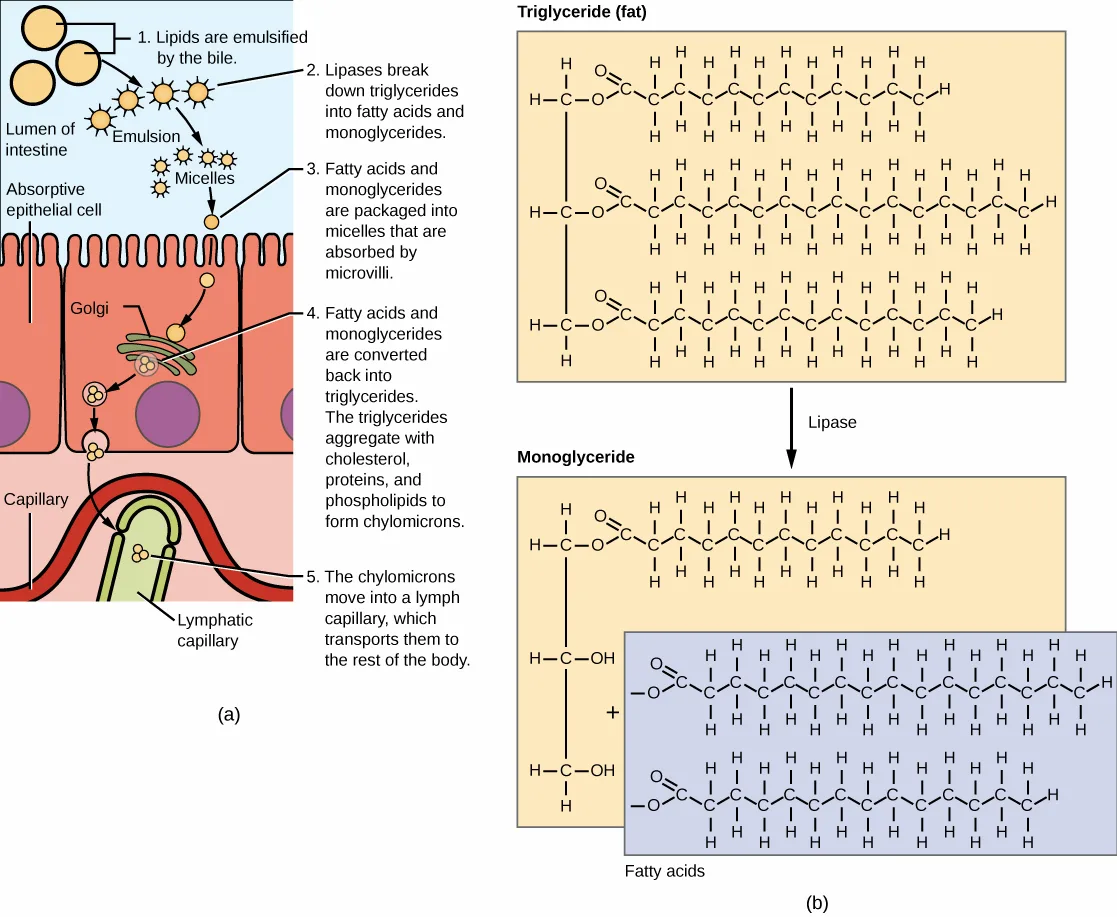
Extended description
Two diagrams sit side by side, labeled (a) and (b). Panel (a) is a cutaway of the intestinal wall, labeled from top to bottom: the ‘Lumen of intestine’ at upper left, a row of ‘Absorptive epithelial cell’ cells with microvilli lining the lumen below it, and a ‘Capillary’ and ‘Lymphatic capillary’ inside the tissue beneath the cells. Five numbered captions trace the process left to right and top to bottom: ‘1. Lipids are emulsified by the bile,’ showing droplets breaking into an ‘Emulsion’ of smaller droplets in the lumen; ‘2. Lipases break down triglycerides into fatty acids and monoglycerides’; ‘3. Fatty acids and monoglycerides are packaged into micelles that are absorbed by microvilli,’ showing small circles labeled ‘Micelles’ at the cell surface; ‘4. Fatty acids and monoglycerides are converted back into triglycerides. The triglycerides aggregate with cholesterol, proteins, and phospholipids to form chylomicrons,’ shown passing through a ‘Golgi’ structure inside the epithelial cell; and ‘5. The chylomicrons move into a lymph capillary, which transports them to the rest of the body,’ shown entering the labeled lymphatic capillary beside the capillary. Panel (b) is a chemical structure diagram. At top, a structure labeled ‘Triglyceride (fat)’ shows a three-carbon glycerol backbone with three long hydrocarbon fatty-acid chains, each attached through an ester linkage. An arrow labeled ‘Lipase’ points down to two product structures: ‘Monoglyceride,’ the same glycerol backbone with only one fatty-acid chain remaining and two free hydroxyl groups where the other two chains were, and, joined to it by a plus sign, two separate ‘Fatty acids’ chains released from the backbone.
Vitamins
Vitamins can be either water-soluble or lipid-soluble. Fat soluble vitamins are absorbed in the same manner as lipids. It is important to consume some amount of dietary lipid to aid the absorption of lipid-soluble vitamins. Water-soluble vitamins can be directly absorbed into the bloodstream from the intestine.
Link to Learning
This overview of the digestion of protein, fat, and carbohydrates walks through each nutrient’s breakdown pathway.
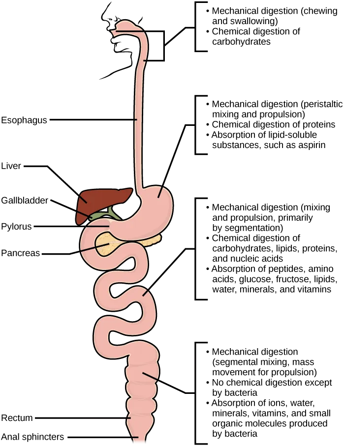
Extended description
The drawing runs top to bottom along a single tube representing the digestive tract, redrawn schematically without other body structures. At the top, an outline of a head in profile shows the mouth and jaw; a tube (the esophagus, labeled at its left) descends from it into a pouch-shaped stomach. The pylorus is labeled where the stomach narrows into the small intestine; the liver and gallbladder are labeled beside the stomach, and the pancreas is labeled just below it, at the top of the small intestine. Below the stomach, the tube coils into the small intestine, then continues into a second, wider coiled tube (the large intestine), ending at the rectum and anal sphincters, both labeled at the bottom. Four bracketed callouts run down the right side, each pointing to one region of the tract: at the mouth and esophagus, ‘Mechanical digestion (chewing and swallowing)’ and ‘Chemical digestion of carbohydrates’; at the stomach, ‘Mechanical digestion (peristaltic mixing and propulsion),’ ‘Chemical digestion of proteins,’ and ‘Absorption of lipid-soluble substances, such as aspirin’; at the small intestine, ‘Mechanical digestion (mixing and propulsion, primarily by segmentation),’ ‘Chemical digestion of carbohydrates, lipids, proteins, and nucleic acids,’ and ‘Absorption of peptides, amino acids, glucose, fructose, lipids, water, minerals, and vitamins’; and at the large intestine, ‘Mechanical digestion (segmental mixing, mass movement for propulsion),’ ‘No chemical digestion except by bacteria,’ and ‘Absorption of ions, water, minerals, vitamins, and small organic molecules produced by bacteria.’
Which of the following statements about digestive processes is true?
Rule out the two statements that misassign where an enzyme acts, and the statement claiming no absorption occurs before the small intestine — the diagram’s stomach panel already lists an absorption event.Elimination
The final step in digestion is the elimination of undigested food content and waste products. The undigested food material enters the colon, where most of the water is reabsorbed. Recall that the colon is also home to the microflora called “intestinal flora” that aid in the digestion process. The semi-solid waste is moved through the colon by peristaltic movements of the muscle and is stored in the rectum. As the rectum expands in response to storage of fecal matter, it triggers the neural signals required to set up the urge to eliminate. The solid waste is eliminated through the anus using peristaltic movements of the rectum.
Common Problems with Elimination
Diarrhea and constipation are some of the most common health concerns that affect digestion. Constipation is a condition where the feces are hardened because of excess water removal in the colon. In contrast, if enough water is not removed from the feces, it results in diarrhea. Many bacteria, including the ones that cause cholera, affect the proteins involved in water reabsorption in the colon and result in excessive diarrhea.
Emesis
Emesis, or vomiting, is elimination of food by forceful expulsion through the mouth. It is often in response to an irritant that affects the digestive tract, including but not limited to viruses, bacteria, emotions, sights, and food poisoning. This forceful expulsion of the food is due to the strong contractions produced by the stomach muscles. The process of emesis is regulated by the medulla.
Summary
Digestion begins with ingestion, where the food is taken in the mouth. Digestion and absorption take place in a series of steps with special enzymes playing important roles in digesting carbohydrates, proteins, and lipids. Elimination describes removal of undigested food contents and waste products from the body. While most absorption occurs in the small intestines, the large intestine is responsible for the final removal of water that remains after the absorptive process of the small intestines. The cells that line the large intestine absorb some vitamins as well as any leftover salts and water. The large intestine (colon) is also where feces is formed.
Key terms
- aminopeptidase — protease that breaks down peptides to single amino acids; secreted by the brush border of small intestine.
- carboxypeptidase — protease that breaks down peptides to single amino acids; produced by the pancreas.
- chylomicron — small lipid globule.
- chymotrypsin — pancreatic protease.
- digestion — mechanical and chemical breakdown of food into small organic fragments.
- dipeptidase — protease that breaks down peptides to single amino acids; secreted by the brush border of small intestine.
- elastase — pancreatic protease.
- ingestion — act of taking in food.
- lactase — enzyme that breaks down lactose into glucose and galactose.
- maltase — enzyme that breaks down maltose into glucose.
- sucrase — enzyme that breaks down sucrose into glucose and fructose.
- trypsin — pancreatic protease that breaks down protein.
Practice
Describe the process of digestion
The act of taking in food through the mouth is called ________.
This is the very first step in obtaining nutrition from food, before any mechanical or chemical breakdown begins.The mechanical and chemical breakdown of food into small organic fragments is called ________.
This step reduces large protein, polysaccharide, and lipid molecules into fragments small enough to cross the digestive epithelium.Which of the following lists the four steps of obtaining nutrition and energy from food in the order the section describes them?
The first step happens at the mouth; the last step removes what the body could not use.Detail the steps involved in digestion and absorption
Where does the majority of protein digestion take place?
Recall which organ pepsin acts in, breaking intact protein down into peptides.Lipases are enzymes that breakdown ________.
Recall what large globules bile salts emulsify before these enzymes can act efficiently.Explain why some dietary lipid is a necessary part of a balanced diet.
Show model answer
Did your answer mention:
Many mammals become ill if they drink milk as adults even though they could consume it as babies. What causes this digestive issue?
Show model answer
Did your answer mention:
The enzyme that breaks down maltose into glucose is called ________.
It acts on the disaccharide produced when amylase breaks down starch and glycogen.The enzyme that breaks down sucrose into glucose and fructose is called ________.
This brush-border enzyme acts on the sugar also known as table sugar.The enzyme that breaks down lactose into glucose and galactose is called ________.
This brush-border enzyme acts on the sugar also known as milk sugar.The pancreatic protease that breaks down protein and acts on peptides in the duodenum is called ________.
It is produced by the pancreas and released into the duodenum along with elastase and chymotrypsin.Define elimination
Which of the following conditions is most likely to cause constipation?
Constipation results when too much water is removed from the feces in the colon.Elimination describes removal of undigested food contents and ________ from the body.
Think about what else the body discards along with undigested food — material the rectum stores until it triggers the urge to eliminate.What triggers the urge to eliminate solid waste, and how is it finally expelled from the body?
Show model answer
Did your answer mention:
Explain the role of both the small and large intestines in absorption
The gut microbiome (the bacterial colonies in the intestines) have become a popular area of study in biomedical research. How could varying gut microbiomes impact a person’s nutrition?
Show model answer
Did your answer mention:
According to the section, which organ absorbs most of the water remaining in undigested food material?
This organ receives what remains of the food only after the small intestine has finished absorbing nutrients.A small lipid globule formed when fatty acids and monoglycerides recombine inside the intestinal absorptive cells is called a ________.
These leave the absorptive cells by exocytosis and enter the lymphatic vessels before reaching the blood.The large intestine (colon) is also where ________ is formed.
Recall what solid waste is called before it collects in the rectum for elimination.This section is adapted from Biology 2e, Section 34.3: Digestive System Processes by Mary Ann Clark, Jung Choi, Matthew Douglas, and OpenStax, © OpenStax, licensed under CC BY-NC-SA 4.0. Access the original for free at openstax.org. Changes: figures re-encoded as WebP; all four figures (Figure_34_03_01, Figure_B34_03_02, Figure_34_03_03, Figure_34_03_04) re-kinded from the manifest’s file-extension “photo” guess (three of them) or its default “diagram” guess to an explicit “diagram” after inspection — all four are drawn illustrations or flow charts, none a captured photograph; a longdesc added to all four figures, walking through the boxes, arrows, numbered steps, and labeled structures their one- or two-sentence captions do not carry; the glossary’s “carboxypeptidase” entry, printed as “secreted by the brush border of the small intestine,” corrected to “produced by the pancreas” to match the section’s own sentence (“Trypsin, elastase, carboxypeptidase, and chymotrypsin are produced by the pancreas”) and Table 34.6 (“Carboxypeptidase | Pancreas”) — reported as a source defect; the Link to Learning note rendered as a callout with its source URL kept; the note wrapping the Visual Connection rendered as its figure followed by a multiple choice, kept in the body, using the identical wording of the note and exercise copies; the end-of-section Review Questions and Critical Thinking Questions adapted into the closing interactive Practice block (multiple choice and self-check respectively); rubric checkpoints added to each self-check, decomposing its model answer (the source solution) into check-off clauses with no new claims; seven key-term recall items (ingestion, digestion, maltase, sucrase, lactase, trypsin, chylomicron) added from the glossary; two cloze recall items (“waste products,” “feces”) added from the section summary to help cover the “define elimination” and “role of the intestines in absorption” objectives, which the glossary alone does not test; one multiple choice on the section’s own stated order of the four digestive steps, and one multiple choice on which organ absorbs most of the remaining water, added locally from the page’s own sentences because those two objective groups were otherwise thin; one self-check on what triggers elimination, added locally from the page’s own sentences for the same reason.