Neurons and Glial Cells
By the end of this section, you will be able to:
- List and describe the functions of the structural components of a neuron
- List and describe the four main types of neurons
- Compare the functions of different types of glial cells
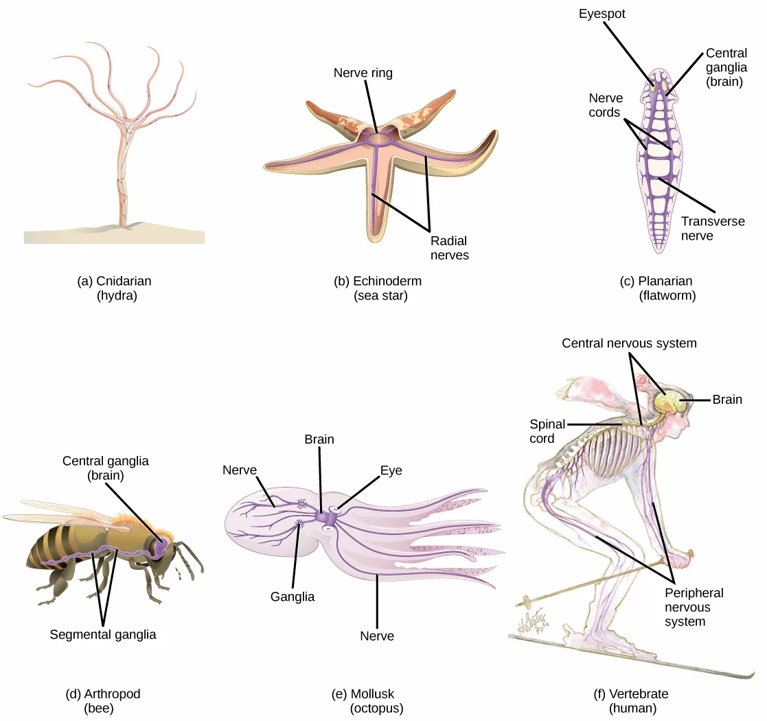
Nervous systems throughout the animal kingdom vary in structure and complexity, as illustrated by the variety of animals shown below. Some organisms, like sea sponges, lack a true nervous system. Others, like jellyfish, lack a true brain and instead have a system of separate but connected nerve cells (neurons) called a “nerve net.” Echinoderms such as sea stars have nerve cells that are bundled into fibers called nerves. Flatworms of the phylum Platyhelminthes have both a central nervous system (CNS), made up of a small “brain” and two nerve cords, and a peripheral nervous system (PNS) containing a system of nerves that extend throughout the body. The insect nervous system is more complex but also fairly decentralized. It contains a brain, ventral nerve cord, and ganglia (clusters of connected neurons). These ganglia can control movements and behaviors without input from the brain. Octopi may have the most complicated of invertebrate nervous systems—they have neurons that are organized in specialized lobes and eyes that are structurally similar to vertebrate species.
Extended description
Six drawings arranged in two rows of three, each captioned with its animal group and example organism. (a) Cnidarian (hydra): an unlabeled pink, tree-like net of nerve fibers spreading up a trunk rooted in the ground. (b) Echinoderm (sea star): two labels, ‘Nerve ring’ pointing to the ring at the body’s center and ‘Radial nerves’ pointing to the nerves running out into the arms. (c) Planarian (flatworm): four labels reading top to bottom — ‘Eyespot’ at the head, ‘Central ganglia (brain)’ just below it, ‘Nerve cords’ pointing to the two parallel cords running the body’s length, and ‘Transverse nerve’ pointing to one of the rungs connecting the two cords. (d) Arthropod (bee): two labels, ‘Central ganglia (brain)’ pointing to the head and ‘Segmental ganglia’ pointing to two swellings spaced along the ventral nerve cord beneath the body. (e) Mollusk (octopus): five labels — ‘Nerve’ at upper left pointing into the mantle, ‘Brain’ and ‘Eye’ pointing to the central body between the eyes, ‘Ganglia’ pointing to a cluster on the opposite side of the brain, and a second ‘Nerve’ label at the bottom pointing down into the arms. (f) Vertebrate (human, drawn skiing): four labels — ‘Central nervous system’ pointing to both the brain and spinal cord, ‘Brain’ pointing to the head, ‘Spinal cord’ pointing down the back, and ‘Peripheral nervous system’ pointing to nerves branching into the arms and legs.
Compared to invertebrates, vertebrate nervous systems are more complex, centralized, and specialized. While there is great diversity among different vertebrate nervous systems, they all share a basic structure: a CNS that contains a brain and spinal cord and a PNS made up of peripheral sensory and motor nerves. One interesting difference between the nervous systems of invertebrates and vertebrates is that the nerve cords of many invertebrates are located ventrally whereas the vertebrate spinal cords are located dorsally. There is debate among evolutionary biologists as to whether these different nervous system plans evolved separately or whether the invertebrate body plan arrangement somehow “flipped” during the evolution of vertebrates.
The nervous system is made up of neurons, specialized cells that can receive and transmit chemical or electrical signals, and glia, cells that provide support functions for the neurons by playing an information processing role that is complementary to neurons. A neuron can be compared to an electrical wire—it transmits a signal from one place to another. Glia can be compared to the workers at the electric company who make sure wires go to the right places, maintain the wires, and take down wires that are broken. Although glia have been compared to workers, recent evidence suggests that they also usurp some of the signaling functions of neurons.
There is great diversity in the types of neurons and glia that are present in different parts of the nervous system. There are four major types of neurons, and they share several important cellular components.
Neurons
The nervous system of the common laboratory fly, Drosophila melanogaster, contains around 100,000 neurons, the same number as a lobster. This number compares to 75 million in the mouse and 300 million in the octopus. A human brain contains around 86 billion neurons. Despite these very different numbers, the nervous systems of these animals control many of the same behaviors—from basic reflexes to more complicated behaviors like finding food and courting mates. The ability of neurons to communicate with each other as well as with other types of cells underlies all of these behaviors.
Most neurons share the same cellular components. But neurons are also highly specialized—different types of neurons have different sizes and shapes that relate to their functional roles.
Parts of a Neuron
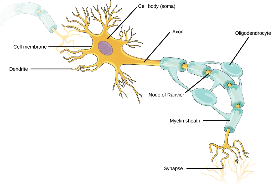
Like other cells, each neuron has a cell body (or soma) that contains a nucleus, smooth and rough endoplasmic reticulum, Golgi apparatus, mitochondria, and other cellular components. Neurons also contain unique structures, illustrated below, for receiving and sending the electrical signals that make neuronal communication possible. Dendrites are tree-like structures that extend away from the cell body to receive messages from other neurons at specialized junctions called synapses. Although some neurons do not have any dendrites, some types of neurons have multiple dendrites. Dendrites can have small protrusions called dendritic spines, which further increase surface area for possible synaptic connections.
Once a signal is received by the dendrite, it then travels passively to the cell body. The cell body contains a specialized structure, the axon hillock that integrates signals from multiple synapses and serves as a junction between the cell body and an axon. An axon is a tube-like structure that propagates the integrated signal to specialized endings called axon terminals. These terminals in turn synapse on other neurons, muscle, or target organs. Chemicals released at axon terminals allow signals to be communicated to these other cells. Neurons usually have one or two axons, but some neurons, like amacrine cells in the retina, do not contain any axons. Some axons are covered with myelin, which acts as an insulator to minimize dissipation of the electrical signal as it travels down the axon, greatly increasing the speed of conduction. This insulation is important as the axon from a human motor neuron can be as long as a meter—from the base of the spine to the toes. The myelin sheath is not actually part of the neuron. Myelin is produced by glial cells. Along the axon there are periodic gaps in the myelin sheath. These gaps are called nodes of Ranvier and are sites where the signal is “recharged” as it travels along the axon.
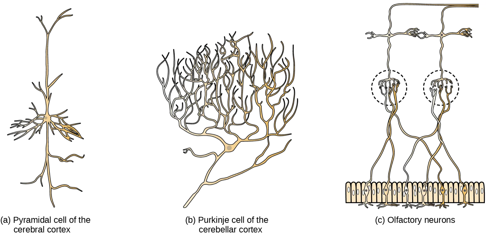
It is important to note that a single neuron does not act alone—neuronal communication depends on the connections that neurons make with one another (as well as with other cells, like muscle cells). Dendrites from a single neuron may receive synaptic contact from many other neurons. For example, dendrites from a Purkinje cell in the cerebellum are thought to receive contact from as many as 200,000 other neurons.
Extended description
A single neuron drawn in gold with eight leader-line labels. At upper left, a separate translucent, myelin-wrapped segment of another axon fades in from off-frame, feeding into this neuron’s soma. The cell body sits left of center: ‘Cell body (soma)’ points to its rounded center holding a purple nucleus, ‘Cell membrane’ points to the body’s outer edge, and ‘Dendrite’ points to one of the branching, tree-like processes radiating from its top and left sides. From the body’s right side, ‘Axon’ points to the tube leading away from the soma. The axon is wrapped in a chain of teal, bead-like myelin segments; ‘Oligodendrocyte’ points to one segment, ‘Node of Ranvier’ points to a gap between two segments, and ‘Myelin sheath’ points to a segment further along the chain. At lower right, the axon ends in branching gold terminals, and ‘Synapse’ points to these terminal branches.
Which of the following statements is false?
Trace what the teal, bead-like covering actually wraps in the figure above — the axon, or the dendrites?Types of Neurons
There are different types of neurons, and the functional role of a given neuron is intimately dependent on its structure. There is an amazing diversity of neuron shapes and sizes found in different parts of the nervous system (and across species), as illustrated by the neurons shown below.
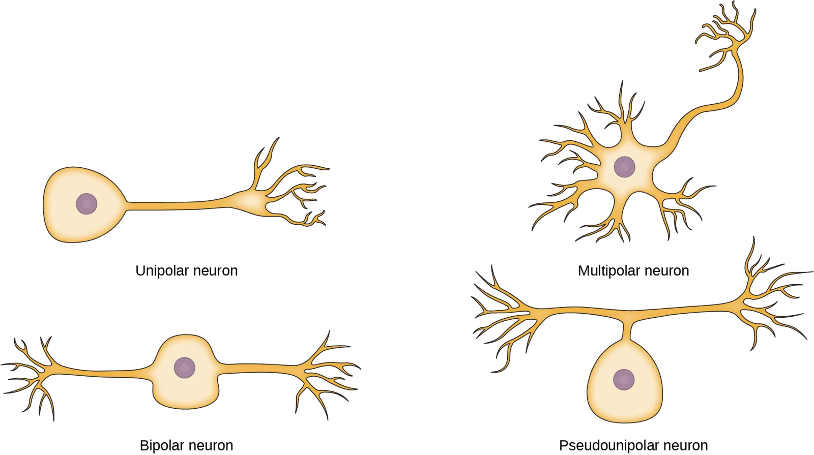
While there are many defined neuron cell subtypes, neurons are broadly divided into four basic types: unipolar, bipolar, multipolar, and pseudounipolar. The figure below illustrates these four basic neuron types. Unipolar neurons have only one structure that extends away from the soma. These neurons are not found in vertebrates but are found in insects where they stimulate muscles or glands. A bipolar neuron has one axon and one dendrite extending from the soma. An example of a bipolar neuron is a retinal bipolar cell, which receives signals from photoreceptor cells that are sensitive to light and transmits these signals to ganglion cells that carry the signal to the brain. Multipolar neurons are the most common type of neuron. Each multipolar neuron contains one axon and multiple dendrites. Multipolar neurons can be found in the central nervous system (brain and spinal cord). An example of a multipolar neuron is a Purkinje cell in the cerebellum, which has many branching dendrites but only one axon. Pseudounipolar cells share characteristics with both unipolar and bipolar cells. A pseudounipolar cell has a single process that extends from the soma, like a unipolar cell, but this process later branches into two distinct structures, like a bipolar cell. Most sensory neurons are pseudounipolar and have an axon that branches into two extensions: one connected to dendrites that receive sensory information and another that transmits this information to the spinal cord.
Everyday Connection. Neurogenesis
At one time, scientists believed that people were born with all the neurons they would ever have. Research performed during the last few decades indicates that neurogenesis, the birth of new neurons, continues into adulthood. Neurogenesis was first discovered in songbirds that produce new neurons while learning songs. For mammals, new neurons also play an important role in learning: about 1000 new neurons develop in the hippocampus (a brain structure involved in learning and memory) each day. While most of the new neurons will die, researchers found that an increase in the number of surviving new neurons in the hippocampus correlated with how well rats learned a new task. Interestingly, both exercise and some antidepressant medications also promote neurogenesis in the hippocampus. Stress has the opposite effect. While neurogenesis is quite limited compared to regeneration in other tissues, research in this area may lead to new treatments for disorders such as Alzheimer’s, stroke, and epilepsy.
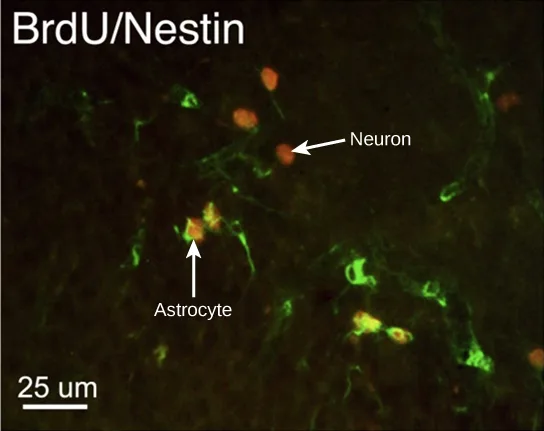
How do scientists identify new neurons? A researcher can inject a compound called bromodeoxyuridine (BrdU) into the brain of an animal. While all cells will be exposed to BrdU, BrdU will only be incorporated into the DNA of newly generated cells that are in S phase. A technique called immunohistochemistry can be used to attach a fluorescent label to the incorporated BrdU, and a researcher can use fluorescent microscopy to visualize the presence of BrdU, and thus new neurons, in brain tissue. The micrograph below shows fluorescently labeled neurons in the hippocampus of a rat.
Glia
While glia are often thought of as the supporting cast of the nervous system, the number of glial cells in the brain actually outnumbers the number of neurons by a factor of ten. Neurons would be unable to function without the vital roles that are fulfilled by these glial cells. Glia guide developing neurons to their destinations, buffer ions and chemicals that would otherwise harm neurons, and provide myelin sheaths around axons. Scientists have recently discovered that they also play a role in responding to nerve activity and modulating communication between nerve cells. When glia do not function properly, the result can be disastrous—most brain tumors are caused by mutations in glia.
Types of Glia
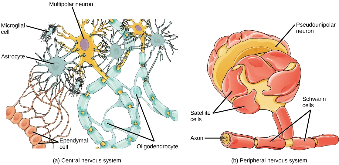
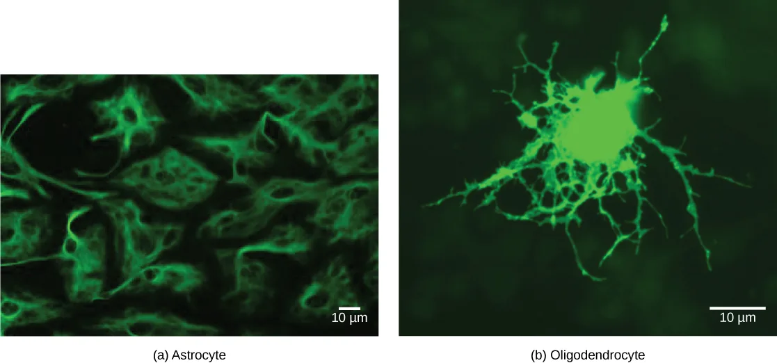
There are several different types of glia with different functions, shown below. Astrocytes, shown in the micrograph below (panel a), make contact with both capillaries and neurons in the CNS. They provide nutrients and other substances to neurons, regulate the concentrations of ions and chemicals in the extracellular fluid, and provide structural support for synapses. Astrocytes also form the blood-brain barrier—a structure that blocks entrance of toxic substances into the brain. Astrocytes, in particular, have been shown through calcium imaging experiments to become active in response to nerve activity, transmit calcium waves between astrocytes, and modulate the activity of surrounding synapses. Satellite glia provide nutrients and structural support for neurons in the PNS. Microglia scavenge and degrade dead cells and protect the brain from invading microorganisms. Oligodendrocytes, shown in the diagram below (panel b), form myelin sheaths around axons in the CNS. One axon can be myelinated by several oligodendrocytes, and one oligodendrocyte can provide myelin for multiple neurons. This is distinctive from the PNS where a single Schwann cell provides myelin for only one axon as the entire Schwann cell surrounds the axon. Radial glia serve as scaffolds for developing neurons as they migrate to their end destinations. Ependymal cells line fluid-filled ventricles of the brain and the central canal of the spinal cord. They are involved in the production of cerebrospinal fluid, which serves as a cushion for the brain, moves the fluid between the spinal cord and the brain, and is a component for the choroid plexus.
Extended description
Two panels, each a colored line drawing with leader-line labels. Panel (a), ‘Central nervous system’: a gold multipolar neuron, labeled ‘Multipolar neuron’, sits at the top center with dendrites branching upward and an axon running right and down through a chain of teal myelin segments labeled ‘Oligodendrocyte’. Bluish-gray star-shaped cells labeled ‘Astrocyte’ contact the neuron and the myelinated axon. Small spiky cells labeled ‘Microglial cell’ are scattered near the astrocytes. At lower left, a row of small round cells labeled ‘Ependymal cell’ sends long extensions toward an astrocyte. Panel (b), ‘Peripheral nervous system’: a red, pseudounipolar neuron labeled ‘Pseudounipolar neuron’ sits at the top, its cell body ringed by flattened cells labeled ‘Satellite cells’. Below it, its axon, labeled ‘Axon’, runs down and is wrapped by a chain of segments labeled ‘Schwann cells’.
Summary
The nervous system is made up of neurons and glia. Neurons are specialized cells that are capable of sending electrical as well as chemical signals. Most neurons contain dendrites, which receive these signals, and axons that send signals to other neurons or tissues. There are four main types of neurons: unipolar, bipolar, multipolar, and pseudounipolar neurons. Glia are non-neuronal cells in the nervous system that support neuronal development and signaling. There are several types of glia that serve different functions.
Key terms
- astrocyte — glial cell in the central nervous system that provide nutrients, extracellular buffering, and structural support for neurons; also makes up the blood-brain barrier.
- axon — tube-like structure that propagates a signal from a neuron’s cell body to axon terminals.
- axon hillock — electrically sensitive structure on the cell body of a neuron that integrates signals from multiple neuronal connections.
- axon terminal — structure on the end of an axon that can form a synapse with another neuron.
- dendrite — structure that extends away from the cell body to receive messages from other neurons.
- ependymal — cell that lines fluid-filled ventricles of the brain and the central canal of the spinal cord; involved in production of cerebrospinal fluid.
- glia — (also, glial cells) cells that provide support functions for neurons.
- microglia — glia that scavenge and degrade dead cells and protect the brain from invading microorganisms.
- myelin — fatty substance produced by glia that insulates axons.
- neuron — specialized cell that can receive and transmit electrical and chemical signals.
- nodes of Ranvier — gaps in the myelin sheath where the signal is recharged.
- oligodendrocyte — glial cell that myelinates central nervous system neuron axons.
- radial glia — glia that serve as scaffolds for developing neurons as they migrate to their final destinations.
- satellite glia — glial cell that provides nutrients and structural support for neurons in the peripheral nervous system.
- Schwann cell — glial cell that creates myelin sheath around a peripheral nervous system neuron axon.
- synapse — junction between two neurons where neuronal signals are communicated.
Practice
List and describe the functions of the structural components of a neuron
Neurons contain ________, which can receive signals from other neurons.
These tree-like structures branch from the cell body specifically to catch messages arriving from other neurons at synapses.How are neurons similar to other cells? How are they unique?
Show model answer
Did your answer mention:
Multiple sclerosis causes demyelination of axons in the brain and spinal cord. Why is this problematic?
Show model answer
Did your answer mention:
Many neurons have only a single axon, but many terminals at the end of the axon. How does this end structure of the axon support its function?
Show model answer
Did your answer mention:
The fatty substance produced by glia that insulates axons is called ________.
This substance forms the segmented sheath that speeds up signal conduction, produced by cells other than the neuron itself.The junction between two neurons where neuronal signals are communicated is called a ________.
Neurotransmitters cross this narrow gap between an axon terminal and the next cell’s receiving surface.List and describe the four main types of neurons
A(n) ________ neuron has one axon and one dendrite extending directly from the cell body.
This type is exemplified by a retinal cell that relays a single signal from a photoreceptor toward ganglion cells.There are four main types of neurons: unipolar, bipolar, multipolar, and ________ neurons.
This type’s single process branches into two extensions a short distance from the soma, one carrying sensory information in and the other passing it on toward the spinal cord.Which type of neuron has one axon and multiple dendrites, and is the most common neuron type found in the central nervous system?
A Purkinje cell in the cerebellum is this type’s textbook example — many branching dendrites, but only one axon.Compare the functions of different types of glial cells
Glia that provide myelin for neurons in the brain are called ________.
This CNS glial cell can wrap myelin around several different axons at once — its PNS counterpart myelinates only one.Meningitis is a viral or bacterial infection of the brain. Which cell type is the first to have its function disrupted during meningitis?
This glial cell forms the blood-brain barrier that a bloodborne infection must cross first.The glial cell that provides nutrients and structural support for neurons in the peripheral nervous system is called ________.
In the PNS, this cell type surrounds the neuron’s cell body rather than its axon.The glia that scavenge and degrade dead cells and protect the brain from invading microorganisms are called ________.
This is the brain’s resident immune cell, small and spiky with many fine projections.The glia that serve as scaffolds for developing neurons as they migrate to their final destinations are called ________.
These cells provide a physical pathway that young neurons climb along on their way to where they will function permanently.This section is adapted from Biology 2e, Section 35.1: Neurons and Glial Cells by Mary Ann Clark, Jung Choi, Matthew Douglas, and OpenStax, © OpenStax, licensed under CC BY-NC-SA 4.0. Access the original for free at openstax.org. Changes: figures re-encoded as WebP; Figure_35_01_01 and Figure_35_01_06 re-kinded from the manifest’s file-extension “photo” guess to “diagram” (a hand-drawn six-panel comparative illustration and a two-panel labeled glial-cell schematic, neither a captured photograph); a longdesc added to the animal-nervous-systems figure (Figure_35_01_01), the labeled neuron diagram (Figure_35_01_02), and the labeled glial-cell diagram (Figure_35_01_06), walking each leader-line label in reading order, since none of their meanings are carried by their one-line captions; the two Link to Learning notes rendered as callouts with descriptive link text in place of the source’s “Access multimedia content” and “This video” anchors, external URLs kept as printed; the Everyday Connection note rendered as a callout with its bold name and italic title, its figure and both paragraphs kept inside the box as printed; in-text pointers to figures (“Figure 35.2,” “Figure 35.3,” “Figure 35.4,” “Figure 35.5,” “Figure 35.6”) replaced with “shown below” or “the figure below,” since Hugo does not number figures; the note wrapping the neuron Visual Connection rendered as its figure followed by a multiple choice, kept in the body; the end-of-section Review Questions and Critical Thinking Questions adapted into the closing interactive Practice block (multiple choice and self-check respectively); rubric checkpoints added to each self-check, decomposing its model answer (the source solution) into check-off clauses with no new claims; five key-term recall items (myelin, synapse, satellite glia, microglia, radial glia) added from the glossary; one summary-derived textin cloze added to the second objective’s group (blanking “pseudounipolar” from the closing summary’s list of the four neuron types); and, because the source keys only one Review Question to the second objective, one multiple choice was written locally for that group, built strictly from the section’s own sentence describing multipolar neurons (“Multipolar neurons are the most common type of neuron. Each multipolar neuron contains one axon and multiple dendrites… can be found in the central nervous system”), its distractors the module’s own three other neuron-type names — both disclosed here and in the source ledger.