The Central Nervous System
By the end of this section, you will be able to:
- Identify the spinal cord, cerebral lobes, and other brain areas on a diagram of the brain
- Describe the basic functions of the spinal cord, cerebral lobes, and other brain areas
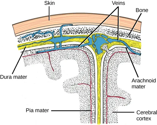
The central nervous system (CNS) is made up of the brain (a part of which is shown below) and spinal cord and is covered with three layers of protective coverings called meninges (from the Greek word for membrane). The outermost layer is the dura mater (Latin for “hard mother”). As the Latin suggests, the primary function for this thick layer is to protect the brain and spinal cord. The dura mater also contains vein-like structures that carry blood from the brain back to the heart. The middle layer is the web-like arachnoid mater. The last layer is the pia mater (Latin for “soft mother”), which directly contacts and covers the brain and spinal cord like plastic wrap. The space between the arachnoid and pia maters is filled with cerebrospinal fluid (CSF). CSF is produced by a tissue called choroid plexus in fluid-filled compartments in the CNS called ventricles. The brain floats in CSF, which acts as a cushion and shock absorber and makes the brain neutrally buoyant. CSF also functions to circulate chemical substances throughout the brain and into the spinal cord.
The entire brain contains only about 8.5 tablespoons of CSF, but CSF is constantly produced in the ventricles. This creates a problem when a ventricle is blocked—the CSF builds up and creates swelling and the brain is pushed against the skull. This swelling condition is called hydrocephalus (“water head”) and can cause seizures, cognitive problems, and even death if a shunt is not inserted to remove the fluid and pressure.
Extended description
The cross-section runs skin to cortex, top to bottom. A wavy strip labeled ‘Skin’ caps the top left, and a stippled strip labeled ‘Bone’ caps the top right, together forming the scalp and skull along the top edge. ‘Veins’ labels a branching blue network that threads through a thick yellow layer just beneath the skin and bone; that yellow layer is labeled ‘Dura mater’ at the lower left. Below it, a thin wavy band, labeled ‘Arachnoid mater’ at the lower right, crosses the middle. A red line just beneath the arachnoid mater, labeled ‘Pia mater’ at the bottom left, follows the folds of the stippled tissue below it, which is labeled ‘Cerebral cortex’ at the bottom right and dips down into a deep central fold.
Brain
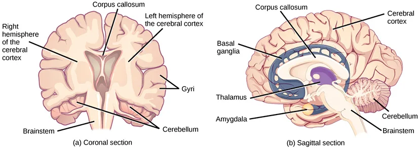
The brain is the part of the central nervous system that is contained in the cranial cavity of the skull. It includes the cerebral cortex, limbic system, basal ganglia, thalamus, hypothalamus, and cerebellum. There are three different ways that a brain can be sectioned in order to view internal structures: a sagittal section cuts the brain left to right, as shown in the sagittal section below, a coronal section cuts the brain front to back, as shown in the coronal section below, and a horizontal section cuts the brain top to bottom.
Cerebral Cortex
The outermost part of the brain is a thick piece of nervous system tissue called the cerebral cortex, which is folded into hills called gyri (singular: gyrus) and valleys called sulci (singular: sulcus). The cortex is made up of two hemispheres—right and left—which are separated by a large sulcus. A thick fiber bundle called the corpus callosum (Latin: “tough body”) connects the two hemispheres and allows information to be passed from one side to the other. Although there are some brain functions that are localized more to one hemisphere than the other, the functions of the two hemispheres are largely redundant. In fact, sometimes (very rarely) an entire hemisphere is removed to treat severe epilepsy. While patients do suffer some deficits following the surgery, they can have surprisingly few problems, especially when the surgery is performed on children who have very immature nervous systems.
Extended description
Panel (a), labeled ‘Coronal section,’ is a front-facing cross-section split down the middle. ‘Right hemisphere of the cerebral cortex’ labels the left side of the image and ‘Left hemisphere of the cerebral cortex’ labels the right side (the brain’s own left and right, as if facing the viewer); ‘Corpus callosum’ labels the pale band arching over the central gap between them; ‘Gyri’ labels the folded ridges on the outer surface of the right-hand hemisphere; ‘Brainstem’ labels the stalk descending from the center; ‘Cerebellum’ labels the wrinkled structures at the base on either side of the brainstem. Panel (b), labeled ‘Sagittal section,’ is a side view. ‘Corpus callosum’ labels the band arching over the central structures near the top; ‘Cerebral cortex’ labels the folded outer tissue above and behind it; ‘Basal ganglia’ labels a ring-shaped structure wrapping around the corpus callosum; ‘Thalamus’ labels an oval structure at the center of that ring; ‘Amygdala’ labels a small round structure below and in front of the thalamus; ‘Cerebellum’ labels the densely wrinkled structure at the back; ‘Brainstem’ labels the stalk connecting the cerebellum to the spinal cord at the bottom.
In other surgeries to treat severe epilepsy, the corpus callosum is cut instead of removing an entire hemisphere. This causes a condition called split-brain, which gives insights into unique functions of the two hemispheres. For example, when an object is presented to patients’ left visual field, they may be unable to verbally name the object (and may claim to not have seen an object at all). This is because the visual input from the left visual field crosses and enters the right hemisphere and cannot then signal to the speech center, which generally is found in the left side of the brain. Remarkably, if a split-brain patient is asked to pick up a specific object out of a group of objects with the left hand, the patient will be able to do so but will still be unable to vocally identify it.
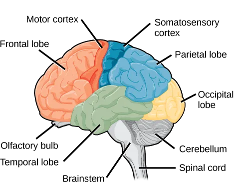
Each cortical hemisphere contains regions called lobes that are involved in different functions. Scientists use various techniques to determine what brain areas are involved in different functions: they examine patients who have had injuries or diseases that affect specific areas and see how those areas are related to functional deficits. They also conduct animal studies where they stimulate brain areas and see if there are any behavioral changes. They use a technique called transcranial magnetic stimulation (TMS) to temporarily deactivate specific parts of the cortex using strong magnets placed outside the head; and they use functional magnetic resonance imaging (fMRI) to look at changes in oxygenated blood flow in particular brain regions that correlate with specific behavioral tasks. These techniques, and others, have given great insight into the functions of different brain regions but have also showed that any given brain area can be involved in more than one behavior or process, and any given behavior or process generally involves neurons in multiple brain areas. That being said, each hemisphere of the mammalian cerebral cortex can be broken down into four functionally and spatially defined lobes: frontal, parietal, temporal, and occipital. The four lobes of the human cerebral cortex are shown below.
Extended description
Reading front to back: ‘Frontal lobe’ labels the orange region at the front of the brain; within it, ‘Motor cortex’ labels a narrow strip along its rear edge, and ‘Olfactory bulb’ labels a small protrusion at its lower front tip. ‘Parietal lobe’ labels the blue region across the top and back; ‘Somatosensory cortex’ labels a strip along its front edge, next to the motor cortex strip. ‘Occipital lobe’ labels the yellow region at the very back. ‘Temporal lobe’ labels the green region along the underside, toward the ear. Below and behind the temporal lobe, ‘Cerebellum’ labels the small wrinkled gray structure at the back base of the brain, ‘Brainstem’ labels the gray stalk beneath the temporal lobe, and ‘Spinal cord’ labels its continuation descending from the brainstem.
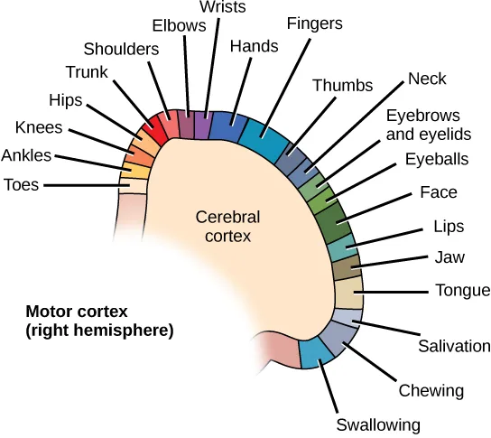
The frontal lobe is located at the front of the brain, over the eyes. This lobe contains the olfactory bulb, which processes smells. The frontal lobe also contains the motor cortex, which is important for planning and implementing movement. Areas within the motor cortex map to different muscle groups, and there is some organization to this map, as shown below. For example, the neurons that control movement of the fingers are next to the neurons that control movement of the hand. Neurons in the frontal lobe also control cognitive functions like maintaining attention, speech, and decision-making. Studies of humans who have damaged their frontal lobes show that parts of this area are involved in personality, socialization, and assessing risk.
Extended description
Twenty-one labeled segments arc from the lower left end of the strip to the lower right end. In order: Toes, Ankles, Knees, Hips, Trunk, Shoulders, Elbows, and Wrists climb up the left side toward the top of the arc; Hands, Fingers, and Thumbs continue near the top; descending the right side: Neck, Eyebrows and eyelids, Eyeballs, Face, Lips, Jaw, Tongue, Salivation, Chewing, and Swallowing. ‘Cerebral cortex’ labels the plain tan region inside the curve of the arc, and ‘Motor cortex (right hemisphere)’ labels the diagram as a whole at lower left.
The parietal lobe is located at the top of the brain. Neurons in the parietal lobe are involved in speech and also reading. Two of the parietal lobe’s main functions are processing somatosensation—touch sensations like pressure, pain, heat, cold—and processing proprioception—the sense of how parts of the body are oriented in space. The parietal lobe contains a somatosensory map of the body similar to the motor cortex.
The occipital lobe is located at the back of the brain. It is primarily involved in vision—seeing, recognizing, and identifying the visual world.
The temporal lobe is located at the base of the brain by your ears and is primarily involved in processing and interpreting sounds. It also contains the hippocampus (Greek for “seahorse”)—a structure that processes memory formation. The hippocampus is illustrated below. The role of the hippocampus in memory was partially determined by studying one famous epileptic patient, HM, who had both sides of his hippocampus removed in an attempt to cure his epilepsy. His seizures went away, but he could no longer form new memories (although he could remember some facts from before his surgery and could learn new motor tasks).
Evolution Connection. Cerebral Cortex.
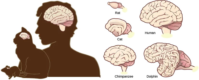
Compared to other vertebrates, mammals have exceptionally large brains for their body size. An entire alligator’s brain, for example, would fill about one and a half teaspoons. This increase in brain to body size ratio is especially pronounced in apes, whales, and dolphins. While this increase in overall brain size doubtlessly played a role in the evolution of complex behaviors unique to mammals, it does not tell the whole story. Scientists have found a relationship between the relatively high surface area of the cortex and the intelligence and complex social behaviors exhibited by some mammals. This increased surface area is due, in part, to increased folding of the cortical sheet (more sulci and gyri). For example, a rat cortex is very smooth with very few sulci and gyri. Cat and sheep cortices have more sulci and gyri. Chimps, humans, and dolphins have even more.
Extended description
At left, a brown silhouette shows a human adult in profile holding a cat, which in turn has a mouse near its paws — the three animals drawn at their relative body sizes, largest to smallest, each with a small inset shape on its head showing its brain at the same relative scale. At right, five separate brain illustrations are arranged in two columns, labeled beneath each. The left column, top to bottom, holds ‘Rat’ (a tiny, nearly smooth brain), ‘Cat’ (a somewhat larger brain with a few shallow folds), and ‘Chimpanzee’ (a substantially larger, more deeply folded brain). The right column holds ‘Human’ at the top (a large, densely folded brain) and ‘Dolphin’ at the bottom (a brain similar in size to the human’s, folded even more densely, shown at an angle).
Basal Ganglia
Interconnected brain areas called the basal ganglia (or basal nuclei), visible in the sagittal section above, play important roles in movement control and posture. Damage to the basal ganglia, as in Parkinson’s disease, leads to motor impairments like a shuffling gait when walking. The basal ganglia also regulate motivation. For example, when a wasp sting led to bilateral basal ganglia damage in a 25-year-old businessman, he began to spend all his days in bed and showed no interest in anything or anybody. But when he was externally stimulated—as when someone asked to play a card game with him—he was able to function normally. Interestingly, he and other similar patients do not report feeling bored or frustrated by their state.
Thalamus
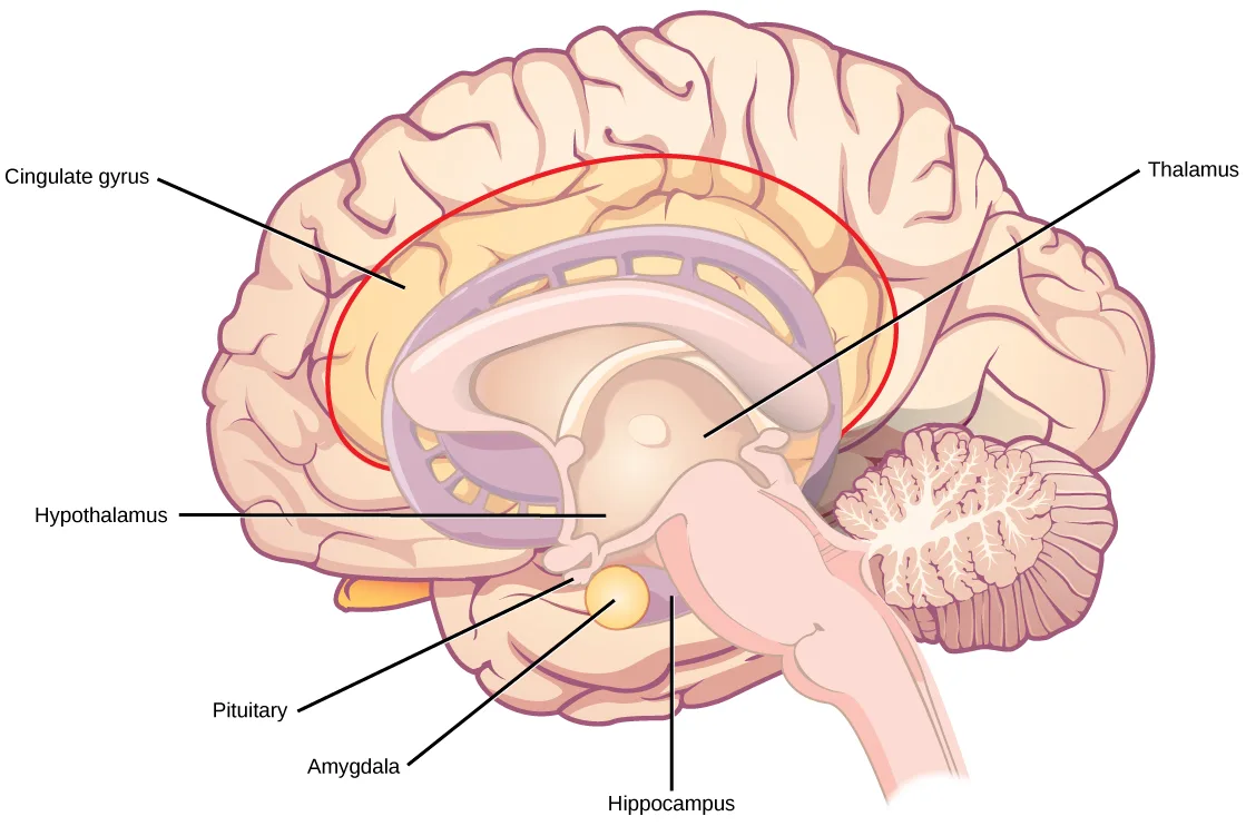
The thalamus (Greek for “inner chamber”), illustrated below, acts as a gateway to and from the cortex. It receives sensory and motor inputs from the body and also receives feedback from the cortex. This feedback mechanism can modulate conscious awareness of sensory and motor inputs depending on the attention and arousal state of the animal. The thalamus helps regulate consciousness, arousal, and sleep states. A rare genetic disorder called fatal familial insomnia causes the degeneration of thalamic neurons and glia. This disorder prevents affected patients from being able to sleep, among other symptoms, and is eventually fatal.
Extended description
‘Cingulate gyrus’ labels the band of cortical tissue outlined in red that arcs over the top of the internal structures, just beneath the outer cortex. ‘Thalamus’ labels the oval structure at the center of that arc. ‘Hypothalamus’ labels the tissue just below and in front of the thalamus. ‘Pituitary’ labels a small round gland hanging beneath the hypothalamus. ‘Amygdala’ labels a small structure just above and in front of the pituitary. ‘Hippocampus’ labels an elongated structure running from beneath the thalamus back toward the wrinkled cerebellum.
Hypothalamus
Below the thalamus is the hypothalamus, shown above. The hypothalamus controls the endocrine system by sending signals to the pituitary gland, a pea-sized endocrine gland that releases several different hormones that affect other glands as well as other cells. This relationship means that the hypothalamus regulates important behaviors that are controlled by these hormones. The hypothalamus is the body’s thermostat—it makes sure key functions like food and water intake, energy expenditure, and body temperature are kept at appropriate levels. Neurons within the hypothalamus also regulate circadian rhythms, sometimes called sleep cycles.
Limbic System
The limbic system is a connected set of structures that regulates emotion, as well as behaviors related to fear and motivation. It plays a role in memory formation and includes parts of the thalamus and hypothalamus as well as the hippocampus. One important structure within the limbic system is a temporal lobe structure called the amygdala (Greek for “almond”), shown above. The two amygdala are important both for the sensation of fear and for recognizing fearful faces. The cingulate gyrus helps regulate emotions and pain.
Cerebellum
The cerebellum (Latin for “little brain”), visible in the diagrams above, sits at the base of the brain on top of the brainstem. The cerebellum controls balance and aids in coordinating movement and learning new motor tasks.
Brainstem
The brainstem, visible in the diagrams above, connects the rest of the brain with the spinal cord. It consists of the midbrain, medulla oblongata, and the pons. Motor and sensory neurons extend through the brainstem allowing for the relay of signals between the brain and spinal cord. Ascending neural pathways cross in this section of the brain allowing the left hemisphere of the cerebrum to control the right side of the body and vice versa. The brainstem coordinates motor control signals sent from the brain to the body. The brainstem controls several important functions of the body including alertness, arousal, breathing, blood pressure, digestion, heart rate, swallowing, walking, and sensory and motor information integration.
Spinal Cord
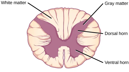
Connecting to the brainstem and extending down the body through the spinal column is the spinal cord, visible in the lobes diagram above. The spinal cord is a thick bundle of nerve tissue that carries information about the body to the brain and from the brain to the body. The spinal cord is contained within the bones of the vertebrate column but is able to communicate signals to and from the body through its connections with spinal nerves (part of the peripheral nervous system). A cross-section of the spinal cord looks like a white oval containing a gray butterfly-shape, as shown below. Myelinated axons make up the “white matter” and neuron and glial cell bodies make up the “gray matter.” Gray matter is also composed of interneurons, which connect two neurons each located in different parts of the body. Axons and cell bodies in the dorsal (facing the back of the animal) spinal cord convey mostly sensory information from the body to the brain. Axons and cell bodies in the ventral (facing the front of the animal) spinal cord primarily transmit signals controlling movement from the brain to the body.
The spinal cord also controls motor reflexes. These reflexes are quick, unconscious movements—like automatically removing a hand from a hot object. Reflexes are so fast because they involve local synaptic connections. For example, the knee reflex that a doctor tests during a routine physical is controlled by a single synapse between a sensory neuron and a motor neuron. While a reflex may only require the involvement of one or two synapses, synapses with interneurons in the spinal column transmit information to the brain to convey what happened (the knee jerked, or the hand was hot).
In the United States, there are around 10,000 spinal cord injuries each year. Because the spinal cord is the information superhighway connecting the brain with the body, damage to the spinal cord can lead to paralysis. The extent of the paralysis depends on the location of the injury along the spinal cord and whether the spinal cord was completely severed. For example, if the spinal cord is damaged at the level of the neck, it can cause paralysis from the neck down, whereas damage to the spinal column further down may limit paralysis to the legs. Spinal cord injuries are notoriously difficult to treat because spinal nerves do not regenerate, although ongoing research suggests that stem cell transplants may be able to act as a bridge to reconnect severed nerves. Researchers are also looking at ways to prevent the inflammation that worsens nerve damage after injury. One such treatment is to pump the body with cold saline to induce hypothermia. This cooling can prevent swelling and other processes that are thought to worsen spinal cord injuries.
Extended description
The gray matter forms a purple X shape at the center of the pale, ridged oval of white matter. ‘Gray matter’ labels the X shape near its upper right arm. ‘Dorsal horn’ labels that same upper arm — the thinner of the X’s two arm pairs. ‘Ventral horn’ labels the lower leg of the X at bottom right — the thicker of the two pairs. ‘White matter’ labels the surrounding pale ring.
Summary
The vertebrate central nervous system contains the brain and the spinal cord, which are covered and protected by three meninges. The brain contains structurally and functionally defined regions. In mammals, these include the cortex (which can be broken down into four primary functional lobes: frontal, temporal, occipital, and parietal), basal ganglia, thalamus, hypothalamus, limbic system, cerebellum, and brainstem—although structures in some of these designations overlap. While functions may be primarily localized to one structure in the brain, most complex functions, like language and sleep, involve neurons in multiple brain regions. The spinal cord is the information superhighway that connects the brain with the rest of the body through its connections with peripheral nerves. It transmits sensory and motor input and also controls motor reflexes.
Key terms
- amygdala — structure within the limbic system that processes fear.
- arachnoid mater — spiderweb-like middle layer of the meninges that cover the central nervous system.
- basal ganglia — interconnected collections of cells in the brain that are involved in movement and motivation; also known as basal nuclei.
- basal nuclei — see basal ganglia.
- brainstem — portion of the brain that connects with the spinal cord; controls basic nervous system functions like breathing, heart rate, and swallowing.
- cerebellum — brain structure involved in posture, motor coordination, and learning new motor actions.
- cerebral cortex — outermost sheet of brain tissue; involved in many higher-order functions.
- choroid plexus — spongy tissue within ventricles that produces cerebrospinal fluid.
- cingulate gyrus — helps regulate emotions and pain; thought to directly drive the body’s conscious response to unpleasant experiences.
- corpus callosum — thick fiber bundle that connects the cerebral hemispheres.
- cerebrospinal fluid (CSF) — clear liquid that surrounds the brain and spinal cord and fills the ventricles and central canal; acts as a shock absorber and circulates material throughout the brain and spinal cord.
- dura mater — tough outermost layer that covers the central nervous system.
- frontal lobe — part of the cerebral cortex that contains the motor cortex and areas involved in planning, attention, and language.
- gyrus — (plural: gyri) ridged protrusions in the cortex.
- hippocampus — brain structure in the temporal lobe involved in processing memories.
- hypothalamus — brain structure that controls hormone release and body homeostasis.
- limbic system — connected brain areas that process emotion and motivation.
- meninge — membrane that covers and protects the central nervous system.
- occipital lobe — part of the cerebral cortex that contains visual cortex and processes visual stimuli.
- parietal lobe — part of the cerebral cortex involved in processing touch and the sense of the body in space.
- pia mater — thin membrane layer directly covering the brain and spinal cord.
- proprioception — sense about how parts of the body are oriented in space.
- somatosensation — sense of touch.
- spinal cord — thick fiber bundle that connects the brain with peripheral nerves; transmits sensory and motor information; contains neurons that control motor reflexes.
- sulcus — (plural: sulci) indents or “valleys” in the cortex.
- temporal lobe — part of the cerebral cortex that processes auditory input; parts of the temporal lobe are involved in speech, memory, and emotion processing.
- thalamus — brain area that relays sensory information to the cortex.
- ventricle — cavity within brain that contains cerebrospinal fluid.
Practice
Identify the spinal cord, cerebral lobes, and other brain areas on a diagram of the brain
The ________ lobe contains the visual cortex.
This lobe sits at the very back of the brain, farthest from the eyes themselves but where what they see is ultimately recognized.The ________ connects the two cerebral hemispheres.
Its Latin name translates to “tough body” — cutting it, rather than removing an entire hemisphere, is what produces a split-brain patient.A membrane that covers and protects the central nervous system is called a ________.
There are three of these layers around the brain and spinal cord, named for how hard, web-like, or soft each one is.The tough outermost layer of the meninges is called the ________.
Its Latin name translates to “hard mother,” and it also carries vein-like structures that return blood from the brain to the heart.The web-like middle layer of the meninges is called the ________.
Its name comes from the Greek word for spider, describing its web-like texture.The thin meningeal layer that directly contacts and covers the brain and spinal cord, like plastic wrap, is called the ________.
Its Latin name translates to “soft mother.”Describe the basic functions of the spinal cord, cerebral lobes, and other brain areas
Neurons in the ________ control motor reflexes.
This structure carries the local synaptic connections responsible for automatic movements, like pulling a hand off a hot object, without needing the brain.Phineas Gage was a 19th-century railroad worker who survived an accident that drove a large iron rod through his head. If the injury resulted in him becoming temperamental and capricious what part of his brain was damaged?
Studies of patients with damage here tie this brain region to personality, socialization, and risk-assessment changes.What methods can be used to determine the function of a particular brain region?
Show model answer
Did your answer mention:
What are the main functions of the spinal cord?
Show model answer
Did your answer mention:
Alzheimer’s disease involves three of the four lobes of the brain. Identify one of the involved lobes and describe the lobe’s symptoms associated with the disease.
Show model answer
Did your answer mention:
Interconnected brain areas involved in movement control, posture, and motivation are called the ________.
Damage to these areas, as in Parkinson’s disease, causes a shuffling gait when walking.The brain area that acts as a gateway, relaying sensory and motor information to and from the cortex, is called the ________.
Its Greek name means “inner chamber”; a rare genetic disorder that degenerates its neurons causes fatal familial insomnia.The connected set of brain structures that regulates emotion and behaviors related to fear and motivation is called the ________.
It includes parts of the thalamus and hypothalamus as well as the hippocampus.This section is adapted from Biology 2e, Section 35.3: The Central Nervous System by Mary Ann Clark, Jung Choi, Matthew Douglas, and OpenStax, © OpenStax, licensed under CC BY-NC-SA 4.0. Access the original for free at openstax.org. Changes: figures re-encoded as WebP; five figures re-kinded from the manifest’s file-extension “photo” guess to “diagram” (Figure_35_03_01, Figure_35_03_02-ddf8, Figure_35_03_04, Figure_35_03_06, and Figure_35_03_07 are all labeled illustrations, not captured photographs); a longdesc added to all seven figures, walking each figure’s leader-line labels in reading order (the meninges cross-section, the coronal/sagittal pair, the four-lobes diagram, the 21-segment motor-cortex map, the five-species brain-size comparison, the limbic-system diagram, and the spinal-cord cross-section); inline references to numbered figures (“Figure 35.19,” “Figure 35.20a/b,” “Figure 35.21,” “Figure 35.22,” “Figure 35.23,” “Figure 35.24,” “Figure 35.25”) changed to “above”/“below” since figures are not numbered here; the Link to Learning and Evolution Connection notes rendered as callouts, the Link to Learning’s source URL kept and the Evolution Connection’s figure kept inside the same callout as in the source; the body paragraph introducing the three sectioning planes, which the module links to “Figure 35.21b” for the sagittal section, corrected to describe the sagittal and coronal panels of the actual two-panel figure (the module’s own Figure 35.21 has no (b) panel; the sagittal view is panel (b) of Figure 35.20) — reported as a source defect; “there around 10,000 spinal cord injuries” corrected to “there are around 10,000 spinal cord injuries” — reported as a source defect; the end-of-chapter Review Questions and Critical Thinking Questions for this section adapted into the closing interactive Practice block (multiple choice and self-check respectively); rubric checkpoints added to each self-check, decomposing its model answer (the source solution) into check-off clauses with no new claims — the Alzheimer’s item’s “Potential answers” solution set as one sentence per lobe, with checkpoints for naming a lobe and describing its symptoms; seven key-term recall items (meninge, dura mater, arachnoid mater, pia mater, basal ganglia, thalamus, limbic system) added from the glossary.