The Peripheral Nervous System
By the end of this section, you will be able to:
- Describe the organization and functions of the sympathetic and parasympathetic nervous systems
- Describe the organization and function of the sensory-somatic nervous system
The peripheral nervous system (PNS) is the connection between the central nervous system and the rest of the body. The CNS is like the power plant of the nervous system. It creates the signals that control the functions of the body. The PNS is like the wires that go to individual houses. Without those “wires,” the signals produced by the CNS could not control the body (and the CNS would not be able to receive sensory information from the body either).
The PNS can be broken down into the autonomic nervous system, which controls bodily functions without conscious control, and the sensory-somatic nervous system, which transmits sensory information from the skin, muscles, and sensory organs to the CNS and sends motor commands from the CNS to the muscles.
Autonomic Nervous System
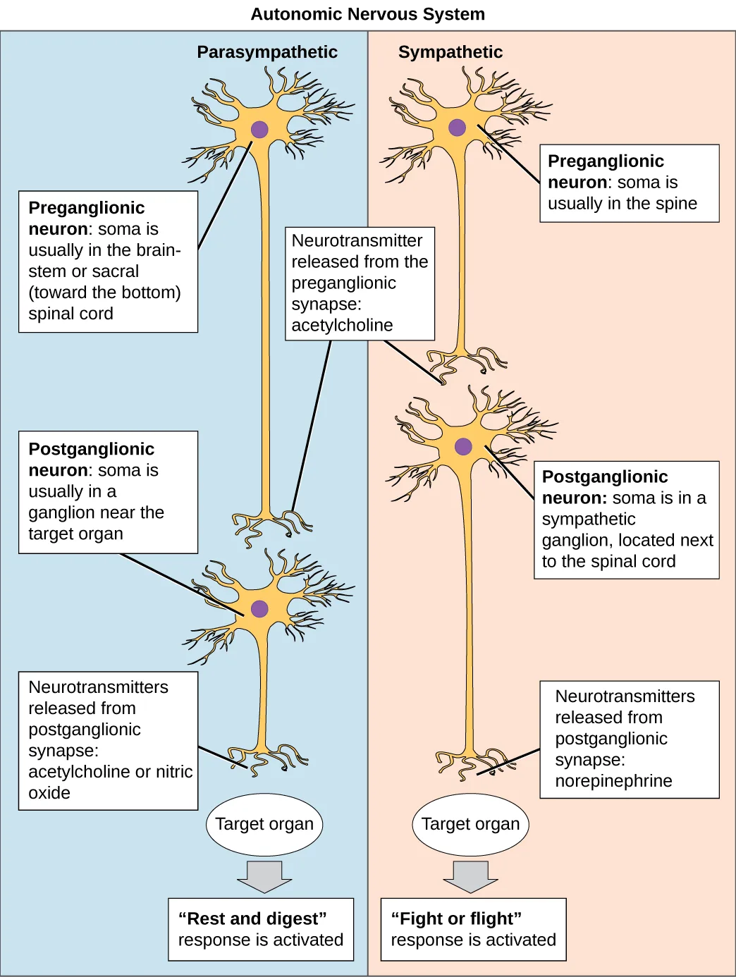
Extended description
The figure is titled ‘Autonomic Nervous System’ and divided into two vertical panels. The left, shaded blue and labeled ‘Parasympathetic’: a preganglionic neuron near the top, labeled ‘Preganglionic neuron: soma is usually in the brainstem or sacral (toward the bottom) spinal cord,’ sends its axon down to a box reading ‘Neurotransmitter released from the preganglionic synapse: acetylcholine,’ then on to a second neuron labeled ‘Postganglionic neuron: soma is usually in a ganglion near the target organ,’ whose axon reaches a box reading ‘Neurotransmitters released from postganglionic synapse: acetylcholine or nitric oxide,’ arriving at an oval labeled ‘Target organ’ and, below it, a box reading ‘“Rest and digest” response is activated.’ The right panel, shaded orange and labeled ‘Sympathetic,’ repeats the same four-box sequence: a preganglionic neuron labeled ‘soma is usually in the spine,’ a box reading ‘Neurotransmitter released from the preganglionic synapse: acetylcholine,’ a postganglionic neuron labeled ‘soma is in a sympathetic ganglion, located next to the spinal cord,’ a box reading ‘Neurotransmitters released from postganglionic synapse: norepinephrine,’ the same ‘Target organ’ oval, and a final box reading ‘“Fight or flight” response is activated.’
Which of the following statements is false?
Check which neurotransmitter each branch actually releases onto its target organ, named in the figure above.The autonomic nervous system serves as the relay between the CNS and the internal organs. It controls the lungs, the heart, smooth muscle, and exocrine and endocrine glands. The autonomic nervous system controls these organs largely without conscious control; it can continuously monitor the conditions of these different systems and implement changes as needed. Signaling to the target tissue usually involves two synapses: a preganglionic neuron (originating in the CNS) synapses to a neuron in a ganglion that, in turn, synapses on the target organ, as illustrated above. There are two divisions of the autonomic nervous system that often have opposing effects: the sympathetic nervous system and the parasympathetic nervous system.
Sympathetic Nervous System
The sympathetic nervous system is responsible for the “fight or flight” response that occurs when an animal encounters a dangerous situation. One way to remember this is to think of the surprise a person feels when encountering a snake (“snake” and “sympathetic” both begin with “s”). Examples of functions controlled by the sympathetic nervous system include an accelerated heart rate and inhibited digestion. These functions help prepare an organism’s body for the physical strain required to escape a potentially dangerous situation or to fend off a predator.
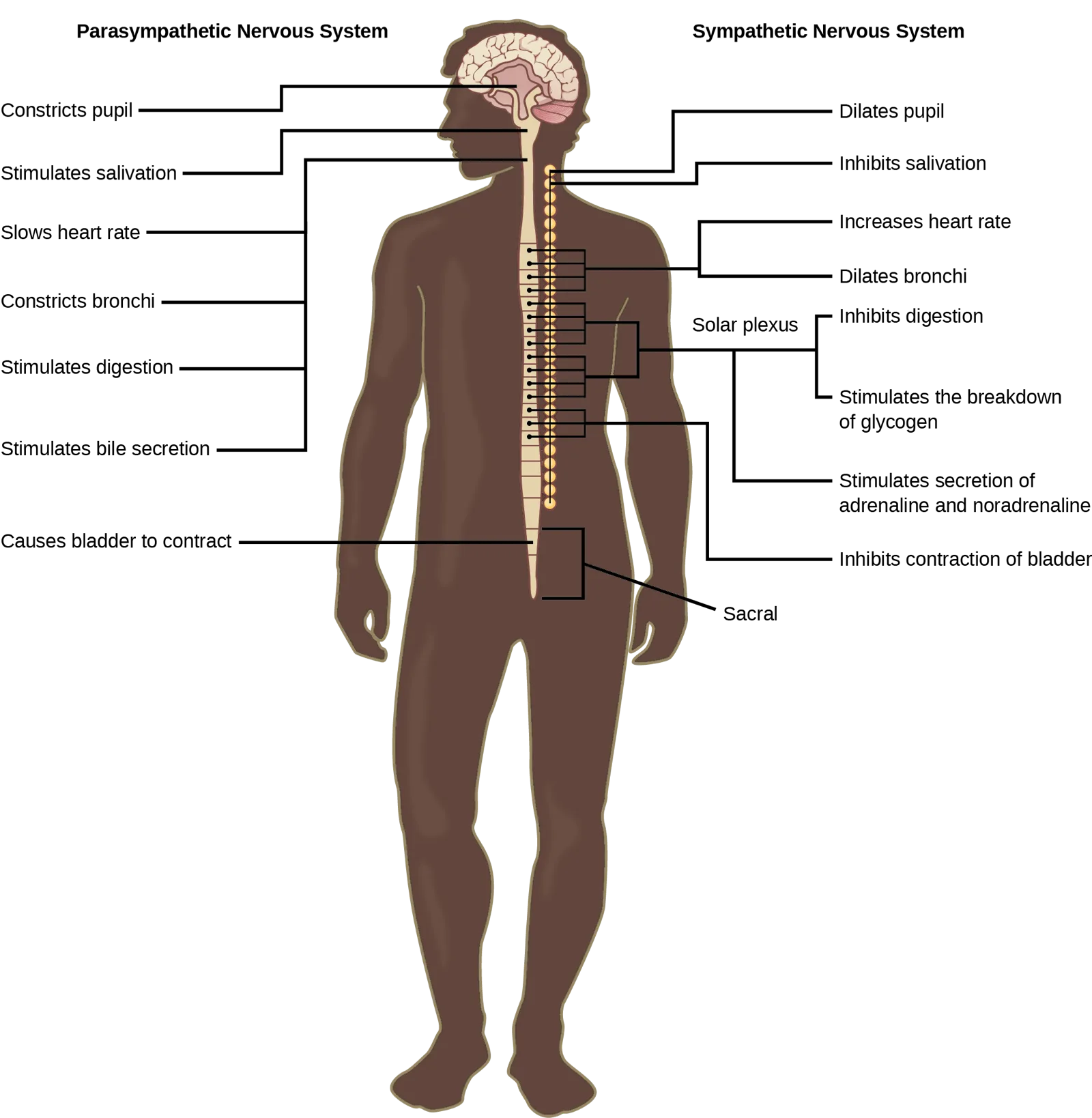
Extended description
Titled with ‘Parasympathetic Nervous System’ above the left column and ‘Sympathetic Nervous System’ above the right column, the figure shows a front-facing human silhouette with the brain, brainstem, and spinal cord drawn down its center. On the left, seven leader lines run from the head and spine to labels reading, top to bottom: ‘Constricts pupil,’ ‘Stimulates salivation,’ ‘Slows heart rate,’ ‘Constricts bronchi,’ ‘Stimulates digestion,’ ‘Stimulates bile secretion,’ and ‘Causes bladder to contract,’ the last pointing to a box near the base of the spine marked ‘Sacral.’ On the right, leader lines run from the head and the chain of ganglia beside the spine to, top to bottom: ‘Dilates pupil,’ ‘Inhibits salivation,’ ‘Increases heart rate,’ ‘Dilates bronchi,’ then a bracket labeled ‘Solar plexus’ joining two lines to ‘Inhibits digestion’ and ‘Stimulates the breakdown of glycogen,’ followed by ‘Stimulates secretion of adrenaline and noradrenaline’ and ‘Inhibits contraction of bladder,’ the last pointing to the same sacral region as the left column’s bladder label.
Most preganglionic neurons in the sympathetic nervous system originate in the spinal cord, as illustrated above. The axons of these neurons release acetylcholine on postganglionic neurons within sympathetic ganglia (the sympathetic ganglia form a chain that extends alongside the spinal cord). The acetylcholine activates the postganglionic neurons. Postganglionic neurons then release norepinephrine onto target organs. As anyone who has ever felt a rush before a big test, speech, or athletic event can attest, the effects of the sympathetic nervous system are quite pervasive. This is both because one preganglionic neuron synapses on multiple postganglionic neurons, amplifying the effect of the original synapse, and because the adrenal gland also releases norepinephrine (and the closely related hormone epinephrine) into the bloodstream. The physiological effects of this norepinephrine release include dilating the trachea and bronchi (making it easier for the animal to breathe), increasing heart rate, and moving blood from the skin to the heart, muscles, and brain (so the animal can think and run). The strength and speed of the sympathetic response helps an organism avoid danger, and scientists have found evidence that it may also increase LTP—allowing the animal to remember the dangerous situation and avoid it in the future.
Parasympathetic Nervous System
While the sympathetic nervous system is activated in stressful situations, the parasympathetic nervous system allows an animal to “rest and digest.” One way to remember this is to think that during a restful situation like a picnic, the parasympathetic nervous system is in control (“picnic” and “parasympathetic” both start with “p”). Parasympathetic preganglionic neurons have cell bodies located in the brainstem and in the sacral (toward the bottom) spinal cord, as shown above. The axons of the preganglionic neurons release acetylcholine on the postganglionic neurons, which are generally located very near the target organs. Most postganglionic neurons release acetylcholine onto target organs, although some release nitric oxide.
The parasympathetic nervous system resets organ function after the sympathetic nervous system is activated (the common adrenaline dump you feel after a ‘fight-or-flight’ event). Effects of acetylcholine release on target organs include slowing of heart rate, lowered blood pressure, and stimulation of digestion.
Sensory-Somatic Nervous System
The sensory-somatic nervous system is made up of cranial and spinal nerves and contains both sensory and motor neurons. Sensory neurons transmit sensory information from the skin, skeletal muscle, and sensory organs to the CNS. Motor neurons transmit messages about desired movement from the CNS to the muscles to make them contract. Without its sensory-somatic nervous system, an animal would be unable to process any information about its environment (what it sees, feels, hears, and so on) and could not control motor movements. Unlike the autonomic nervous system, which has two synapses between the CNS and the target organ, sensory and motor neurons have only one synapse—one ending of the neuron is at the organ and the other directly contacts a CNS neuron. Acetylcholine is the main neurotransmitter released at these synapses.
Humans have 12 cranial nerves, nerves that emerge from or enter the skull (cranium), as opposed to the spinal nerves, which emerge from the vertebral column. Each cranial nerve is accorded a name, which are detailed below. Some cranial nerves transmit only sensory information. For example, the olfactory nerve transmits information about smells from the nose to the brain. (Source note: the source says “to the brainstem”; this book’s Taste and Smell section has olfactory axons end in the olfactory bulb of the forebrain, the one sensory pathway that bypasses the brainstem and thalamus.) Other cranial nerves transmit almost solely motor information. For example, the oculomotor nerve controls the opening and closing of the eyelid and some eye movements. Other cranial nerves contain a mix of sensory and motor fibers. For example, the glossopharyngeal nerve has a role in both taste (sensory) and swallowing (motor).
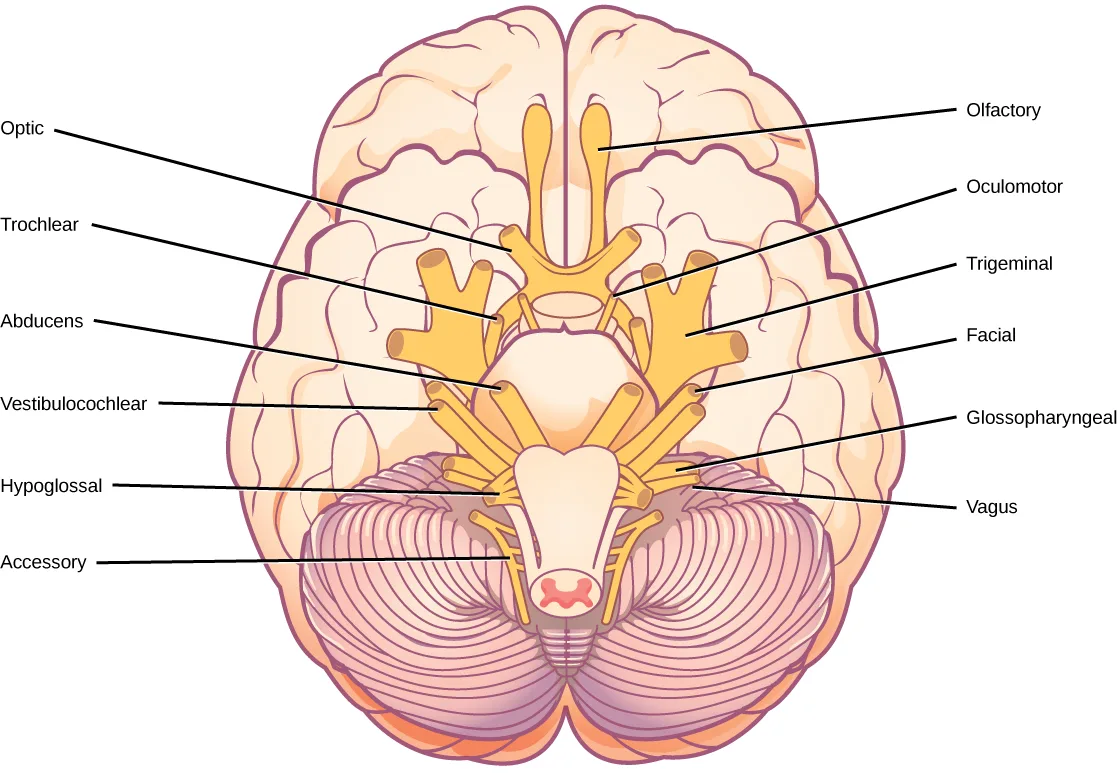
Extended description
The diagram shows the underside of the brain, with the two olfactory bulbs at the top center and the cerebellum’s ridged surface below. Left column, top to bottom: ‘Optic,’ pointing to the nerve just behind the olfactory bulbs; ‘Trochlear,’ a thin nerve beside the thick trigeminal; ‘Abducens,’ emerging near the brainstem; ‘Vestibulocochlear,’ just behind it; ‘Hypoglossal,’ lower on the brainstem; and ‘Accessory,’ the nerve farthest back, at the top of the spinal cord. Right column, top to bottom: ‘Olfactory,’ the short bulb at the very front; ‘Oculomotor,’ just behind it; ‘Trigeminal,’ the thickest nerve, branching into three; ‘Facial,’ behind the trigeminal; ‘Glossopharyngeal,’ lower on the brainstem; and ‘Vagus,’ the lowest of the labeled nerves before the spinal cord.
Spinal nerves transmit sensory and motor information between the spinal cord and the rest of the body. Each of the 31 spinal nerves (in humans) contains both sensory and motor axons. The sensory neuron cell bodies are grouped in structures called dorsal root ganglia and are shown below. Each sensory neuron has one projection—with a sensory receptor ending in skin, muscle, or sensory organs—and another that synapses with a neuron in the dorsal spinal cord. Motor neurons have cell bodies in the ventral gray matter of the spinal cord that project to muscle through the ventral root. These neurons are usually stimulated by interneurons within the spinal cord but are sometimes directly stimulated by sensory neurons.
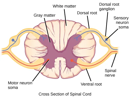
Extended description
Labels point to the following structures. At upper left, ‘Gray matter’ marks the purple butterfly-shaped center and ‘White matter’ the paler tissue surrounding it. At upper right, ‘Dorsal root’ marks the yellow nerve bundle entering the upper back of the cord, ‘Dorsal root ganglion’ the swelling on that root just outside the cord, and ‘Sensory neuron soma’ a blue dot within that ganglion. At lower left, ‘Motor neuron soma’ marks a point within the gray matter’s lower horn. At lower right, ‘Ventral root’ marks the nerve bundle leaving the underside of the cord, and ‘Spinal nerve’ marks the point where the dorsal and ventral roots merge into a single nerve. A red line traces the motor pathway from the gray matter out through the ventral root, and a blue line traces the sensory pathway from the sensory neuron soma into the dorsal root and gray matter.
Summary
The peripheral nervous system contains both the autonomic and sensory-somatic nervous systems. The autonomic nervous system provides unconscious control over visceral functions and has two divisions: the sympathetic and parasympathetic nervous systems. The sympathetic nervous system is activated in stressful situations to prepare the animal for a “fight or flight” response. The parasympathetic nervous system is active during restful periods. The sensory-somatic nervous system is made of cranial and spinal nerves that transmit sensory information from skin and muscle to the CNS and motor commands from the CNS to the muscles.
Key terms
- acetylcholine — neurotransmitter released by neurons in the central nervous system and peripheral nervous system.
- autonomic nervous system — part of the peripheral nervous system that controls bodily functions.
- cranial nerve — sensory and/or motor nerve that emanates from the brain.
- norepinephrine — neurotransmitter and hormone released by activation of the sympathetic nervous system.
- parasympathetic nervous system — division of autonomic nervous system that regulates visceral functions during rest and digestion.
- sensory-somatic nervous system — system of sensory and motor nerves.
- spinal nerve — nerve projecting between skin or muscle and spinal cord.
- sympathetic nervous system — division of autonomic nervous system activated during stressful “fight or flight” situations.
Practice
Describe the organization and functions of the sympathetic and parasympathetic nervous systems
Activation of the sympathetic nervous system causes:
Sympathetic activation prepares the body to escape danger — consider whether escaping requires more or less blood reaching the muscles and brain, and more or less digestion.Where are parasympathetic preganglionic cell bodies located?
Rule out the sensory-neuron location (dorsal root ganglia), a movement-coordination structure (cerebellum), and any non-neural tissue (skin) — parasympathetic preganglionic somas sit within the CNS, in one of the two locations the section names for this pathway.What are the main differences between the sympathetic and parasympathetic branches of the autonomic nervous system?
Show model answer
Did your answer mention:
Scientists have suggested that the autonomic nervous system is not well-adapted to modern human life. How is the sympathetic nervous system an ineffective response to the everyday challenges faced by modern humans?
Show model answer
Did your answer mention:
The division of the autonomic nervous system that regulates visceral functions during rest and digestion is called the ________.
This branch of the autonomic nervous system is active during calm situations like a picnic — the mnemonic the text gives pairs the situation’s first letter with the system’s own first letter.The division of the autonomic nervous system activated during stressful “fight or flight” situations is called the ________.
This branch activates during a dangerous encounter — the mnemonic the text gives pairs a hissing animal’s name with the system’s own name, both beginning with the same letter.Describe the organization and function of the sensory-somatic nervous system
________ is released by motor nerve endings onto muscle.
This is the same neurotransmitter released at both synapses of the autonomic pathway’s parasympathetic branch — not the one sympathetic postganglionic neurons release onto their target organs.What are the main functions of the sensory-somatic nervous system?
Show model answer
Did your answer mention:
Describe how the sensory-somatic nervous system reacts by reflex to a person touching something hot. How does this allow for rapid responses in potentially dangerous situations?
Show model answer
Did your answer mention:
A sensory and/or motor nerve that emanates from the brain is called a ________.
Humans have twelve of these, each with its own name, and they differ from nerves that instead emerge from the vertebral column.A nerve that projects between skin or muscle and the spinal cord is called a ________.
Humans have 31 of these, and unlike its cranial counterpart, it emerges from the vertebral column rather than the skull.This section is adapted from Biology 2e, Section 35.4: The Peripheral Nervous System by Mary Ann Clark, Jung Choi, Matthew Douglas, and OpenStax, © OpenStax, licensed under CC BY-NC-SA 4.0. Access the original for free at openstax.org. Changes: figures re-encoded as WebP; Figure_35_04_03 and Figure_35_04_04 re-kinded from the manifest’s file-extension “photo” guess to “diagram” (both are labeled line illustrations, not captured photographs); a longdesc added to all four figures (the autonomic-pathway flow diagram, the labeled sympathetic/parasympathetic body diagram, the cranial-nerve diagram, and the spinal-cord cross-section) whose labels and structure are not carried by their one- or two-sentence captions; the note wrapping the Autonomic Nervous System Visual Connection rendered as its figure followed by a multiple choice, kept in the body — the module prints this item twice with different wording for option (a): the note copy reads “responsible for resting the body,” the numbered Visual Connection Questions copy (used here, matching the printed exercise and its keyed solution “D”) reads “responsible for relaxing the body” — reported as a source defect; the end-of-section Review Questions and Critical Thinking Questions adapted into the closing interactive Practice block (multiple choice and self-check respectively); rubric checkpoints added to each self-check, decomposing its model answer (the source solution) into check-off clauses with no new claims; four key-term recall items (parasympathetic nervous system, sympathetic nervous system, cranial nerve, spinal nerve) added from the glossary. One claim is corrected with a visible Source note: the olfactory nerve ends in the olfactory bulb, so it carries smell to the brain rather than “to the brainstem” (erratum 444).