Sensory Processes
By the end of this section, you will be able to:
- Identify the general and special senses in humans
- Describe three important steps in sensory perception
- Explain the concept of just-noticeable difference in sensory perception
Senses provide information about the body and its environment. Humans have five special senses: olfaction (smell), gustation (taste), equilibrium (balance and body position), vision, and hearing. Additionally, we possess general senses, also called somatosensation, which respond to stimuli like temperature, pain, pressure, and vibration. Vestibular sensation, which is an organism’s sense of spatial orientation and balance, proprioception (position of bones, joints, and muscles), and the sense of limb position that is used to track kinesthesia (limb movement) are part of somatosensation. Although the sensory systems associated with these senses are very different, they all share a common function: to convert a stimulus (such as light, or sound, or the position of the body) into an electrical signal in the nervous system. This process is called sensory transduction.
There are two broad types of cellular systems that perform sensory transduction. In one, a neuron works with a sensory receptor, a cell, or cell process that is specialized to engage with and detect a specific stimulus. Stimulation of the sensory receptor activates the associated afferent neuron, which carries information about the stimulus to the central nervous system. In the second type of sensory transduction, a sensory nerve ending responds to a stimulus in the internal or external environment: this neuron constitutes the sensory receptor. Free nerve endings can be stimulated by several different stimuli, thus showing little receptor specificity. For example, pain receptors in your gums and teeth may be stimulated by temperature changes, chemical stimulation, or pressure.
Reception
The first step in sensation is reception, which is the activation of sensory receptors by stimuli such as mechanical stimuli (being bent or squished, for example), chemicals, or temperature. The receptor can then respond to the stimuli. The region in space in which a given sensory receptor can respond to a stimulus, be it far away or in contact with the body, is that receptor’s receptive field. Think for a moment about the differences in receptive fields for the different senses. For the sense of touch, a stimulus must come into contact with the body. For the sense of hearing, a stimulus can be a moderate distance away (some baleen whale sounds can propagate for many kilometers). For vision, a stimulus can be very far away; for example, the visual system perceives light from stars at enormous distances.
Transduction
The most fundamental function of a sensory system is the translation of a sensory signal to an electrical signal in the nervous system. This takes place at the sensory receptor, and the change in electrical potential that is produced is called the receptor potential. How is sensory input, such as pressure on the skin, changed to a receptor potential? In this example, a type of receptor called a mechanoreceptor (shown below) possesses specialized membranes that respond to pressure. Disturbance of these dendrites by compressing them or bending them opens gated ion channels in the plasma membrane of the sensory neuron, changing its electrical potential. Recall that in the nervous system, a positive change of a neuron’s electrical potential (also called the membrane potential), depolarizes the neuron. Receptor potentials are graded potentials: the magnitude of these graded (receptor) potentials varies with the strength of the stimulus. If the magnitude of depolarization is sufficient (that is, if membrane potential reaches a threshold), the neuron will fire an action potential. In most cases, the correct stimulus impinging on a sensory receptor will drive membrane potential in a positive direction, although for some receptors, such as those in the visual system, this is not always the case.
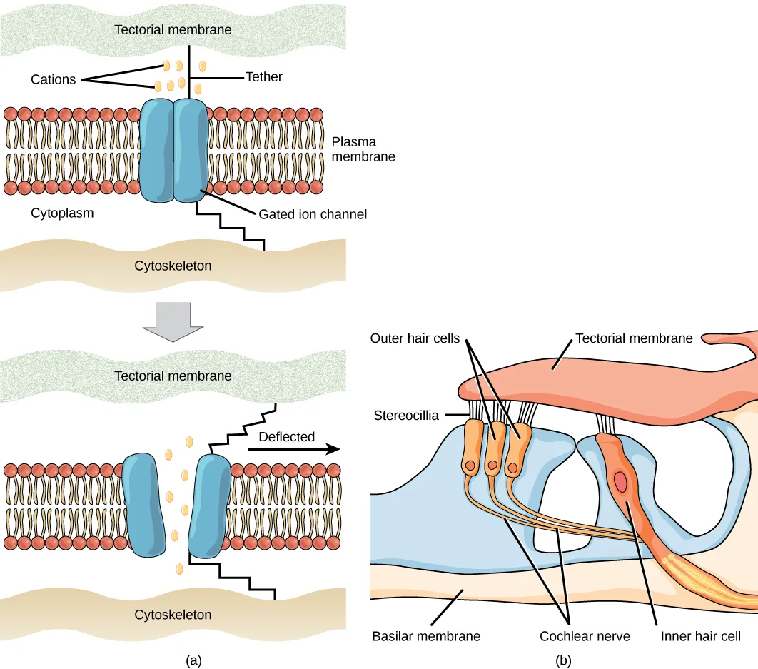
Extended description
Panel (a) stacks two versions of the same cross-section, joined by a downward arrow. In the top version, from top to bottom: a wavy band labeled ‘Tectorial membrane’; below it seven small ovals labeled ‘Cations’ (a row of three above a row of four) flanking a vertical line labeled ‘Tether’ that runs from the tectorial membrane down to a pair of joined blue cylinders labeled ‘Gated ion channel’, set in a band of red-headed phospholipids labeled ‘Plasma membrane’; below the membrane, open space labeled ‘Cytoplasm’; and at the bottom a wavy band labeled ‘Cytoskeleton’, connected to the channel by a stair-step line. In the bottom version, the tectorial membrane (labeled again) has shifted right, labeled ‘Deflected’ beside a rightward arrow; the two halves of the gated ion channel have pulled apart, and the cations now sit in the gap between them; the cytoskeleton is labeled again at the bottom. Panel (b), left to right: ‘Outer hair cells’ labels three orange cells whose ‘Stereocilia’ touch the overlying pink ‘Tectorial membrane’; nerve fibers from the outer hair cells run down to the ‘Cochlear nerve’; the ‘Basilar membrane’ is labeled beneath the cells; and at the right, a separate, larger orange cell labeled ‘Inner hair cell’ has its own stereocilia touching the tectorial membrane.
Sensory receptors for different senses are very different from each other, and they are specialized according to the type of stimulus they sense: they have receptor specificity. For example, touch receptors, light receptors, and sound receptors are each activated by different stimuli. Touch receptors are not sensitive to light or sound; they are sensitive only to touch or pressure. However, stimuli may be combined at higher levels in the brain, as happens with olfaction, contributing to our sense of taste.
Encoding and Transmission of Sensory Information
Four aspects of sensory information are encoded by sensory systems: the type of stimulus, the location of the stimulus in the receptive field, the duration of the stimulus, and the relative intensity of the stimulus. Thus, action potentials transmitted over a sensory receptor’s afferent axons encode one type of stimulus, and this segregation of the senses is preserved in other sensory circuits. For example, auditory receptors transmit signals over their own dedicated system, and electrical activity in the axons of the auditory receptors will be interpreted by the brain as an auditory stimulus—a sound.
The intensity of a stimulus is often encoded in the rate of action potentials produced by the sensory receptor. Thus, an intense stimulus will produce a more rapid train of action potentials, and reducing the stimulus will likewise slow the rate of production of action potentials. A second way in which intensity is encoded is by the number of receptors activated. An intense stimulus might initiate action potentials in a large number of adjacent receptors, while a less intense stimulus might stimulate fewer receptors. Integration of sensory information begins as soon as the information is received in the CNS, and the brain will further process incoming signals.
Perception
Perception is an individual’s interpretation of a sensation. Although perception relies on the activation of sensory receptors, perception happens not at the level of the sensory receptor, but at higher levels in the nervous system, in the brain. The brain distinguishes sensory stimuli through a sensory pathway: action potentials from sensory receptors travel along neurons that are dedicated to a particular stimulus. These neurons are dedicated to that particular stimulus and synapse with particular neurons in the brain or spinal cord.
All sensory signals, except those from the olfactory system, are transmitted through the central nervous system and are routed to the thalamus and to the appropriate region of the cortex. Recall that the thalamus is a structure in the forebrain that serves as a clearinghouse and relay station for sensory (as well as motor) signals. When the sensory signal exits the thalamus, it is conducted to the specific area of the cortex (shown below) dedicated to processing that particular sense.
How are neural signals interpreted? Interpretation of sensory signals between individuals of the same species is largely similar, owing to the inherited similarity of their nervous systems; however, there are some individual differences. A good example of this is individual tolerances to a painful stimulus, such as dental pain, which certainly differ.
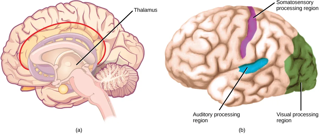
Extended description
Panel (a) is a single label: a line from the word ‘Thalamus’ points to a small pale oval near the center of a mid-sagittal brain section, which is encircled by a red curved outline running around the inner limbic structures. Panel (b) is a lateral view of the brain carrying three labeled colored regions: a purple band near the top, running down toward the middle of the brain, labeled ‘Somatosensory processing region’; a blue, oval patch lower down over the temporal lobe, labeled ‘Auditory processing region’; and a green region covering the back of the brain, labeled ‘Visual processing region’.
Scientific Method Connection. Just-Noticeable Difference
It is easy to differentiate between a one-pound bag of rice and a two-pound bag of rice. There is a one-pound difference, and one bag is twice as heavy as the other. However, would it be as easy to differentiate between a 20- and a 21-pound bag?
Question: What is the smallest detectible weight difference between a one-pound bag of rice and a larger bag? What is the smallest detectible difference between a 20-pound bag and a larger bag? In both cases, at what weights are the differences detected? This smallest detectible difference in stimuli is known as the just-noticeable difference (JND).
Background: Research background literature on JND and on Weber’s Law, a description of a proposed mathematical relationship between the overall magnitude of the stimulus and the JND. You will be testing JND of different weights of rice in bags. Choose a convenient increment that is to be stepped through while testing. For example, you could choose 10 percent increments between one and two pounds (1.1, 1.2, 1.3, 1.4, and so on) or 20 percent increments (1.2, 1.4, 1.6, and 1.8).
Hypothesis: Develop a hypothesis about JND in terms of percentage of the whole weight being tested (such as “the JND between the two small bags and between the two large bags is proportionally the same,” or “. . . is not proportionally the same.”) So, for the first hypothesis, if the JND between the one-pound bag and a larger bag is 0.2 pounds (that is, 20 percent; 1.0 pound feels the same as 1.1 pounds, but 1.0 pound feels less than 1.2 pounds), then the JND between the 20-pound bag and a larger bag will also be 20 percent. (So, 20 pounds feels the same as 22 pounds or 23 pounds, but 20 pounds feels less than 24 pounds.)
Test the hypothesis: Enlist 24 participants, and split them into two groups of 12. To set up the demonstration, assuming a 10 percent increment was selected, have the first group be the one-pound group. As a counter-balancing measure against a systematic error, however, six of the first group will compare one pound to two pounds, and step down in weight (1.0 to 2.0, 1.0 to 1.9, and so on), while the other six will step up (1.0 to 1.1, 1.0 to 1.2, and so on). Apply the same principle to the 20-pound group (20 to 40, 20 to 38, and so on, and 20 to 22, 20 to 24, and so on). Given the large difference between 20 and 40 pounds, you may wish to use 30 pounds as your larger weight. In any case, use two weights that are easily detectable as different.
Record the observations: Record the data in a table similar to the table below. For the one-pound and 20-pound groups (base weights) record a plus sign (+) for each participant that detects a difference between the base weight and the step weight. Record a minus sign (-) for each participant that finds no difference. If one-tenth steps were not used, then replace the steps in the “Step Weight” columns with the step you are using.
Results of JND Testing (+ = difference; – = no difference)
| Step Weight | One pound | 20 pounds | Step Weight |
|---|---|---|---|
| 1.1 | 22 | ||
| 1.2 | 24 | ||
| 1.3 | 26 | ||
| 1.4 | 28 | ||
| 1.5 | 30 | ||
| 1.6 | 32 | ||
| 1.7 | 34 | ||
| 1.8 | 36 | ||
| 1.9 | 38 | ||
| 2.0 | 40 |
Analyze the data/report the results: What step weight did all participants find to be equal with one-pound base weight? What about the 20-pound group?
Draw a conclusion: Did the data support the hypothesis? Are the final weights proportionally the same? If not, why not? Do the findings adhere to Weber’s Law? Weber’s Law states that the concept that a just-noticeable difference in a stimulus is proportional to the magnitude of the original stimulus.
Summary
A sensory activation occurs when a physical or chemical stimulus is processed into a neural signal (sensory transduction) by a sensory receptor. Perception is an individual interpretation of a sensation and is a brain function. Humans have special senses: olfaction, gustation, equilibrium, and hearing, plus the general senses of somatosensation.
Sensory receptors are either specialized cells associated with sensory neurons or the specialized ends of sensory neurons that are a part of the peripheral nervous system, and they are used to receive information about the environment (internal or external). Each sensory receptor is modified for the type of stimulus it detects. For example, neither gustatory receptors nor auditory receptors are sensitive to light. Each sensory receptor is responsive to stimuli within a specific region in space, which is known as that receptor’s receptive field. The most fundamental function of a sensory system is the translation of a sensory signal to an electrical signal in the nervous system.
All sensory signals, except those from the olfactory system, enter the central nervous system and are routed to the thalamus. When the sensory signal exits the thalamus, it is conducted to the specific area of the cortex dedicated to processing that particular sense.
Key terms
- kinesthesia — sense of body movement.
- mechanoreceptor — sensory receptor modified to respond to mechanical disturbance such as being bent, touch, pressure, motion, and sound.
- perception — individual interpretation of a sensation; a brain function.
- proprioception — sense of limb position; used to track kinesthesia.
- reception — receipt of a signal (such as light or sound) by sensory receptors.
- receptive field — region in space in which a stimulus can activate a given sensory receptor.
- receptor potential — membrane potential in a sensory receptor in response to detection of a stimulus.
- sensory receptor — specialized neuron or other cells associated with a neuron that is modified to receive specific sensory input.
- sensory transduction — conversion of a sensory stimulus into electrical energy in the nervous system by a change in the membrane potential.
- vestibular sense — sense of spatial orientation and balance.
Practice
Identify the general and special senses in humans
Many people experience motion sickness while traveling in a car. This sensation results from contradictory inputs arising from which senses?
Think about which two senses can send contradictory signals about motion — the eyes report one thing about the body’s motion, and the inner ear’s balance organs report another.Describe the difference in the localization of the sensory receptors for general and special senses in humans.
Show model answer
Did your answer mention:
The sense of body movement is called ________.
This sense works together with the sense of limb position to track how a limb is moving, not just where it currently sits.The sense of the position of bones, joints, and muscles is called ________.
This sense reports where a limb is right now, distinct from the sense that tracks how it is moving.An organism’s sense of spatial orientation and balance is called the ________.
This sense combines with sight to keep you oriented — when the two disagree in a moving vehicle, motion sickness can follow.Describe three important steps in sensory perception
Where does perception occur?
This is the specific area the sensory signal is routed to only after leaving the thalamus, not the relay station itself.If a person’s cold receptors no longer convert cold stimuli into sensory signals, that person has a problem with the process of ________.
This is the step where a stimulus itself is converted into an electrical signal — not simply detected (reception) or carried onward (transmission).After somatosensory transduction, the sensory signal travels through the brain as a(n) _____ signal.
Neurons signal using changes in membrane voltage, regardless of whether the original stimulus was pressure, light, or temperature.If a person sustains damage to axons leading from sensory receptors to the central nervous system, which step or steps of sensory perception will be affected?
Show model answer
Did your answer mention:
The receipt of a signal, such as light or sound, by sensory receptors is called ________.
This is the very first of the three steps in sensory perception, before any conversion to an electrical signal takes place.The conversion of a sensory stimulus into electrical energy in the nervous system, by a change in membrane potential, is called ________.
This step happens at the sensory receptor itself and produces what this section calls a receptor potential.The individual interpretation of a sensation, which is a brain function, is called ________.
This step happens at higher levels of the nervous system, in the brain, not at the sensory receptor itself.The membrane potential in a sensory receptor that arises in response to detecting a stimulus is called the ________.
It is produced right at the sensory receptor, and its magnitude is graded with the strength of the stimulus.A specialized neuron, or another cell associated with a neuron, that is modified to receive a specific kind of sensory input is called a ________.
Stimulating this cell activates an associated afferent neuron, which carries information about the stimulus toward the central nervous system.A sensory receptor modified to respond to mechanical disturbance, such as being bent, touch, pressure, motion, or sound, is called a ________.
This receptor type opens gated ion channels in its membrane when its dendrites are compressed or bent, shown in the figure above.The region in space in which a stimulus can activate a given sensory receptor is called that receptor’s ________.
For touch this region must be in contact with the body; for hearing or vision it can be very far away.Explain the concept of just-noticeable difference in sensory perception
In what way does the overall magnitude of a stimulus affect the just-noticeable difference in the perception of that stimulus?
Show model answer
Did your answer mention:
The smallest difference in a stimulus that a person can detect is called the ________.
This concept is illustrated by the rice-bag experiment above, and follows Weber’s Law relating it to a stimulus’s overall magnitude.According to Weber’s Law, a just-noticeable difference in a stimulus is ________ to the magnitude of the original stimulus.
In the rice-bag example, a 20-pound bag needs a much bigger added weight to notice a difference than a 1-pound bag does — but is that added weight a similar share of the original in both cases?This section is adapted from Biology 2e, Section 36.1: Sensory Processes by Mary Ann Clark, Jung Choi, Matthew Douglas, and OpenStax, © OpenStax, licensed under CC BY-NC-SA 4.0. Access the original for free at openstax.org. Changes: figures re-encoded as WebP; both figures (Figure_36_01_01f, Figure_36_01_02) re-kinded from the manifest’s file-extension “photo” guess to “diagram” after inspection — both are hand-drawn, labeled line illustrations, not captured photographs; a longdesc added to both, walking the mechanoreceptor/stereocilia diagram’s tethered-channel and ear-hair-cell labels and the brain diagram’s thalamus and cortical-region labels in reading order; the two in-text figure pointers (“as shown in Figure 36.2,” “the specific area of the cortex (Figure 36.3)”) replaced with “shown below,” since Hugo does not number figures; the Scientific Method Connection note rendered as a callout with its bold name and italic title, its bold procedural labels (“Question,” “Background,” “Hypothesis,” “Test the hypothesis,” “Record the observations,” “Analyze the data/report the results,” “Draw a conclusion”) kept as the source repeats them, and its embedded data-collection table (title “Results of JND Testing,” a title row spanning the columns in the source) transcribed as a Markdown table with the title kept as a bold line above it, blank data cells left blank as printed; the end-of-section Review Questions and Critical Thinking Questions adapted into the closing interactive Practice block (multiple choice and self-check, respectively); rubric checkpoints added to each self-check, decomposing its model answer (the source solution) into check-off clauses with no new claims; all ten key-term recall items added from the glossary, covering every glossary term; because the section has no Review Question or glossary term for just-noticeable difference (JND) itself, its objective group also carries two locally written items disclosed here: a text-recall item built strictly from the Scientific Method Connection note’s own defining sentence (“This smallest detectible difference in stimuli is known as the just-noticeable difference (JND)”), and a multiple choice built strictly from the note’s own closing sentence on Weber’s Law. This module has no Visual Connection.