Somatosensation
By the end of this section, you will be able to:
- Describe four important mechanoreceptors in human skin
- Describe the topographical distribution of somatosensory receptors between glabrous and hairy skin
- Explain why the perception of pain is subjective
Somatosensation is a mixed sensory category and includes all sensation received from the skin and mucous membranes, as well as from the limbs and joints. Somatosensation is also known as tactile sense, or more familiarly, as the sense of touch. Somatosensation occurs all over the exterior of the body and at some interior locations as well. A variety of receptor types—embedded in the skin, mucous membranes, muscles, joints, internal organs, and cardiovascular system—play a role.
Recall that the epidermis is the outermost layer of skin in mammals. It is relatively thin, is composed of keratin-filled cells, and has no blood supply. The epidermis serves as a barrier to water and to invasion by pathogens. Below this, the much thicker dermis contains blood vessels, sweat glands, hair follicles, lymph vessels, and lipid-secreting sebaceous glands (shown below). Below the epidermis and dermis is the subcutaneous tissue, or hypodermis, the fatty layer that contains blood vessels, connective tissue, and the axons of sensory neurons. The hypodermis, which holds about 50 percent of the body’s fat, attaches the dermis to the bone and muscle, and supplies nerves and blood vessels to the dermis.
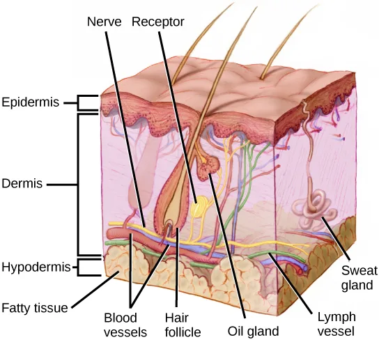
Extended description
Bracket labels along the left edge mark three horizontal layers, top to bottom: a thin ‘Epidermis,’ a much thicker ‘Dermis,’ and a ‘Hypodermis,’ with ‘Fatty tissue’ labeling the pale globular tissue within the hypodermis. Several hair shafts emerge from the block’s top surface. Two labels at the top, ‘Nerve’ and ‘Receptor,’ each follow a line down into the dermis: the nerve line reaches a branching nerve ending, and the receptor line reaches a bulb-shaped structure beside the base of a hair follicle. Across the bottom of the block, five more labels point to structures cut through by its front face, left to right: ‘Blood vessels’ (vessels running along the dermis-hypodermis boundary), ‘Hair follicle’ (the tube-shaped structure the hair shaft grows from), ‘Oil gland’ (a small gland attached to the follicle), ‘Lymph vessel’ (a vessel beside the blood vessels), and ‘Sweat gland’ (a coiled tube near the block’s right edge, extending from the dermis up through the epidermis).
Somatosensory Receptors
Somatosensory receptors are classified into four categories: mechanoreceptors, thermoreceptors, pain receptors (nociceptors), and chemoreceptors. These categories are based on the nature of stimuli each receptor class transduces. What is commonly referred to as “touch” involves more than one kind of stimulus and more than one kind of receptor. Mechanoreceptors in the skin are described as encapsulated (that is, surrounded by a capsule) or unencapsulated (a group that includes free nerve endings). A free nerve ending, as its name implies, is an unencapsulated dendrite of a sensory neuron. Free nerve endings are the most common nerve endings in skin, and they extend into the middle of the epidermis. Free nerve endings are sensitive to painful stimuli, to hot and cold, and to light touch. They are slow to adjust to a stimulus and so are less sensitive to abrupt changes in stimulation.
There are three classes of mechanoreceptors: tactile, proprioceptors, and baroreceptors. Mechanoreceptors sense stimuli due to physical deformation of their plasma membranes. They contain mechanically gated ion channels whose gates open or close in response to pressure, touch, stretching, and sound. There are four primary tactile mechanoreceptors in human skin: Merkel’s disks, Meissner’s corpuscles, Ruffini endings, and Pacinian corpuscles; two are located toward the surface of the skin and two are located deeper. A fifth type of mechanoreceptor, Krause end bulbs, are found only in specialized regions. Merkel’s disks (shown below) are found in the upper layers of skin near the base of the epidermis, both in skin that has hair and on glabrous skin, that is, the hairless skin found on the palms and fingers, the soles of the feet, and the lips of humans and other primates. Merkel’s disks are densely distributed in the fingertips and lips. They are slow-adapting, unencapsulated nerve endings, and they respond to light touch. Light touch, also known as discriminative touch, is a light pressure that allows the location of a stimulus to be pinpointed. The receptive fields of Merkel’s disks are small with well-defined borders. That makes them finely sensitive to edges and they come into use in tasks such as typing on a keyboard.
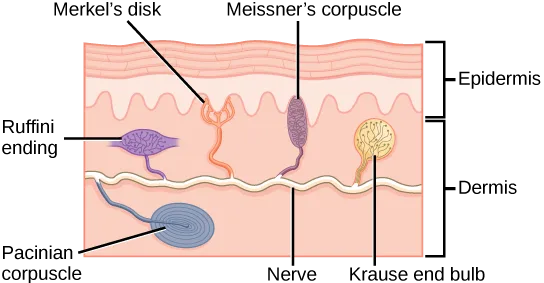
Extended description
The cross-section is bracketed on the right into an upper ‘Epidermis’ band and a lower ‘Dermis’ band, with a horizontal wavy nerve line running through the middle of the dermis; every receptor connects to it by a thin stalk. From left to right along the upper dermis, just below the epidermis boundary: ‘Ruffini ending’ labels a purple, fan-shaped receptor; ‘Merkel’s disk’ labels an orange, finger-like structure reaching up to the underside of the epidermis; ‘Meissner’s corpuscle’ labels a purple, vertical oval also touching the underside of the epidermis; ‘Krause end bulb’ labels a small, round, yellow-orange structure. Below the nerve line, in the lower dermis, ‘Pacinian corpuscle’ labels a blue, tightly coiled oval — the only receptor drawn beneath the nerve. ‘Nerve’ labels the horizontal line itself.
Which of the following statements about mechanoreceptors is false?
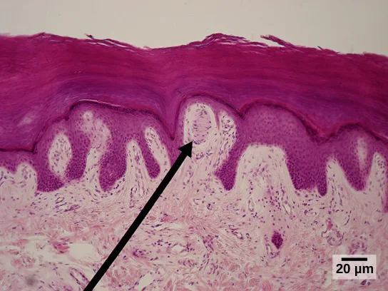
Compare each statement’s claim against where the figure and the passage place that receptor — one of the four moves a receptor into a layer the section doesn’t put it in.Meissner’s corpuscles, (shown below) also known as tactile corpuscles, are found in the upper dermis, but they project into the epidermis. They, too, are found primarily in the glabrous skin on the fingertips and eyelids. They respond to fine touch and pressure, but they also respond to low-frequency vibration or flutter. They are rapidly adapting, fluid-filled, encapsulated neurons with small, well-defined borders and are responsive to fine details. Like Merkel’s disks, Meissner’s corpuscles are not as plentiful in the palms as they are in the fingertips.
Deeper in the epidermis, near the base, are Ruffini endings, which are also known as bulbous corpuscles. They are found in both glabrous and hairy skin. These are slow-adapting, encapsulated mechanoreceptors that detect skin stretch and deformations within joints, so they provide valuable feedback for gripping objects and controlling finger position and movement. Thus, they also contribute to proprioception and kinesthesia. Ruffini endings also detect warmth. Note that these warmth detectors are situated deeper in the skin than are the cold detectors. It is not surprising, then, that humans detect cold stimuli before they detect warm stimuli.
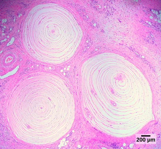
Pacinian corpuscles (seen below) are located deep in the dermis of both glabrous and hairy skin and are structurally similar to Meissner’s corpuscles; they are found in the bone periosteum, joint capsules, pancreas and other viscera, breast, and genitals. They are rapidly adapting mechanoreceptors that sense deep transient (but not prolonged) pressure and high-frequency vibration. Pacinian receptors detect pressure and vibration by being compressed, stimulating their internal dendrites. There are fewer Pacinian corpuscles and Ruffini endings in skin than there are Merkel’s disks and Meissner’s corpuscles.
In proprioception, proprioceptive and kinesthetic signals travel through myelinated afferent neurons running from the spinal cord to the medulla. Neurons are not physically connected, but communicate via neurotransmitters secreted into synapses or “gaps” between communicating neurons. Once in the medulla, the neurons continue carrying the signals to the thalamus.
Muscle spindles are stretch receptors that detect the amount of stretch, or lengthening of muscles. Related to these are Golgi tendon organs, which are tension receptors that detect the force of muscle contraction. Proprioceptive and kinesthetic signals come from limbs. Unconscious proprioceptive signals run from the spinal cord to the cerebellum, the brain region that coordinates muscle contraction, rather than to the thalamus, like most other sensory information.
Baroreceptors detect pressure changes in an organ. They are found in the walls of the carotid artery and the aorta where they monitor blood pressure, and in the lungs where they detect the degree of lung expansion. Stretch receptors are found at various sites in the digestive and urinary systems.
In addition to these two types of deeper receptors, there are also rapidly adapting hair receptors, which are found on nerve endings that wrap around the base of hair follicles. There are a few types of hair receptors that detect slow and rapid hair movement, and they differ in their sensitivity to movement. Some hair receptors also detect skin deflection, and certain rapidly adapting hair receptors allow detection of stimuli that have not yet touched the skin.
Integration of Signals from Mechanoreceptors
The configuration of the different types of receptors working in concert in human skin results in a very refined sense of touch. The nociceptive receptors—those that detect pain—are located near the surface. Small, finely calibrated mechanoreceptors—Merkel’s disks and Meissner’s corpuscles—are located in the upper layers and can precisely localize even gentle touch. The large mechanoreceptors—Pacinian corpuscles and Ruffini endings—are located in the lower layers and respond to deeper touch. (Consider that the deep pressure that reaches those deeper receptors would not need to be finely localized.) Both the upper and lower layers of the skin hold rapidly and slowly adapting receptors. Both primary somatosensory cortex and secondary cortical areas are responsible for processing the complex picture of stimuli transmitted from the interplay of mechanoreceptors.
Density of Mechanoreceptors
The distribution of touch receptors in human skin is not consistent over the body. In humans, touch receptors are less dense in skin covered with any type of hair, such as the arms, legs, torso, and face. Touch receptors are denser in glabrous skin (the type found on human fingertips and lips, for example), which is typically more sensitive and is thicker than hairy skin (4 to 5 mm versus 2 to 3 mm).
How is receptor density estimated in a human subject? The relative density of pressure receptors in different locations on the body can be demonstrated experimentally using a two-point discrimination test. In this demonstration, two sharp points, such as two thumbtacks, are brought into contact with the subject’s skin (though not hard enough to cause pain or break the skin). The subject reports if they feel one point or two points. If the two points are felt as one point, it can be inferred that the two points are both in the receptive field of a single sensory receptor. If two points are felt as two separate points, each is in the receptive field of two separate sensory receptors. The points could then be moved closer and retested until the subject reports feeling only one point, and the size of the receptive field of a single receptor could be estimated from that distance.
Thermoreception
In addition to Krause end bulbs that detect cold and Ruffini endings that detect warmth, there are different types of cold receptors on some free nerve endings: thermoreceptors, located in the dermis, skeletal muscles, liver, and hypothalamus, that are activated by different temperatures. Their pathways into the brain run from the spinal cord through the thalamus to the primary somatosensory cortex. Warmth and cold information from the face travels through one of the cranial nerves to the brain. You know from experience that a tolerably cold or hot stimulus can quickly progress to a much more intense stimulus that is no longer tolerable. Any stimulus that is too intense can be perceived as pain because temperature sensations are conducted along the same pathways that carry pain sensations.
Pain
Pain is the name given to nociception, which is the neural processing of injurious stimuli in response to tissue damage. Pain is caused by true sources of injury, such as contact with a heat source that causes a thermal burn or contact with a corrosive chemical. But pain also can be caused by harmless stimuli that mimic the action of damaging stimuli, such as contact with capsaicins, the compounds that cause peppers to taste hot and which are used in self-defense pepper sprays and certain topical medications. Peppers taste “hot” because the protein receptors that bind capsaicin open the same calcium channels that are activated by warm receptors.
Nociception starts at the sensory receptors, but pain, inasmuch as it is the perception of nociception, does not start until it is communicated to the brain. There are several nociceptive pathways to and through the brain. Most axons carrying nociceptive information into the brain from the spinal cord project to the thalamus (as do other sensory neurons) and the neural signal undergoes final processing in the primary somatosensory cortex. Interestingly, one nociceptive pathway projects not to the thalamus but directly to the hypothalamus in the forebrain, which modulates the cardiovascular and neuroendocrine functions of the autonomic nervous system. Recall that threatening—or painful—stimuli stimulate the sympathetic branch of the visceral sensory system, readying a fight-or-flight response.
Link to Learning
View a video that animates the five phases of nociceptive pain.
Summary
Somatosensation includes all sensation received from the skin and mucous membranes, as well as from the limbs and joints. Somatosensation occurs all over the exterior of the body and at some interior locations as well, and a variety of receptor types, embedded in the skin and mucous membranes, play a role.
There are several types of specialized sensory receptors. Rapidly adapting free nerve endings detect nociception, hot and cold, and light touch. Slowly adapting, encapsulated Merkel’s disks are found in fingertips and lips, and respond to light touch. Meissner’s corpuscles, found in glabrous skin, are rapidly adapting, encapsulated receptors that detect touch, low-frequency vibration, and flutter. Ruffini endings are slowly adapting, encapsulated receptors that detect skin stretch, joint activity, and warmth. Hair receptors are rapidly adapting nerve endings wrapped around the base of hair follicles that detect hair movement and skin deflection. Finally, Pacinian corpuscles are encapsulated, rapidly adapting receptors that detect transient pressure and high-frequency vibration.
Key terms
- free nerve ending — ending of an afferent neuron that lacks a specialized structure for detection of sensory stimuli; some respond to touch, pain, or temperature.
- glabrous — describes the non-hairy skin found on palms and fingers, soles of feet, and lips of humans and other primates.
- Golgi tendon organ — muscular proprioceptive tension receptor that provides the sensory component of the Golgi tendon reflex.
- Meissner’s corpuscle — (also, tactile corpuscle) encapsulated, rapidly-adapting mechanoreceptor in the skin that responds to light touch.
- Merkel’s disk — unencapsulated, slowly-adapting mechanoreceptor in the skin that responds to touch.
- muscle spindle — proprioceptive stretch receptor that lies within a muscle and that, through the stretch reflex it triggers, returns the muscle to an optimal length for efficient contraction. (Source note: the source glossary says the spindle “shortens the muscle”; the spindle senses stretch, and the shortening is done by the muscle’s own fibers under the reflex the spindle drives (Purves et al., Neuroscience, 2nd ed., chapter 16).)
- nociception — neural processing of noxious (such as damaging) stimuli.
- Pacinian corpuscle — encapsulated mechanoreceptor in the skin that responds to deep pressure and vibration.
- Ruffini ending — (also, bulbous corpuscle) slowly-adapting mechanoreceptor in the skin that responds to skin stretch and joint position.
Practice
Describe four important mechanoreceptors in human skin
Many diabetic patients are warned by their doctors to test their glucose levels by pricking the sides of their fingers rather than the pads. Pricking the sides avoids stimulating which receptor?
Think about which receptor type is most densely packed in the glabrous skin of the fingertip pad, and which of the four is best suited to fine touch discrimination rather than deep pressure or pain.If you were to burn your epidermis, what receptor type would you most likely burn?
This unencapsulated receptor type is the most common nerve ending in skin and extends the furthest into the epidermis of any receptor discussed in this section.An unencapsulated, slowly-adapting mechanoreceptor in the skin that responds to touch is called a ________.
This receptor is densely packed in the fingertips and lips, with small, well-defined receptive fields well suited to sensing edges.A slowly-adapting, encapsulated mechanoreceptor in the skin that responds to skin stretch and joint position is called a ________.
This receptor sits deep in the epidermis near its base, and detects warmth from a position deeper than the skin’s cold detectors.An encapsulated mechanoreceptor in the skin that responds to deep pressure and vibration is called a ________.
This receptor sits deep in the dermis of both glabrous and hairy skin and is structurally similar to Meissner’s corpuscles, but rapidly adapts to high-frequency vibration rather than fine, sustained touch.Describe the topographical distribution of somatosensory receptors between glabrous and hairy skin
_____ are found only in _____ skin, and detect skin deflection.
This receptor type wraps around the base of hair follicles, so it can only be present where hair follicles are.What can be inferred about the relative sizes of the areas of cortex that process signals from skin not densely innervated with sensory receptors and skin that is densely innervated with sensory receptors?
Show model answer
Did your answer mention:
The non-hairy skin found on the palms and fingers, the soles of the feet, and the lips of humans and other primates is described as ________.
Merkel’s disks and Meissner’s corpuscles are both especially dense in this type of skin, particularly at the fingertips.Explain why the perception of pain is subjective
Many studies have demonstrated that females are able to tolerate the same painful stimuli for longer than males. Why don’t all people experience pain the same way?
Show model answer
Did your answer mention:
The neural processing of noxious, or damaging, stimuli is called ________.
This process is distinct from pain itself, which is the brain’s perception of it once the signal is communicated there.According to the section, nociception starts at the sensory receptors, but pain does not start until nociceptive signals are communicated to ________.
This is the step where the signal is actually perceived and interpreted, rather than just relayed — the reason pain is subjective rather than a fixed reading off the sensory receptors.This section is adapted from Biology 2e, Section 36.2: Somatosensation by Mary Ann Clark, Jung Choi, Matthew Douglas, and OpenStax, © OpenStax, licensed under CC BY-NC-SA 4.0. Access the original for free at openstax.org. Changes: figures re-encoded as WebP; Figure_36_02_01 re-kinded from the manifest’s file-extension “photo” guess to “diagram” (a hand-drawn labeled cross-section, not a captured photograph); alts rewritten from the manifest’s walk-through-style source text to plain visual descriptions, with the walk-through moved into a longdesc for the skin cross-section and the mechanoreceptor-location diagram, whose meaning is not carried by their captions; in-text figure pointers (“Figure 36.4” through “Figure 36.7”) replaced with “shown below,” “seen below,” or “below,” since Hugo does not number figures; the note wrapping the mechanoreceptor Visual Connection rendered as its figure followed by a multiple choice, kept in the body; the Link to Learning note rendered as a callout with descriptive link text in place of the source’s “this video” anchor, external URL kept as printed; the end-of-section Review Questions and Critical Thinking Questions adapted into the closing interactive Practice block (multiple choice and self-check respectively); rubric checkpoints added to each self-check, decomposing its model answer (the source solution) into check-off clauses with no new claims; five key-term recall items (Merkel’s disk, Ruffini ending, Pacinian corpuscle, glabrous, nociception) added from the glossary; one multiple choice built locally from the Pain section’s own sentence (“pain … does not start until it is communicated to the brain”) to give the third objective a third item, since neither the source’s keyed exercises nor the summary supply a third; the Visual Connection’s exercise copy (fs-idp18848768) prints option (a) as “Pacini corpuscles,” while the note copy printed in the body reads “Pacinian corpuscles” — the body’s spelling “Pacinian corpuscles,” used everywhere else in the module, is kept in the option text; reported as a source defect; the Review Question “_____ are found only in _____ skin, and detect skin deflection.” (fs-idm75363216) keeps its source options but is keyed “hair receptors; hairy” rather than the source’s key B (“Merkel’s disks; glabrous”), because the section’s own text says Merkel’s disks are found “both in skin that has hair and on glabrous skin” (not only glabrous) and never credits them with detecting skin deflection, while hair receptors are explicitly described as “found on nerve endings that wrap around the base of hair follicles” and as detecting skin deflection — reported as a source defect; the opening paragraph’s “as well from as the limbs and joints,” printed the same way in both the CNXML and the PDF, corrected to “as well as from the limbs and joints” — reported as a source defect. One key term is corrected with a visible Source note: the muscle spindle senses stretch and triggers the reflex that shortens the muscle, rather than shortening it itself (erratum 445).