Taste and Smell
By the end of this section, you will be able to:
- Explain in what way smell and taste stimuli differ from other sensory stimuli
- Identify the five primary tastes that can be distinguished by humans
- Explain in anatomical terms why a dog’s sense of smell is more acute than a human’s
Taste, also called gustation, and smell, also called olfaction, are the most interconnected senses in that both involve molecules of the stimulus entering the body and bonding to receptors. Smell lets an animal sense the presence of food or other animals—whether potential mates, predators, or prey—or other chemicals in the environment that can impact their survival. Similarly, the sense of taste allows animals to discriminate between types of foods. While the value of a sense of smell is obvious, what is the value of a sense of taste? Different tasting foods have different attributes, both helpful and harmful. For example, sweet-tasting substances tend to be highly caloric, which could be necessary for survival in lean times. Bitterness is associated with toxicity, and sourness is associated with spoiled food. Salty foods are valuable in maintaining homeostasis by helping the body retain water and by providing ions necessary for cells to function.
Tastes and Odors
Both taste and odor stimuli are molecules taken in from the environment. The primary tastes detected by humans are sweet, sour, bitter, salty, and umami. The first four tastes need little explanation. The identification of umami as a fundamental taste occurred fairly recently—it was identified in 1908 by Japanese scientist Kikunae Ikeda while he worked with seaweed broth, but it was not widely accepted as a taste that could be physiologically distinguished until many years later. The taste of umami, also known as savoriness, is attributable to the taste of the amino acid L-glutamate. In fact, monosodium glutamate, or MSG, is often used in cooking to enhance the savory taste of certain foods. What is the adaptive value of being able to distinguish umami? Savory substances tend to be high in protein.
All odors that we perceive are molecules in the air we breathe. If a substance does not release molecules into the air from its surface, it has no smell. And if a human or other animal does not have a receptor that recognizes a specific molecule, then that molecule has no smell. Humans have about 350 olfactory receptor subtypes that work in various combinations to allow us to sense about 10,000 different odors. Compare that to mice, for example, which have about 1,300 olfactory receptor types, and therefore probably sense more odors. Both odors and tastes involve molecules that stimulate specific chemoreceptors. Although humans commonly distinguish taste as one sense and smell as another, they work together to create the perception of flavor. A person’s perception of flavor is reduced if they have congested nasal passages.
Reception and Transduction
Odorants (odor molecules) enter the nose and dissolve in the olfactory epithelium, the mucosa at the back of the nasal cavity, as illustrated below. The olfactory epithelium is a collection of specialized olfactory receptors in the back of the nasal cavity that spans an area about 5 cm² in humans. Recall that sensory cells are neurons. An olfactory receptor, which is a dendrite of a specialized neuron, responds when it binds certain molecules inhaled from the environment by sending impulses directly to the olfactory bulb of the brain. Humans have about 12 million olfactory receptors, distributed among hundreds of different receptor types that respond to different odors. Twelve million seems like a large number of receptors, but compare that to other animals: rabbits have about 100 million, most dogs have about 1 billion, and bloodhounds—dogs selectively bred for their sense of smell—have about 4 billion. The overall size of the olfactory epithelium also differs between species, with that of bloodhounds, for example, being many times larger than that of humans.
Olfactory neurons are bipolar neurons (neurons with two processes extending from the cell body). Each neuron has a single dendrite buried in the olfactory epithelium, and extending from this dendrite are 5 to 20 receptor-laden, hair-like cilia that trap odorant molecules. The sensory receptors on the cilia are proteins, and it is the variations in their amino acid chains that make the receptors sensitive to different odorants. Each olfactory sensory neuron has only one type of receptor on its cilia, and the receptors are specialized to detect specific odorants, so the bipolar neurons themselves are specialized. When an odorant binds with a receptor that recognizes it, the sensory neuron associated with the receptor is stimulated. Olfactory stimulation is the only sensory information that directly reaches the cerebral cortex, whereas other sensations are relayed through the thalamus.
Many of the details of the sense of smell were discovered relatively recently by Linda B. Buck and Richard Axel. In 1991, they were the first to pinpoint the olfactory receptors in the back of the nasal cavity of mice. Buck later proposed the way that these receptors could detect so many different smells: some receptors detect more than one odor, and many odors can be detected by more than one receptor. These detection abilities are applied in different combinations of detection to create a pattern that can both be recognized and remembered. The exact nature of this scent identification (sometimes called odor coding) is still being researched and debated. Buck and Axel later shared the Nobel Prize in medicine for solving the next piece of the olfactory puzzle: the way that genes code the olfactory receptors, essentially showing the genetic nature of the sense of smell.
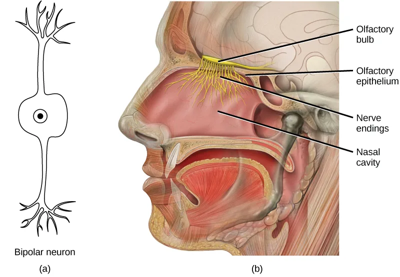
Extended description
Panel (a), on the left, is a simple outline drawing of a bipolar neuron: branching dendrites fan out from the top of an oval cell body, which contains a small dot marking the nucleus, and a mirroring set of branching processes extends from the bottom; the drawing is labeled ‘Bipolar neuron’ beneath it. Panel (b), on the right, is a color side-view illustration of a human head, cut away to show the nasal cavity. A cluster of yellow, thread-like nerve fibers fans down from a horizontal band at the top of the nasal cavity into the mucosa lining its inner wall. Four leader lines run from the illustration to labels at the right margin, top to bottom: ‘Olfactory bulb’ (the horizontal band the fibers emerge from), ‘Olfactory epithelium’ (the fibers’ upper branching region), ‘Nerve endings’ (the finer branches lower in the nasal cavity), and ‘Nasal cavity’ (the open space itself).
Evolution Connection. Pheromones.
A pheromone is a chemical released by an animal that affects the behavior or physiology of animals of the same species. Pheromonal signals can have profound effects on animals that inhale them, but pheromones apparently are not consciously perceived in the same way as other odors. There are several different types of pheromones, which are released in urine or as glandular secretions. Certain pheromones are attractants to potential mates, others are repellents to potential competitors of the same sex, and still others play roles in mother-infant attachment. Some pheromones can also influence the timing of puberty, modify reproductive cycles, and even prevent embryonic implantation. While the roles of pheromones in many nonhuman species are important, pheromones have become less important in human behavior over evolutionary time compared to their importance to organisms with more limited behavioral repertoires.
The vomeronasal organ (VNO, or Jacobson’s organ) is a tubular, fluid-filled, olfactory organ present in many vertebrate animals that sits adjacent to the nasal cavity. It is very sensitive to pheromones and is connected to the nasal cavity by a duct. When molecules dissolve in the mucosa of the nasal cavity, they then enter the VNO where the pheromone molecules among them bind with specialized pheromone receptors. Upon exposure to pheromones from their own species or others, many animals, including cats, may display the flehmen response, shown below, a curling of the upper lip that helps pheromone molecules enter the VNO.
Pheromonal signals are sent, not to the main olfactory bulb, but to a different neural structure that projects directly to the amygdala (recall that the amygdala is a brain center important in emotional reactions, such as fear). The pheromonal signal then continues to areas of the hypothalamus that are key to reproductive physiology and behavior. While some scientists assert that the VNO is apparently functionally vestigial in humans, even though there is a similar structure located near human nasal cavities, others are researching it as a possible functional system that may, for example, contribute to synchronization of menstrual cycles in people living in close proximity.

Taste
Detecting a taste (gustation) is fairly similar to detecting an odor (olfaction), given that both taste and smell rely on chemical receptors being stimulated by certain molecules. The primary organ of taste is the taste bud. A taste bud is a cluster of gustatory receptors (taste cells) that are located within the bumps on the tongue called papillae (singular: papilla), illustrated below. There are several structurally distinct papillae. Filiform papillae, which are located across the tongue, are tactile, providing friction that helps the tongue move substances, and contain no taste cells. In contrast, fungiform papillae, which are located mainly on the anterior two-thirds of the tongue, each contain one to eight taste buds and also have receptors for pressure and temperature.
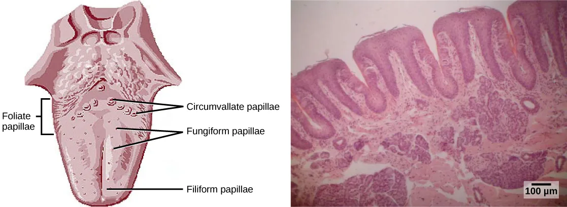
Extended description
Panel (a) shows a tongue viewed from above, widest at the back and tapering slightly at the tip, its surface stippled with small round bumps throughout the front two-thirds. At the left margin, a bracket spans the back-left edge of the tongue and is labeled ‘Foliate papillae.’ Near the back of the tongue, in an inverted-V arrangement, three leader lines converge on a row of larger ringed bumps, labeled ‘Circumvallate papillae.’ Two leader lines point to smaller bumps scattered across the middle of the tongue, labeled ‘Fungiform papillae.’ One leader line points down to the finely stippled front two-thirds of the tongue’s surface, labeled ‘Filiform papillae.’ Panel (b), to the right, is a magenta-and-purple light micrograph showing several tall, finger-like tissue folds rising side by side from a denser base layer, matching the foliate papillae’s ridged folds.
In addition to those two types of chemically and mechanically sensitive papillae are foliate papillae—leaf-like papillae located in parallel folds along the edges and toward the back of the tongue, as seen in the micrograph above. Foliate papillae contain about 1,300 taste buds within their folds. Finally, there are circumvallate papillae, which are wall-like papillae in the shape of an inverted “V” at the back of the tongue. Each of these papillae is surrounded by a groove and contains about 250 taste buds.
Each taste bud’s taste cells are replaced every 10 to 14 days. These are elongated cells with hair-like processes called microvilli at the tips that extend into the taste bud pore, illustrated below. Food molecules (tastants) are dissolved in saliva, and they bind with and stimulate the receptors on the microvilli. The receptors for tastants are located across the outer portion and front of the tongue, outside of the middle area where the filiform papillae are most prominent.
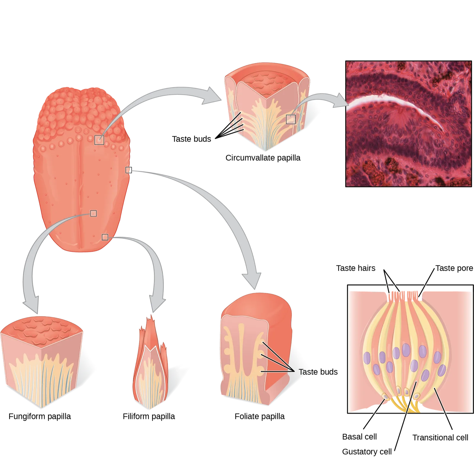
Extended description
At upper left, a tongue viewed from above, its surface stippled with small bumps, carries four small square markers. The uppermost marker, near the back of the tongue, connects by a gray arrow curving up and right to a wedge-shaped cutaway block labeled ‘Circumvallate papilla’: a dome-shaped bump ringed by a groove, with a fan of four fine leader lines converging on a ‘Taste buds’ label pointing into the block’s cut face. A second gray arrow leads from this block rightward into a magenta-and-purple light micrograph at the top right. A second marker, right of center on the tongue, connects by an arrow curving down and right to a wedge-shaped block at the bottom right labeled ‘Foliate papilla’: a block with vertical ridged folds, also labeled ‘Taste buds’ by converging leader lines. A third marker, lower-left on the tongue, connects by an arrow curving down and left to a bottom-left block labeled ‘Fungiform papilla’: a block with a bumpy dome top and no taste-bud leader lines drawn. A fourth marker, just below the third, connects by an arrow curving straight down to a bottom-middle block labeled ‘Filiform papilla’: a slender, pointed, bristle-like projection, also with no taste-bud leader lines. Separate from these four arrows, at the bottom right, a standalone cutaway shows a single onion-shaped taste bud: a bundle of parallel spindle-shaped cells studded with oval purple nuclei, capped at the surface by short hair-like projections. Three leader lines from the top: two fan from ‘Taste hairs’ to the two clusters of projections, and one runs from ‘Taste pore’ to the gap between them where the bundle meets the surface; three leader lines from the lower part of the bundle point to ‘Basal cell,’ ‘Gustatory cell,’ and ‘Transitional cell’ among the small oval cells at its base.
In humans, there are five primary tastes, and each taste has only one corresponding type of receptor. Thus, like olfaction, each receptor is specific to its stimulus (tastant). Transduction of the five tastes happens through different mechanisms that reflect the molecular composition of the tastant. A salty tastant (containing NaCl) provides the sodium ions (Na⁺) that enter the taste neurons and excite them directly. Sour tastants are acids and belong to the thermoreceptor protein family. Binding of an acid or other sour-tasting molecule triggers a change in the ion channel and these increase hydrogen ion (H⁺) concentrations in the taste neurons, thus depolarizing them. Sweet, bitter, and umami tastants require a G-protein coupled receptor. These tastants bind to their respective receptors, thereby exciting the specialized neurons associated with them.
Both tasting abilities and sense of smell change with age. In humans, the senses decline dramatically by age 50 and continue to decline. A child may find a food to be too spicy, whereas an elderly person may find the same food to be bland and unappetizing.
Link to Learning
View this animation, which shows how the sense of taste works.
Smell and Taste in the Brain
Olfactory neurons project from the olfactory epithelium to the olfactory bulb as thin, unmyelinated axons. The olfactory bulb is composed of neural clusters called glomeruli, and each glomerulus receives signals from one type of olfactory receptor, so each glomerulus is specific to one odorant. From glomeruli, olfactory signals travel directly to the olfactory cortex and then to the frontal cortex and the thalamus. Recall that this is a different path from most other sensory information, which is sent directly to the thalamus before ending up in the cortex. Olfactory signals also travel directly to the amygdala, thereafter reaching the hypothalamus, thalamus, and frontal cortex. The last structure that olfactory signals directly travel to is a cortical center in the temporal lobe structure important in spatial, autobiographical, declarative, and episodic memories. Olfaction is finally processed by areas of the brain that deal with memory, emotions, reproduction, and thought.
Taste neurons project from taste cells in the tongue, esophagus, and palate to the medulla, in the brainstem. From the medulla, taste signals travel to the thalamus and then to the primary gustatory cortex. Information from different regions of the tongue is segregated in the medulla, thalamus, and cortex.
Summary
There are five primary tastes in humans: sweet, sour, bitter, salty, and umami. Each taste has its own receptor type that responds only to that taste. Tastants enter the body and are dissolved in saliva. Taste cells are located within taste buds, which are found on three of the four types of papillae in the mouth.
Regarding olfaction, there are many thousands of odorants, but humans detect only about 10,000. Like taste receptors, olfactory receptors are each responsive to only one odorant. Odorants dissolve in nasal mucosa, where they excite their corresponding olfactory sensory cells. When these cells detect an odorant, they send their signals to the main olfactory bulb and then to other locations in the brain, including the olfactory cortex.
Key terms
- bipolar neuron — neuron with two processes extending from the cell body, typically in opposite directions.
- glomerulus — in the olfactory bulb, one of the two neural clusters that receives signals from one type of olfactory receptor.
- gustation — sense of taste.
- odorant — airborne molecule that stimulates an olfactory receptor.
- olfaction — sense of smell.
- olfactory bulb — neural structure in the vertebrate brain that receives signals from olfactory receptors.
- olfactory epithelium — specialized tissue in the nasal cavity where olfactory receptors are located.
- olfactory receptor — dendrite of a specialized neuron.
- papilla — one of the small bump-like projections from the tongue.
- pheromone — substance released by an animal that can affect the physiology or behavior of other animals.
- tastant — food molecule that stimulates gustatory receptors.
- taste bud — clusters of taste cells.
- umami — one of the five basic tastes, which is described as “savory” and which may be largely the taste of L-glutamate.
Practice
Explain in what way smell and taste stimuli differ from other sensory stimuli
All sensory signals except _____ travel to the _____ in the brain before the cerebral cortex.
Recall which of the special senses sends its signal directly to the cerebral cortex, bypassing the structure that relays every other sense first.From the perspective of the recipient of the signal, in what ways do pheromones differ from other odorants?
Show model answer
Did your answer mention:
The sense of taste is also called ________.
This term shares a Latin root with words like ‘gustatory’ and ‘degustation.’The sense of smell is also called ________.
This term shares a root with ‘olfactory,’ the adjective used throughout this section for the nose’s receptors, bulb, and epithelium.Identify the five primary tastes that can be distinguished by humans
Which of the following has the fewest taste receptors?
This is the papilla type the section describes as purely tactile, providing friction rather than sensing anything chemically.How many different taste molecules do taste cells each detect?
Compare this to how many receptor types respond to a given odorant — the module states the same rule for taste cells.Salty foods activate the taste cells by _____.
Reread how sodium ions from a salty tastant interact with the taste neuron, and compare it to how sour tastants — which involve a different ion — are described as working instead.How is the ability to recognize the umami taste an evolutionary advantage?
Reread what molecule the taste of umami is attributed to, and what class of nutrient that molecule belongs to.What might be the effect on an animal of not being able to perceive taste?
Show model answer
Did your answer mention:
Explain in anatomical terms why a dog’s sense of smell is more acute than a human’s
A few recent cancer detection studies have used trained dogs to detect lung cancer in urine samples. What is the hypothesis behind this study? Why are dogs a better choice of detectors in this study than humans?
Show model answer
Did your answer mention:
A dendrite of a specialized neuron that responds to specific inhaled molecules is called an ________.
Humans have about 12 million of these; bloodhounds have about 4 billion.The specialized tissue in the back of the nasal cavity where olfactory receptors are located is called the ________.
This tissue spans about 5 cm² in humans but is many times larger in bloodhounds.A neuron with two processes extending from the cell body, typically in opposite directions, is called a ________.
Every olfactory sensory neuron in the nose is built this way, with a single dendrite buried in the epithelium and an axon leading to the olfactory bulb.This section is adapted from Biology 2e, Section 36.3: Taste and Smell by Mary Ann Clark, Jung Choi, Matthew Douglas, and OpenStax, © OpenStax, licensed under CC BY-NC-SA 4.0. Access the original for free at openstax.org. Changes: figures re-encoded as WebP; two figures re-kinded from the manifest’s file-extension “photo” guess to “diagram” (Figure_36_03_01, a bipolar-neuron schematic paired with a labeled anatomical illustration, and Figure_36_03_03, a labeled tongue diagram paired with a micrograph — neither a captured photograph on its own); alts rewritten as plain visual descriptions in place of the manifest’s bare source alts; a longdesc added to every diagram whose meaning is not carried by its one-line caption — the bipolar-neuron/olfactory-anatomy figure, the papillae-location figure, and the taste-bud composite figure — transcribing each drawing’s labels and leader lines in reading order and counting its panels and arrows; in-text pointers to figures (“as illustrated in”, specific figure numbers) replaced with “illustrated below,” “shown below,” or “above,” since Hugo does not number figures; the Pheromones note rendered as an Evolution Connection callout with its bold name and italic title, its three paragraphs and figure kept inside the box as printed; the Link to Learning note rendered as a callout with descriptive link text in place of the source’s bare “animation” anchor, the external URL kept as printed; the end-of-section Review Questions and Critical Thinking Questions (drawn from the chapter’s back-of-book exercise sets, which key to this section) adapted into the closing interactive Practice block (multiple choice and self-check respectively); rubric checkpoints added to each self-check, decomposing its model answer (the source solution) into check-off clauses with no new claims; five key-term recall items added from the glossary (gustation, olfaction, olfactory receptor, olfactory epithelium, bipolar neuron); ion notation set as Unicode superscripts (Na⁺, H⁺) and the area unit as a Unicode superscript (5 cm²) per house notation; the source’s “detected by more than more receptor” corrected to “more than one receptor” — reported as a source defect.