Hearing and Vestibular Sensation
By the end of this section, you will be able to:
- Describe the relationship of amplitude and frequency of a sound wave to attributes of sound
- Trace the path of sound through the auditory system to the site of transduction of sound
- Identify the structures of the vestibular system that respond to gravity
Audition, or hearing, is important to humans and to other animals for many different interactions. It enables an organism to detect and receive information about danger, such as an approaching predator, and to participate in communal exchanges like those concerning territories or mating. On the other hand, although it is physically linked to the auditory system, the vestibular system is not involved in hearing. Instead, an animal’s vestibular system detects its own movement, both linear and angular acceleration and deceleration, and balance.
Sound
Auditory stimuli are sound waves, which are mechanical, pressure waves that move through a medium, such as air or water. There are no sound waves in a vacuum since there are no air molecules to move in waves. The speed of sound waves differs, based on altitude, temperature, and medium, but at sea level and a temperature of 20 °C (68 °F), sound waves travel in the air at about 343 meters per second.
As is true for all waves, there are four main characteristics of a sound wave: frequency, wavelength, period, and amplitude. Frequency is the number of waves per unit of time, and in sound is heard as pitch. High-frequency (≥15,000 Hz) sounds are higher-pitched (short wavelength) than low-frequency (long wavelengths; ≤100 Hz) sounds. For sound frequency, the most commonly used unit is hertz (Hz), or cycles per second. Most humans can perceive sounds with frequencies between 30 and 20,000 Hz. Everyone’s ability to hear high frequencies decreases with age. Dogs detect up to about 40,000 Hz; cats, 60,000 Hz; bats, 100,000 Hz; and dolphins 150,000 Hz, and American shad (Alosa sapidissima), a fish, can hear 180,000 Hz. Those frequencies above the human range are called ultrasound.
Amplitude, or the dimension of a wave from the resting position (the equilibrium) to the peak, in sound is heard as volume and is illustrated below. The sound waves of louder sounds have greater amplitude than those of softer sounds. For sound, volume is measured in decibels (dB). The softest sound that a human can hear is the zero point. Humans speak normally at 60 decibels.
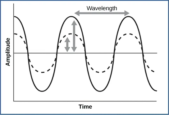
Extended description
A horizontal centerline (equilibrium) runs through the middle of the graph. A solid wave traces three full crests and troughs above and below it; a dashed wave of the same wavelength and in phase with the solid wave traces smaller crests and troughs that stay inside the solid wave’s envelope. Above the second and third solid crests, a horizontal double-headed arrow labeled ‘Wavelength’ spans from one crest to the next. Between the first and second crests, a shorter vertical double-headed arrow labeled ‘Amplitude’ spans from the centerline up to the dashed wave’s crest.
Reception of Sound
In mammals, sound waves are collected by the external, cartilaginous part of the ear called the auricle, then travel through the auditory canal and cause vibration of the thin diaphragm called the tympanum or ear drum, the innermost part of the outer ear (illustrated below). Interior to the tympanum is the middle ear. The middle ear holds three small bones called the ossicles, which transfer energy from the moving tympanum to the inner ear. The three ossicles are the malleus (also known as the hammer), the incus (the anvil), and stapes (the stirrup). The aptly named stapes looks very much like a stirrup. The three ossicles are unique to mammals, and each plays a role in hearing. The malleus attaches at three points to the interior surface of the tympanic membrane. The incus attaches the malleus to the stapes. In humans, the stapes is not long enough to reach the tympanum. If we did not have the malleus and the incus, then the vibrations of the tympanum would never reach the inner ear. These bones also function to collect force and amplify sounds. The ear ossicles are homologous to bones in a fish mouth: the bones that support gills in fish are thought to be adapted for use in the vertebrate ear over evolutionary time. Many animals (frogs, reptiles, and birds, for example) use the stapes of the middle ear to transmit vibrations to the inner ear.
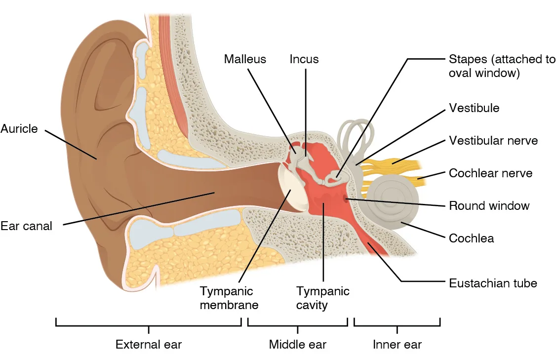
Extended description
Left to right: ‘Auricle’ labels the outer visible fold of the ear, and ‘Ear canal’ labels the tube leading inward to the ‘Tympanic membrane,’ a pale oval sealing the canal’s inner end. Behind it, in the air-filled ‘Tympanic cavity,’ two small bones are labeled ‘Malleus’ and ‘Incus’; a third is captioned ‘Stapes (attached to oval window)’ at the border with the inner ear. Continuing right, ‘Vestibule’ and ‘Vestibular nerve’ label the looping canal structure and its nerve, ‘Cochlear nerve’ labels a separate nerve bundle, ‘Round window’ labels a small membrane below the cochlea, ‘Cochlea’ labels the coiled snail-shell structure, and ‘Eustachian tube’ labels a duct running down and back from the tympanic cavity toward the throat. Three brackets beneath the drawing span, left to right: ‘External ear’ (auricle through tympanic membrane), ‘Middle ear’ (the tympanic cavity), and ‘Inner ear’ (vestibule through cochlea).
Transduction of Sound
Vibrating objects, such as vocal cords, create sound waves or pressure waves in the air. When these pressure waves reach the ear, the ear transduces this mechanical stimulus (pressure wave) into a nerve impulse (electrical signal) that the brain perceives as sound. The pressure waves strike the tympanum, causing it to vibrate. The mechanical energy from the moving tympanum transmits the vibrations to the three bones of the middle ear. The stapes transmits the vibrations to a thin diaphragm called the oval window, which is the outermost structure of the inner ear. The structures of the inner ear are found in the labyrinth, a bony, hollow structure that is the most interior portion of the ear. Here, the energy from the sound wave is transferred from the stapes through the flexible oval window and to the fluid of the cochlea. The vibrations of the oval window create pressure waves in the fluid (perilymph) inside the cochlea. The cochlea is a whorled structure, like the shell of a snail, and it contains receptors for transduction of the mechanical wave into an electrical signal (illustrated below). Inside the cochlea, the basilar membrane is a mechanical analyzer that runs the length of the cochlea, curling toward the cochlea’s center.
The mechanical properties of the basilar membrane change along its length, such that it is thicker, tauter, and narrower at the outside of the whorl (where the cochlea is largest), and thinner, floppier, and broader toward the apex, or center, of the whorl (where the cochlea is smallest). Different regions of the basilar membrane vibrate according to the frequency of the sound wave conducted through the fluid in the cochlea. For these reasons, the fluid-filled cochlea detects different wave frequencies (pitches) at different regions of the membrane. When the sound waves in the cochlear fluid contact the basilar membrane, it flexes back and forth in a wave-like fashion. Above the basilar membrane is the tectorial membrane.
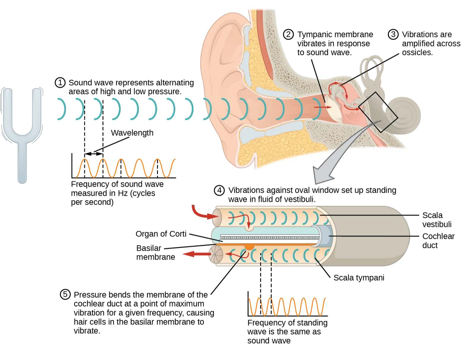
Extended description
Five numbered captions trace the sequence. ① ‘Sound wave represents alternating areas of high and low pressure,’ beside a tuning fork at far left and a row of teal wave-crest arcs traveling right toward the ear; below this, a small graph plots ‘Frequency of sound wave measured in Hz (cycles per second),’ with ‘Wavelength’ labeling the span between two crests. ② ‘Tympanic membrane vibrates in response to sound wave,’ with an arrow into the tympanic membrane inside the cutaway ear. ③ ‘Vibrations are amplified across ossicles,’ with an arrow crossing the three middle-ear bones. ④ ‘Vibrations against oval window set up standing wave in fluid of vestibuli,’ where a large gray arrow points down from the ear into a cutaway cylinder labeled, top to bottom, ‘Scala vestibuli,’ ‘Cochlear duct’ (containing ‘Organ of Corti’ and ‘Basilar membrane’), and ‘Scala tympani.’ ⑤ ‘Pressure bends the membrane of the cochlear duct at a point of maximum vibration for a given frequency, causing hair cells in the basilar membrane to vibrate,’ beside a second small graph plotting ‘Frequency of standing wave is the same as sound wave.’
Cochlear implants can restore hearing in people who have a nonfunctional cochlea. The implant consists of a microphone that picks up sound. A speech processor selects sounds in the range of human speech, and a transmitter converts these sounds to electrical impulses, which are then sent to the auditory nerve. Which of the following types of hearing loss would not be restored by a cochlear implant?
A cochlear implant sends its electrical signal straight onto a nerve, bypassing the cochlea entirely — think about what still has to be working for that signal to reach the brain at all.The site of transduction is in the organ of Corti (spiral organ). It is composed of hair cells held in place above the basilar membrane like flowers projecting up from soil, with their exposed short, hair-like stereocilia contacting or embedded in the tectorial membrane above them. The inner hair cells are the primary auditory receptors and exist in a single row, numbering approximately 3,500. The stereocilia from inner hair cells extend into small dimples on the tectorial membrane’s lower surface. The outer hair cells are arranged in three or four rows. They number approximately 12,000, and they function to fine tune incoming sound waves. The longer stereocilia that project from the outer hair cells actually attach to the tectorial membrane. All of the stereocilia are mechanoreceptors, and when bent by vibrations they respond by opening a gated ion channel (refer to the hair cell diagram below). As a result, the hair cell membrane is depolarized, and a signal is transmitted to the cochlear nerve. Intensity (volume) of sound is determined by how many hair cells at a particular location are stimulated.
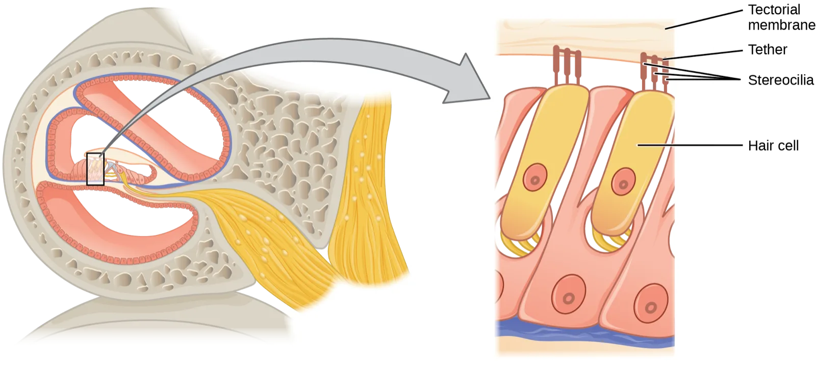
Extended description
In the right, zoomed panel, top to bottom: ‘Tectorial membrane’ labels the pale horizontal band across the top. Below it, two tall yellow hair cells stand side by side, each topped with a cluster of finger-like projections that reach up and touch the tectorial membrane; ‘Tether’ labels the short connecting strands linking the tips of neighboring projections together, and ‘Stereocilia’ labels the projections themselves. ‘Hair cell’ labels the tall cell body beneath the stereocilia bundle, each with a round nucleus visible inside it.
The hair cells are arranged on the basilar membrane in an orderly way. The basilar membrane vibrates in different regions, according to the frequency of the sound waves impinging on it. Likewise, the hair cells that lay above it are most sensitive to a specific frequency of sound waves. Hair cells can respond to a small range of similar frequencies, but they require stimulation of greater intensity to fire at frequencies outside of their optimal range. The difference in response frequency between adjacent inner hair cells is about 0.2 percent. Compare that to adjacent piano strings, which are about six percent different. Place theory, which is the model for how biologists think pitch detection works in the human ear, states that high frequency sounds selectively vibrate the basilar membrane of the inner ear near the entrance port (the oval window). Lower frequencies travel farther along the membrane before causing appreciable excitation of the membrane. The basic pitch-determining mechanism is based on the location along the membrane where the hair cells are stimulated. The place theory is the first step toward an understanding of pitch perception. Considering the extreme pitch sensitivity of the human ear, it is thought that there must be some auditory “sharpening” mechanism to enhance the pitch resolution.
When sound waves produce fluid waves inside the cochlea, the basilar membrane flexes, bending the stereocilia that attach to the tectorial membrane. Their bending results in action potentials in the hair cells, and auditory information travels along the neural endings of the bipolar neurons of the hair cells (collectively, the auditory nerve) to the brain. When the hairs bend, they release an excitatory neurotransmitter at a synapse with a sensory neuron, which then conducts action potentials to the central nervous system. The cochlear branch of the vestibulocochlear cranial nerve sends information on hearing. The auditory system is very refined, and there is some modulation or “sharpening” built in. The brain can send signals back to the cochlea, resulting in a change of length in the outer hair cells, sharpening or dampening the hair cells’ response to certain frequencies.
Higher Processing
The inner hair cells are most important for conveying auditory information to the brain. About 90 percent of the afferent neurons carry information from inner hair cells, with each hair cell synapsing with 10 or so neurons. Outer hair cells connect to only 10 percent of the afferent neurons, and each afferent neuron innervates many hair cells. The afferent, bipolar neurons that convey auditory information travel from the cochlea to the medulla, through the pons and midbrain in the brainstem, finally reaching the primary auditory cortex in the temporal lobe.
Vestibular Information
The stimuli associated with the vestibular system are linear acceleration (gravity) and angular acceleration and deceleration. Gravity, acceleration, and deceleration are detected by evaluating the inertia on receptive cells in the vestibular system. Gravity is detected through head position. Angular acceleration and deceleration are expressed through turning or tilting of the head.
The vestibular system has some similarities with the auditory system. It utilizes hair cells just like the auditory system, but it excites them in different ways. There are five vestibular receptor organs in the inner ear: the utricle, the saccule, and three semicircular canals. Together, they make up what’s known as the vestibular labyrinth that is shown below. The utricle and saccule respond to acceleration in a straight line, such as gravity. The roughly 30,000 hair cells in the utricle and 16,000 hair cells in the saccule lie below a gelatinous layer, with their stereocilia projecting into the gelatin. Embedded in this gelatin are calcium carbonate crystals—like tiny rocks. When the head is tilted, the crystals continue to be pulled straight down by gravity, but the new angle of the head causes the gelatin to shift, thereby bending the stereocilia. The bending of the stereocilia stimulates the neurons, and they signal to the brain that the head is tilted, allowing the maintenance of balance. It is the vestibular branch of the vestibulocochlear cranial nerve that deals with balance.
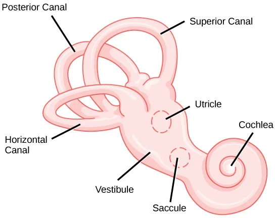
Extended description
At upper left, ‘Posterior Canal’ labels one looping tube; at upper right, ‘Superior Canal’ labels a second loop that interlocks with it near the top. At lower left, ‘Horizontal Canal’ labels a third, smaller loop crossing beneath the other two. All three canals converge into the central ‘Vestibule,’ inside which two small dashed-outline circles are labeled ‘Utricle’ (upper) and ‘Saccule’ (lower, closer to the cochlea). At the right end of the vestibule, the structure narrows and coils into the snail-shell-shaped ‘Cochlea.’
The fluid-filled semicircular canals are tubular loops set at oblique angles. They are arranged in three spatial planes. The base of each canal has a swelling that contains a cluster of hair cells. The hairs project into a gelatinous cap called the cupula and monitor angular acceleration and deceleration from rotation. They would be stimulated by driving your car around a corner, turning your head, or falling forward. One canal lies horizontally, while the other two lie at about 45 degree angles to the horizontal axis, as illustrated above. When the brain processes input from all three canals together, it can detect angular acceleration or deceleration in three dimensions. When the head turns, the fluid in the canals shifts, thereby bending stereocilia and sending signals to the brain. Upon cessation of accelerating or decelerating—or just moving—the movement of the fluid within the canals slows or stops. For example, imagine holding a glass of water. When moving forward, water may splash backwards onto the hand, and when motion has stopped, water may splash forward onto the fingers. While in motion, the water settles in the glass and does not splash. Note that the canals are not sensitive to velocity itself, but to changes in velocity, so moving forward at 60 mph with your eyes closed would not give the sensation of movement, but suddenly accelerating or braking would stimulate the receptors.
Higher Processing
Hair cells from the utricle, saccule, and semicircular canals also communicate through bipolar neurons to the cochlear nucleus in the medulla. Cochlear neurons send descending projections to the spinal cord and ascending projections to the pons, thalamus, and cerebellum. Connections to the cerebellum are important for coordinated movements. There are also projections to the temporal cortex, which account for feelings of dizziness; projections to autonomic nervous system areas in the brainstem, which account for motion sickness; and projections to the primary somatosensory cortex, which monitors subjective measurements of the external world and self-movement. People with lesions in the vestibular area of the somatosensory cortex see vertical objects in the world as being tilted. Finally, the vestibular signals project to certain optic muscles to coordinate eye and head movements.
Link to Learning
Click through this interactive tutorial to review the parts of the ear and how they function to process sound.
Summary
Audition is important for territory defense, predation, predator defense, and communal exchanges. The vestibular system, which is not auditory, detects linear acceleration and angular acceleration and deceleration. Both the auditory system and vestibular system use hair cells as their receptors.
Auditory stimuli are sound waves. The sound wave energy reaches the outer ear (auricle, canal, tympanum), and vibrations of the tympanum send the energy to the middle ear. The middle ear bones shift and the stapes transfers mechanical energy to the oval window of the fluid-filled inner ear cochlea. Once in the cochlea, the energy causes the basilar membrane to flex, thereby bending the stereocilia on receptor hair cells. This activates the receptors, which send their auditory neural signals to the brain.
The vestibular system has five parts that work together to provide the sense of direction, thus helping to maintain balance. The utricle and saccule measure head orientation: their calcium carbonate crystals shift when the head is tilted, thereby activating hair cells. The semicircular canals work similarly, such that when the head is turned, the fluid in the canals bends stereocilia on hair cells. The vestibular hair cells also send signals to the thalamus and to the somatosensory cortex, but also to the cerebellum, the structure above the brainstem that plays a large role in timing and coordination of movement.
Key terms
- audition — sense of hearing.
- auricle — cartilaginous outer ear.
- basilar membrane — stiff structure in the cochlea that indirectly anchors auditory receptors.
- cochlea — whorled structure that contains receptors for transduction of the mechanical wave into an electrical signal.
- incus — (also, anvil) second of the three bones of the middle ear.
- inner ear — innermost part of the ear; consists of the cochlea and the vestibular system.
- labyrinth — bony, hollow structure that is the most internal part of the ear; contains the sites of transduction of auditory and vestibular information.
- malleus — (also, hammer) first of the three bones of the middle ear.
- middle ear — part of the hearing apparatus that functions to transfer energy from the tympanum to the oval window of the inner ear.
- organ of Corti — in the basilar membrane, the site of the transduction of sound, a mechanical wave, to a neural signal.
- ossicle — one of the three bones of the middle ear.
- outer ear — part of the ear that consists of the auricle, ear canal, and tympanum and which conducts sound waves into the middle ear.
- oval window — thin diaphragm between the middle and inner ears that receives sound waves from contact with the stapes bone of the middle ear.
- semicircular canal — one of three half-circular, fluid-filled tubes in the vestibular labyrinth that monitors angular acceleration and deceleration.
- stapes — (also, stirrup) third of the three bones of the middle ear.
- stereocilia — in the auditory system, hair-like projections from hair cells that help detect sound waves.
- tectorial membrane — cochlear structure that lies above the hair cells and participates in the transduction of sound at the hair cells.
- tympanum — (also, tympanic membrane or ear drum) thin diaphragm between the outer and middle ears.
- ultrasound — sound frequencies above the human detectable ceiling of approximately 20,000 Hz.
Practice
Describe the relationship of amplitude and frequency of a sound wave to attributes of sound
In sound, pitch is measured in _____, and volume is measured in _____.
This section names the wave-cycles-per-second unit when it first describes frequency, and it names the loudness unit later, when it says how many of them normal human speech registers.How would a rise in altitude likely affect the speed of a sound transmitted through air? Why?
Show model answer
Did your answer mention:
Sound frequencies above the human detectable ceiling of approximately 20,000 Hz are called ________.
Several other animals in this section — dogs, cats, bats, and dolphins among them — can hear frequencies in this range that stay silent to human ears.Trace the path of sound through the auditory system to the site of transduction of sound
Auditory hair cells are indirectly anchored to the _____.
The glossary describes this structure as the one that ‘indirectly anchors’ the receptors — distinguish it from the membrane the stereocilia merely touch or embed into just above it.How does the structure of the ear allow a person to determine where a sound originates?
Show model answer
Did your answer mention:
The sense of hearing is called ________.
This is the term the section uses throughout, rather than just saying ‘hearing.’The cartilaginous outer ear is called the ________.
Sound waves are collected by this structure before traveling through the auditory canal to the eardrum.The whorled structure that contains receptors for transduction of the mechanical wave into an electrical signal is called the ________.
This structure is shaped like the shell of a snail.In the basilar membrane, the site of the transduction of sound, a mechanical wave, to a neural signal is called the ________.
This structure sits above the basilar membrane and holds the hair cells whose stereocilia are embedded in or touch the membrane above them.One of the three bones of the middle ear is called an ________.
These three tiny bones—nicknamed the hammer, anvil, and stirrup—transfer energy from the tympanum to the inner ear.The thin diaphragm between the outer and middle ears is called the ________.
Incoming sound waves cause this diaphragm to vibrate first, before three tiny bones pass the vibration onward.Identify the structures of the vestibular system that respond to gravity
Benign Paroxysmal Positional Vertigo is a disorder where some of the calcium carbonate crystals in the utricle migrate into the semicircular canals. Why does this condition cause periods of dizziness?
Semicircular canals normally detect turning and rotation, not the straight-down pull that the utricle and saccule respond to — think about what the migrated crystals now do inside them.Which of the following are found both in the auditory system and the vestibular system?
Both systems transduce a mechanical stimulus into a nerve signal using the same kind of receptor cell — one option is found only in the cochlea, one only in the vestibular labyrinth, and one is shared by both.How might being in a place with less gravity than Earth has (such as Earth’s moon) affect vestibular sensation, and why?
Show model answer
Did your answer mention:
One of three half-circular, fluid-filled tubes in the vestibular labyrinth that monitors angular acceleration and deceleration is called a ________.
One of these three tubes lies horizontally, while the other two sit at about 45-degree angles to it.The ________ and saccule measure head orientation: their calcium carbonate crystals shift when the head is tilted, thereby activating hair cells.
This vestibular receptor organ, along with the saccule, lies in the vestibule and responds to straight-line acceleration such as gravity — not to rotation.This section is adapted from Biology 2e, Section 36.4: Hearing and Vestibular Sensation by Mary Ann Clark, Jung Choi, Matthew Douglas, and OpenStax, © OpenStax, licensed under CC BY-NC-SA 4.0. Access the original for free at openstax.org. Changes: figures re-encoded as WebP; two figures re-kinded from the manifest’s file-extension “photo” guess to “diagram” (Figure_36_04_01 and Figure_36_04_04-bbc7 are both hand-drawn line graphics — a wavelength/amplitude graph and a labeled anatomical illustration — neither a captured photograph); a longdesc added to every diagram whose meaning is not carried by its one-line caption — the wavelength/amplitude graph, the labeled ear cross-section, the cochlear-implant Visual Connection’s five-step sound-transduction diagram, the hair-cell diagram, and the vestibular-labyrinth illustration — transcribing each drawing’s own printed labels and counting its panels, arrows, or numbered steps; in-text pointers to figures (“Figure 36.12” through “Figure 36.16”) replaced with “shown below,” “illustrated below,” or “illustrated above,” since Hugo does not number figures; a corrupted sentence in the pinned CNXML — “High-frequency (≥15.,ds.” — present identically in the source PDF and the live openstax.org page, restored to “High-frequency (≥15,000 Hz) sounds are higher-pitched (short wavelength) than low-frequency (long wavelengths; ≤100 Hz) sounds,” matching an independent mirror of the same OpenStax text (courses.lumenlearning.com) — reported as a source defect; the note wrapping the cochlear-implant Visual Connection rendered as its figure followed by a multiple choice, kept in the body in the Transduction of Sound section — the note copy and the <exercise> copy print identical question and option wording, so no adjudication was needed; the end-of-section Review Questions and Critical Thinking Questions adapted into the closing interactive Practice block (multiple choice and self-check respectively); rubric checkpoints added to each self-check, decomposing its model answer (the source solution) into check-off clauses with no new claims; eight key-term recall items added from the glossary (audition, auricle, cochlea, organ of Corti, ossicle, tympanum, ultrasound, semicircular canal), covering a representative subset of the section’s nineteen glossary terms rather than all of them, the other eleven (basilar membrane, incus, inner ear, labyrinth, malleus, middle ear, outer ear, oval window, stapes, stereocilia, tectorial membrane) appear only in the Key terms list and the prose, basilar membrane is also the key of a Review Question, and malleus/incus/stapes are near-duplicates of the ossicle item; one summary-derived textin cloze added to the vestibular objective’s group (blanking “utricle” from the closing summary’s sentence “The utricle and saccule measure head orientation…”).