How Hormones Work
By the end of this section, you will be able to:
- Explain how hormones work
- Discuss the role of different types of hormone receptors
Hormones mediate changes in target cells by binding to specific hormone receptors. In this way, even though hormones circulate throughout the body and come into contact with many different cell types, they only affect cells that possess the necessary receptors. Receptors for a specific hormone may be found on many different cells or may be limited to a small number of specialized cells. For example, thyroid hormones act on many different tissue types, stimulating metabolic activity throughout the body. Cells can have many receptors for the same hormone but often also possess receptors for different types of hormones. The number of receptors that respond to a hormone determines the cell’s sensitivity to that hormone, and the resulting cellular response. Additionally, the number of receptors that respond to a hormone can change over time, resulting in increased or decreased cell sensitivity. In up-regulation, the number of receptors increases in response to prolonged low hormone levels, making the cell more sensitive to the hormone and allowing for more cellular activity. When the number of receptors decreases in response to rising hormone levels, called down-regulation, cellular activity is reduced.
Receptor binding alters cellular activity and results in an increase or decrease in normal body processes. Depending on the location of the protein receptor on the target cell and the chemical structure of the hormone, hormones can mediate changes directly by binding to intracellular hormone receptors and modulating gene transcription, or indirectly by binding to cell surface receptors and stimulating signaling pathways.
Intracellular Hormone Receptors
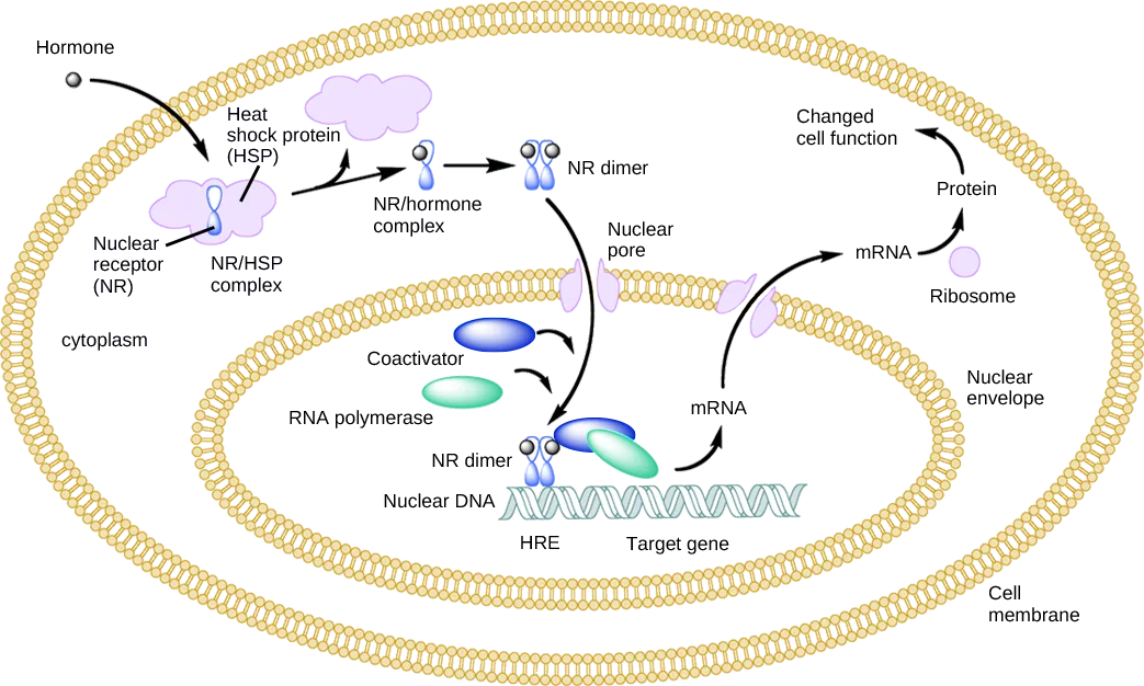
Lipid-derived (soluble) hormones such as steroid hormones diffuse across the membranes of the endocrine cell. Once outside the cell, they bind to transport proteins that keep them soluble in the bloodstream. At the target cell, the hormones are released from the carrier protein and diffuse across the lipid bilayer of the plasma membrane of cells. The steroid hormones pass through the plasma membrane of a target cell and adhere to intracellular receptors residing in the cytoplasm or in the nucleus. The cell signaling pathways induced by the steroid hormones regulate specific genes on the cell’s DNA. The hormones and receptor complex act as transcription regulators by increasing or decreasing the synthesis of mRNA molecules of specific genes. This, in turn, determines the amount of corresponding protein that is synthesized by altering gene expression. This protein can be used either to change the structure of the cell or to produce enzymes that catalyze chemical reactions. In this way, the steroid hormone regulates specific cell processes as illustrated below.
Extended description
The illustration is a cutaway of a cell inside a cell: an outer double ring of beaded phospholipids labeled ‘Cell membrane’ encloses the ‘cytoplasm’, which in turn encloses a second double ring labeled ‘Nuclear envelope’ around the nucleus. Reading left to right: a small ball labeled ‘Hormone’ has an arrow pointing to a pink cloud shape labeled ‘Heat shock protein (HSP)’ that encloses a blue Y-shaped ‘Nuclear receptor (NR)’; together they are labeled ‘NR/HSP complex’. An arrow leads to a smaller blue receptor still holding the hormone ball, labeled ‘NR/hormone complex’. An arrow leads to two of those complexes paired side by side, labeled ‘NR dimer’. A curved arrow carries that NR dimer through a purple pore shape in the nuclear envelope labeled ‘Nuclear pore’ and into the nucleus, where the same paired shape is labeled ‘NR dimer’ again, sitting beside a blue-green DNA double helix labeled ‘Nuclear DNA’, with ‘HRE’ and ‘Target gene’ labeling two points along it. Two ovals, a blue ‘Coactivator’ and a green ‘RNA polymerase’, have arrows pointing into that same NR dimer/DNA site. An arrow leads from there to a small oval labeled ‘mRNA’ inside the nucleus; another arrow carries a second ‘mRNA’ oval out through a second, unlabeled pore shape in the nuclear envelope into the cytoplasm, where an arrow leads to a ‘Ribosome’, then to ‘Protein’, then to ‘Changed cell function’ at the top right.
Heat shock proteins (HSP) are so named because they help refold misfolded proteins. In response to increased temperature (a “heat shock”), heat shock proteins are activated by release from the NR/HSP complex. At the same time, transcription of HSP genes is activated. Why do you think the cell responds to a heat shock by increasing the activity of proteins that help refold misfolded proteins?
Show model answer
Did your answer mention:
Other lipid-soluble hormones that are not steroid hormones, such as vitamin D and thyroxine, have receptors located in the nucleus. While thyroxine is mostly hydrophobic, its passage across the membrane is dependent on transporter protein. Vitamin D diffuses across both the plasma membrane and the nuclear envelope. Once in the cell, both hormones bind to receptors in the nucleus. The hormone-receptor complex stimulates transcription of specific genes.
Plasma Membrane Hormone Receptors
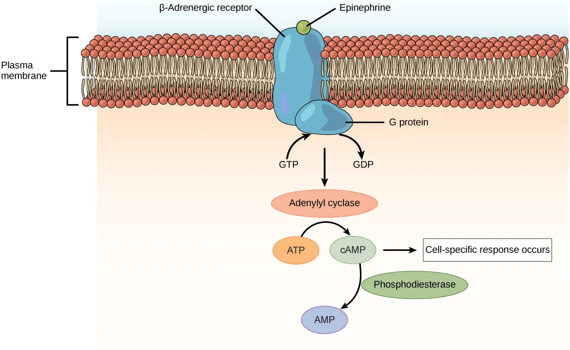
Amino acid-derived hormones (with the exception of thyroxine) and polypeptide hormones are not lipid-derived (lipid-soluble) and therefore cannot diffuse through the plasma membrane of cells. Lipid insoluble hormones bind to receptors on the outer surface of the plasma membrane, via plasma membrane hormone receptors. Unlike steroid hormones, lipid insoluble hormones do not directly affect the target cell because they cannot enter the cell and act directly on DNA. Binding of these hormones to a cell surface receptor results in activation of a signaling pathway; this triggers intracellular activity and carries out the specific effects associated with the hormone. In this way, nothing passes through the cell membrane; the hormone that binds at the surface remains at the surface of the cell while the intracellular product remains inside the cell. The hormone that initiates the signaling pathway is called a first messenger, which activates a second messenger in the cytoplasm, as illustrated below.
Extended description
A bracket at left labels the lipid bilayer ‘Plasma membrane’. Spanning it is a blue receptor labeled ‘β-Adrenergic receptor’, with a small green ball labeled ‘Epinephrine’ bound at its outer, extracellular end. Below the membrane, the receptor connects to a second blue shape labeled ‘G protein’, with one arrow labeled ‘GTP’ pointing into it and a second arrow labeled ‘GDP’ pointing away from it. An arrow leads down from the G protein to an orange oval labeled ‘Adenylyl cyclase’. A curved arrow connects an orange oval labeled ‘ATP’ to a green oval labeled ‘cAMP’, passing by the adenylyl cyclase oval. From ‘cAMP’, one arrow points right to a boxed label reading ‘Cell-specific response occurs’, and a second arrow curves down past a green oval labeled ‘Phosphodiesterase’ to a purple oval labeled ‘AMP’.
One very important second messenger is cyclic AMP (cAMP). When a hormone binds to its membrane receptor, a G-protein that is associated with the receptor is activated; G-proteins are proteins separate from receptors that are found in the cell membrane. When a hormone is not bound to the receptor, the G-protein is inactive and is bound to guanosine diphosphate, or GDP. When a hormone binds to the receptor, the G-protein is activated by binding guanosine triphosphate, or GTP, in place of GDP. After binding, GTP is hydrolysed by the G-protein into GDP and becomes inactive.
The activated G-protein in turn activates a membrane-bound enzyme called adenylyl cyclase. Adenylyl cyclase catalyzes the conversion of ATP to cAMP. cAMP, in turn, activates a group of proteins called protein kinases, which transfer a phosphate group from ATP to a substrate molecule in a process called phosphorylation. The phosphorylation of a substrate molecule changes its structural orientation, thereby activating it. These activated molecules can then mediate changes in cellular processes.
The effect of a hormone is amplified as the signaling pathway progresses. The binding of a hormone at a single receptor causes the activation of many G-proteins, which activates adenylyl cyclase. Each molecule of adenylyl cyclase then triggers the formation of many molecules of cAMP. Further amplification occurs as protein kinases, once activated by cAMP, can catalyze many reactions. In this way, a small amount of hormone can trigger the formation of a large amount of cellular product. To stop hormone activity, cAMP is deactivated by the cytoplasmic enzyme phosphodiesterase, or PDE. PDE is always present in the cell and breaks down cAMP to control hormone activity, preventing overproduction of cellular products.
The specific response of a cell to a lipid insoluble hormone depends on the type of receptors that are present on the cell membrane and the substrate molecules present in the cell cytoplasm. Cellular responses to hormone binding of a receptor include altering membrane permeability and metabolic pathways, stimulating synthesis of proteins and enzymes, and activating hormone release.
Summary
Hormones cause cellular changes by binding to receptors on target cells. The number of receptors on a target cell can increase or decrease in response to hormone activity. Hormones can affect cells directly through intracellular hormone receptors or indirectly through plasma membrane hormone receptors.
Lipid-derived (soluble) hormones can enter the cell by diffusing across the plasma membrane and binding to DNA to regulate gene transcription and to change the cell’s activities by inducing production of proteins that affect, in general, the long-term structure and function of the cell. Lipid insoluble hormones bind to receptors on the plasma membrane surface and trigger a signaling pathway to change the cell’s activities by inducing production of various cell products that affect the cell in the short-term. The hormone is called a first messenger and the cellular component is called a second messenger. G-proteins activate the second messenger (cyclic AMP), triggering the cellular response. Response to hormone binding is amplified as the signaling pathway progresses. Cellular responses to hormones include the production of proteins and enzymes and altered membrane permeability.
Key terms
- adenylate cyclase — an enzyme that catalyzes the conversion of ATP to cyclic AMP.
- down-regulation — a decrease in the number of hormone receptors in response to increased hormone levels.
- first messenger — the hormone that binds to a plasma membrane hormone receptor to trigger a signal transduction pathway.
- G-protein — a membrane protein activated by the hormone first messenger to activate formation of cyclic AMP.
- hormone receptor — the cellular protein that binds to a hormone.
- intracellular hormone receptor — a hormone receptor in the cytoplasm or nucleus of a cell.
- phosphodiesterase (PDE) — enzyme that deactivates cAMP, stopping hormone activity.
- plasma membrane hormone receptor — a hormone receptor on the surface of the plasma membrane of a cell.
- up-regulation — an increase in the number of hormone receptors in response to prolonged low hormone levels.
Practice
Explain how hormones work
What effect will a cAMP inhibitor have on a peptide hormone-mediated signaling pathway?
cAMP sits partway through this pathway — think about which step happens right after cAMP forms, not before it.When insulin binds to its receptor, the complex is endocytosed into the cell. This is an example of ______ in response to hormone signaling.
The receptor disappears from the cell surface once it’s brought inside — think about which listed process describes a reduction in receptor availability rather than a new signaling event.Name two important functions of hormone receptors.
Show model answer
Did your answer mention:
How can hormones mediate changes?
Show model answer
Did your answer mention:
An increase in the number of hormone receptors in response to prolonged low hormone levels is called ________.
This change makes a cell more sensitive to a hormone it isn’t seeing much of, by adding more places for that hormone to bind — its opposite happens when hormone levels climb instead.A decrease in the number of hormone receptors in response to increased hormone levels is called ________.
This change reduces how strongly a cell responds once a hormone has been unusually plentiful for a while — it is the opposite of the process triggered by prolonged low hormone levels.A membrane protein activated by the hormone first messenger to activate formation of cyclic AMP is called a ________.
This protein sits in the cell membrane bound to GDP until a hormone binds its receptor, at which point it swaps GDP for GTP and switches on the enzyme that makes the second messenger.The enzyme that deactivates cAMP, stopping hormone activity, is called ________.
This enzyme is always present in the cell, breaking down the second messenger once its job is done so cellular product isn’t overproduced; the section also gives it a two-letter short form.The enzyme that catalyzes the conversion of ATP to cyclic AMP is called ________.
This membrane-bound enzyme is switched on by the activated G-protein, one step before the second messenger appears.Discuss the role of different types of hormone receptors
A new antagonist molecule has been discovered that binds to and blocks plasma membrane receptors. What effect will this antagonist have on testosterone, a steroid hormone?
Testosterone is lipid-soluble — think about which kind of receptor it actually uses, and whether that receptor is the one this antagonist blocks.Why is cAMP-mediated signal amplification not required in steroid hormone signaling? Describe how steroid signaling is amplified instead.
Show model answer
Did your answer mention:
The cellular protein that binds to a hormone is called a ________.
Only cells carrying this molecule for a given hormone are affected by it, even though the hormone itself circulates throughout the whole body.A hormone receptor located in the cytoplasm or nucleus of a cell is called a(n) ________.
Only a lipid-soluble hormone that can cross the plasma membrane ever reaches this kind of receptor.A hormone receptor on the surface of the plasma membrane of a cell is called a(n) ________.
A hormone that cannot dissolve in the lipid bilayer binds here instead, at the cell’s outer surface, and never actually enters the cell.The hormone that binds to a plasma membrane hormone receptor to trigger a signal transduction pathway is called the ________.
This is the hormone itself, named for its position at the very start of the signaling chain — a second one takes over once the pathway reaches inside the cell.This section is adapted from Biology 2e, Section 37.2: How Hormones Work by Mary Ann Clark, Jung Choi, Matthew Douglas, and OpenStax, © OpenStax, licensed under CC BY-NC-SA 4.0. Access the original for free at openstax.org. Changes: figures re-encoded as WebP; Figure_37_02_02 re-kinded from the manifest’s file-extension “photo” guess to “diagram” (it is a hand-drawn signaling-cascade illustration, not a captured photograph); both figures’ source alts were letter-spaced screen-reader spellings (“N R slash H S P”, “G D P”, “lower case c upper case A M P”) and were rewritten as plain descriptions, with the label-by-label walk-throughs moved into a longdesc on each; the note wrapping the heat-shock-protein Visual Connection rendered as its figure followed by a self-check, kept in the body in the Intracellular Hormone Receptors section — the note copy and the <exercise> copy print identical question wording, so no adjudication was needed; the glossary’s own definition of up-regulation (“an increase in the number of hormone receptors in response to increased hormone levels”) is contradicted by the section’s defining sentence for the term (“In up-regulation, the number of receptors increases in response to prolonged low hormone levels…”) and by the parallel, correctly worded down-regulation definition — corrected on the page to “in response to prolonged low hormone levels,” reported as a source defect; the Critical Thinking solution for “Name two important functions of hormone receptors” repeats the same “rising hormone levels” wording for up-regulation, so its self-check model answer was corrected to match the section’s own defining sentence and disclosed the same way; the end-of-section Review Questions and Critical Thinking Questions adapted into the closing interactive Practice block (multiple choice and self-check respectively); rubric checkpoints added to each self-check, decomposing its model answer into check-off clauses with no new claims; nine key-term recall items added from the glossary, covering every glossary term in this short section; the textin keyed to “adenylate cyclase” (the glossary’s own spelling) accepts “adenylyl cyclase,” the compound form the body prose uses at its defining occurrence.