Regulation of Body Processes
By the end of this section, you will be able to:
- Explain how hormones regulate the excretory system
- Discuss the role of hormones in the reproductive system
- Describe how hormones regulate metabolism
- Explain the role of hormones in different diseases
Hormones have a wide range of effects and modulate many different body processes. The key regulatory processes that will be examined here are those affecting the excretory system, the reproductive system, metabolism, blood calcium concentrations, growth, and the stress response.
Hormonal Regulation of the Excretory System
Maintaining a proper water balance in the body is important to avoid dehydration or over-hydration (hyponatremia). The water concentration of the body is monitored by osmoreceptors in the hypothalamus, which detect the concentration of electrolytes in the extracellular fluid. The concentration of electrolytes in the blood rises when there is water loss caused by excessive perspiration, inadequate water intake, or low blood volume due to blood loss. An increase in blood electrolyte levels results in a neuronal signal being sent from the osmoreceptors in hypothalamic nuclei. The pituitary gland has two components: anterior and posterior. The anterior pituitary is composed of glandular cells that secrete protein hormones. The posterior pituitary is an extension of the hypothalamus. It is composed largely of neurons that are continuous with the hypothalamus.
The hypothalamus produces a polypeptide hormone known as antidiuretic hormone (ADH), which is transported to and released from the posterior pituitary gland. The principal action of ADH is to regulate the amount of water excreted by the kidneys. As ADH (which is also known as vasopressin) causes direct water reabsorption from the kidney tubules, salts and wastes are concentrated in what will eventually be excreted as urine. The hypothalamus controls the mechanisms of ADH secretion, either by regulating blood volume or the concentration of water in the blood. Dehydration or physiological stress can cause an increase of osmolarity above 300 mOsm/L, which in turn, raises ADH secretion and causes water to be retained, causing an increase in blood pressure. ADH travels in the bloodstream to the kidneys. Once at the kidneys, ADH changes the kidneys to become more permeable to water by temporarily inserting water channels, aquaporins, into the kidney tubules. Water moves out of the kidney tubules through the aquaporins, reducing urine volume. The water is reabsorbed into the capillaries lowering blood osmolarity back toward normal. As blood osmolarity decreases, a negative feedback mechanism reduces osmoreceptor activity in the hypothalamus, and ADH secretion is reduced. ADH release can be reduced by certain substances, including alcohol, which can cause increased urine production and dehydration.
Chronic underproduction of ADH or a mutation in the ADH receptor results in diabetes insipidus. If the posterior pituitary does not release enough ADH, water cannot be retained by the kidneys and is lost as urine. This causes increased thirst, but water taken in is lost again and must be continually consumed. If the condition is not severe, dehydration may not occur, but severe cases can lead to electrolyte imbalances due to dehydration.
Another hormone responsible for maintaining electrolyte concentrations in extracellular fluids is aldosterone, a steroid hormone that is produced by the adrenal cortex. In contrast to ADH, which promotes the reabsorption of water to maintain proper water balance, aldosterone maintains proper water balance by enhancing Na⁺ reabsorption and K⁺ secretion from extracellular fluid of the cells in kidney tubules. Because it is produced in the cortex of the adrenal gland and affects the concentrations of minerals Na⁺ and K⁺, aldosterone is referred to as a mineralocorticoid, a corticosteroid that affects ion and water balance. Aldosterone release is stimulated by a decrease in blood sodium levels, blood volume, or blood pressure, or an increase in blood potassium levels. It also prevents the loss of Na⁺ from sweat, saliva, and gastric juice. The reabsorption of Na⁺ also results in the osmotic reabsorption of water, which alters blood volume and blood pressure.
Aldosterone production can be stimulated by low blood pressure, which triggers a sequence of chemical release, as illustrated below. When blood pressure drops, the renin-angiotensin-aldosterone system (RAAS) is activated. Cells in the juxtaglomerular apparatus, which regulates the functions of the nephrons of the kidney, detect this and release renin. Renin, an enzyme, circulates in the blood and reacts with a plasma protein produced by the liver called angiotensinogen. When angiotensinogen is cleaved by renin, it produces angiotensin I, which is then converted into angiotensin II in the lungs. Angiotensin II functions as a hormone and then causes the release of the hormone aldosterone by the adrenal cortex, resulting in increased Na⁺ reabsorption, water retention, and an increase in blood pressure. Angiotensin II in addition to being a potent vasoconstrictor also causes an increase in ADH and increased thirst, both of which help to raise blood pressure.
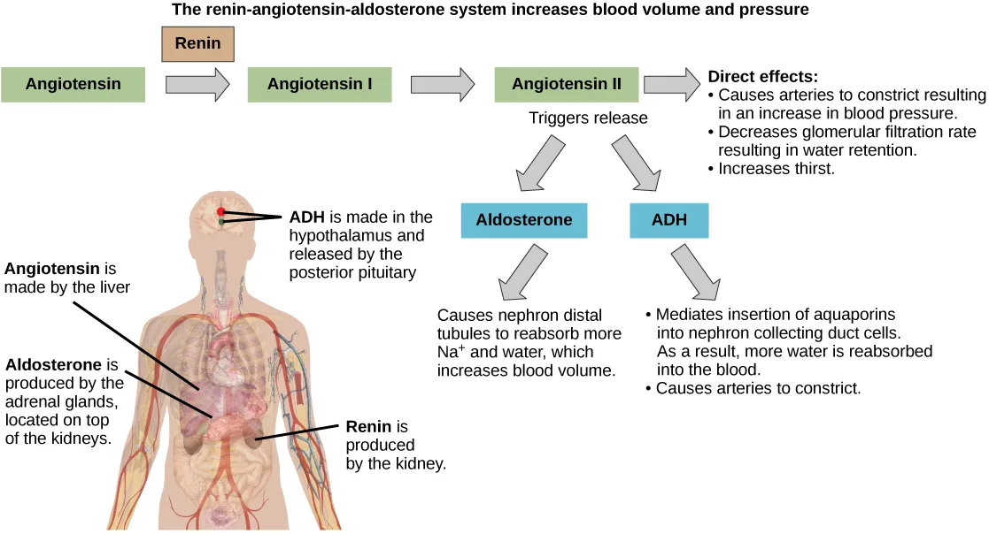
Extended description
A left-to-right chain of boxes reads ‘Angiotensin’ → ‘Angiotensin I’ → ‘Angiotensin II,’ with a tan box labeled ‘Renin’ positioned above the gap between ‘Angiotensin’ and ‘Angiotensin I.’ An arrow from ‘Angiotensin II’ points right to a bulleted list headed ‘Direct effects:’, which lists three effects: arterial constriction that increases blood pressure, a decreased glomerular filtration rate that causes water retention, and increased thirst. Below ‘Angiotensin II,’ two arrows under the label ‘Triggers release’ point down to two boxes, ‘Aldosterone’ and ‘ADH.’ An arrow below ‘Aldosterone’ points to the text: ‘Causes nephron distal tubules to reabsorb more Na⁺ and water, which increases blood volume.’ An arrow below ‘ADH’ points to two bullets: ‘Mediates insertion of aquaporins into nephron collecting duct cells. As a result, more water is reabsorbed into the blood,’ and ‘Causes arteries to constrict.’ Below this chain, a body outline carries four leader lines: one to the liver, labeled ‘Angiotensin is made by the liver’; one to the adrenal glands atop the kidneys, labeled ‘Aldosterone is produced by the adrenal glands, located on top of the kidneys’; one to a kidney, labeled ‘Renin is produced by the kidney’; and one to a red dot in the brain, labeled ‘ADH is made in the hypothalamus and released by the posterior pituitary.’
Hormonal Regulation of the Reproductive System
Regulation of the reproductive system is a process that requires the action of hormones from the pituitary gland, the adrenal cortex, and the gonads. During puberty in both males and females, the hypothalamus produces gonadotropin-releasing hormone (GnRH), which stimulates the production and release of follicle-stimulating hormone (FSH) and luteinizing hormone (LH) from the anterior pituitary gland. These hormones regulate the gonads (testes in males and ovaries in females) and therefore are called gonadotropins. In both males and females, FSH stimulates gamete production and LH stimulates production of hormones by the gonads. An increase in gonad hormone levels inhibits GnRH production through a negative feedback loop.
Regulation of the Testicular Reproductive System
In the testes, FSH stimulates the maturation of sperm cells. FSH production is inhibited by the hormone inhibin, which is released by the testes. LH stimulates production of the sex hormones (androgens) by the interstitial cells of the testes and therefore is also called interstitial cell-stimulating hormone.
The most widely known androgen in males is testosterone. Testosterone promotes the production of sperm and a suite of secondary sex characteristics, such as the growth and development of the testes and penis, increased skeletal and muscular growth, enlargement of the larynx, increased growth and redistribution of body hair, and increased sexual drive. The adrenal cortex also produces small amounts of testosterone precursor, although the role of this additional hormone production is not fully understood. Testosterone secretion is regulated by both the hypothalamus and the anterior pituitary gland. The hypothalamus sends releasing hormones that stimulate the release of gonadotropins from the anterior pituitary gland.
Everyday Connection. The Dangers of Synthetic Hormones.

Some athletes attempt to boost their performance by using artificial hormones that enhance muscle performance. Anabolic steroids, a form of testosterone, are one of the most widely known performance-enhancing drugs. Steroids are used in sports to help build muscle mass. Other hormones that are used to enhance athletic performance include erythropoietin, which triggers the production of red blood cells, and human growth hormone, which can help in building muscle mass. Most performance enhancing drugs are illegal for nonmedical purposes. They are also banned by national and international governing bodies including the International Olympic Committee, the U.S. Olympic Committee, the National Collegiate Athletic Association, the Major League Baseball, and the National Football League.
The side effects of synthetic hormones are often significant and nonreversible, and in some cases, fatal. Androgens can produce several complications such as liver dysfunctions and liver tumors, prostate gland enlargement, difficulty urinating, premature closure of epiphyseal cartilages, testicular atrophy, infertility, and immune system depression. The physiological strain caused by these substances is often greater than what the body can handle, leading to unpredictable and dangerous effects and linking their use to heart attacks, strokes, and impaired cardiac function.
Regulation of the Ovarian Reproductive System
In the ovaries, FSH stimulates development of egg cells, called ova, which develop in structures called follicles. Follicle cells produce the hormone inhibin, which inhibits FSH production. LH also plays a role in the development of ova, induction of ovulation, and stimulation of estradiol and progesterone production by the ovaries (as well as testosterone production by the testes), as illustrated below. Estradiol and progesterone are steroid hormones that serve several functions in the human body. Estradiol causes the egg to mature and release during the menstrual cycle, and thickens the uterine lining prior to egg implantation. Estradiol also helps with bone health, nitric oxide production, and brain function. (Low levels of estradiol have been connected to osteoporosis, mood swings, weight gain, and interrupted menstrual cycle. High levels of estradiol correlate with an increased risk of uterine and breast cancer, and cardiovascular disease.) During puberty, estradiol produces a suite of characteristics such as the increased development of breast tissue, redistribution of fat towards hips, legs, and breast, and the maturation of the uterus and vagina. Both estradiol and progesterone regulate the menstrual cycle.
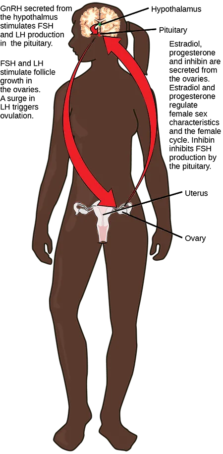
Extended description
At the upper left, text reads ‘GnRH secreted from the hypothalamus stimulates FSH and LH production in the pituitary,’ with leader lines from a brain cross-section labeling ‘Hypothalamus’ and, just below it, ‘Pituitary.’ Below that, text reads ‘FSH and LH stimulate follicle growth in the ovaries. A surge in LH triggers ovulation.’ Two overlapping red arrows form a crossing loop between the head and the pelvis: one curves down from the brain to the uterus and ovary, its arrowhead pointing down into the pelvis; the other curves up from the pelvis to the brain, its arrowhead pointing up into the head. At the lower body, leader lines label ‘Uterus’ and, beside it, ‘Ovary.’ To the right, text reads ‘Estradiol, progesterone and inhibin are secreted from the ovaries. Estradiol and progesterone regulate female sex characteristics and the female cycle. Inhibin inhibits FSH production by the pituitary.’
In addition to producing FSH and LH, the anterior portion of the pituitary gland also produces the hormone prolactin (PRL). Prolactin stimulates the production of milk by the mammary glands following childbirth. Prolactin release inhibits the release of GnRH from the hypothalamus, resulting in a loss of FSH and LH release from the anterior pituitary. Prolactin levels are regulated by the hypothalamic hormones prolactin-releasing hormone (PRH) and prolactin-inhibiting hormone (PIH), which is now known to be dopamine. PRH stimulates the release of prolactin and PIH inhibits it.
The posterior pituitary releases the hormone oxytocin, which stimulates uterine contractions during childbirth. The uterine smooth muscles are not very sensitive to oxytocin until late in pregnancy when the number of oxytocin receptors in the uterus peaks. Stretching of tissues in the uterus and cervix stimulates oxytocin release during childbirth. Contractions increase in intensity as blood levels of oxytocin rise via a positive feedback mechanism until the birth is complete. Oxytocin also stimulates the contraction of myoepithelial cells around the milk-producing mammary glands. As these cells contract, milk is forced from the secretory alveoli into milk ducts and is ejected from the breasts in milk ejection (“let-down”) reflex. Oxytocin release is stimulated by the suckling of an infant, which triggers the synthesis of oxytocin in the hypothalamus and its release into circulation at the posterior pituitary.
Hormonal Regulation of Metabolism
Blood glucose levels vary widely over the course of a day as periods of food consumption alternate with periods of fasting. Insulin and glucagon are the two hormones primarily responsible for maintaining homeostasis of blood glucose levels. Additional regulation is mediated by the thyroid hormones.
Regulation of Blood Glucose Levels by Insulin and Glucagon
Cells of the body require nutrients in order to function, and these nutrients are obtained through feeding. In order to manage nutrient intake, storing excess intake and utilizing reserves when necessary, the body uses hormones to moderate energy stores. Insulin is produced by the beta cells of the pancreas, which are stimulated to release insulin as blood glucose levels rise (for example, after a meal is consumed). Insulin lowers blood glucose levels by enhancing the rate of glucose uptake and utilization by target cells, which use glucose for ATP production. It also stimulates the liver to convert glucose to glycogen, which is then stored by cells for later use. Insulin also increases glucose transport into certain cells, such as muscle cells and the liver. This results from an insulin-mediated increase in the number of glucose transporter proteins in cell membranes, which remove glucose from circulation by facilitated diffusion. As insulin binds to its target cell via insulin receptors and signal transduction, it triggers the cell to incorporate glucose transport proteins into its membrane. This allows glucose to enter the cell, where it can be used as an energy source. However, this does not occur in all cells: some cells, including those in the kidneys and brain, can access glucose without the use of insulin. Insulin also stimulates the conversion of glucose to fat in adipocytes and the synthesis of proteins. These actions mediated by insulin cause blood glucose concentrations to fall, called a hypoglycemic “low sugar” effect, which inhibits further insulin release from beta cells through a negative feedback loop.
Link to Learning
Watch an animation describing the role of insulin and the pancreas in diabetes.
Impaired insulin function can lead to a condition called diabetes mellitus, the main symptoms of which are illustrated below. This can be caused by low levels of insulin production by the beta cells of the pancreas, or by reduced sensitivity of tissue cells to insulin. This prevents glucose from being absorbed by cells, causing high levels of blood glucose, or hyperglycemia (high sugar). High blood glucose levels make it difficult for the kidneys to recover all the glucose from nascent urine, resulting in glucose being lost in urine. High glucose levels also result in less water being reabsorbed by the kidneys, causing high amounts of urine to be produced; this may result in dehydration. Over time, high blood glucose levels can cause nerve damage to the eyes and peripheral body tissues, as well as damage to the kidneys and cardiovascular system. Oversecretion of insulin can cause hypoglycemia, low blood glucose levels. This causes insufficient glucose availability to cells, often leading to muscle weakness, and can sometimes cause unconsciousness or death if left untreated.
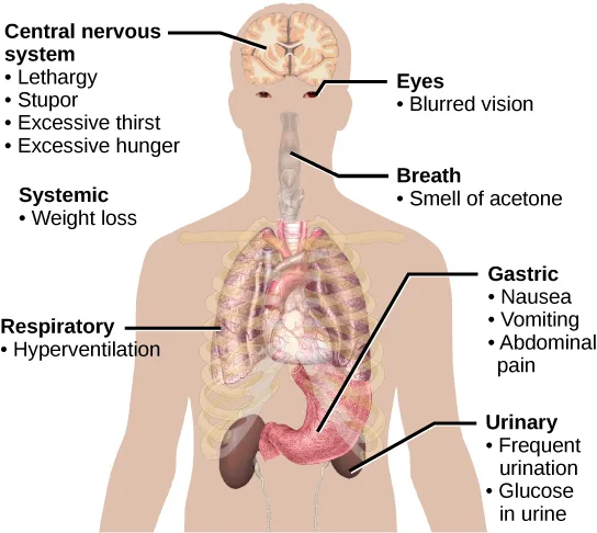
Extended description
Leader lines from a body outline point to six of seven labeled regions; the seventh, ‘Systemic,’ sits near the torso with no leader line. Reading top to bottom: ‘Central nervous system’ (a line to the head) lists lethargy, stupor, excessive thirst, and excessive hunger. ‘Eyes’ lists blurred vision. ‘Breath’ (a line to the throat) lists smell of acetone. ‘Systemic’ lists weight loss. ‘Respiratory’ (a line to the lungs) lists hyperventilation. ‘Gastric’ (a line to the stomach) lists nausea, vomiting, and abdominal pain. ‘Urinary’ (a line to the kidney and bladder area) lists frequent urination and glucose in urine.
When blood glucose levels decline below normal levels, for example between meals or when glucose is utilized rapidly during exercise, the hormone glucagon is released from the alpha cells of the pancreas. Glucagon raises blood glucose levels, eliciting what is called a hyperglycemic effect, by stimulating the breakdown of glycogen to glucose in skeletal muscle cells and liver cells in a process called glycogenolysis. Glucose can then be utilized as energy by muscle cells and released into circulation by the liver cells. Glucagon also stimulates absorption of amino acids from the blood by the liver, which then converts them to glucose. This process of glucose synthesis is called gluconeogenesis. Glucagon also stimulates adipose cells to release fatty acids into the blood. These actions mediated by glucagon result in an increase in blood glucose levels to normal homeostatic levels. Rising blood glucose levels inhibit further glucagon release by the pancreas via a negative feedback mechanism. In this way, insulin and glucagon work together to maintain homeostatic glucose levels, as shown below.
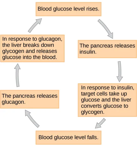
Extended description
Six boxes are arranged in a ring and connected by curved arrows that run clockwise. Starting at the top and following the arrows clockwise: ‘Blood glucose level rises.’ leads to ‘The pancreas releases insulin.’ (upper right), which leads to ‘In response to insulin, target cells take up glucose and the liver converts glucose to glycogen.’ (lower right), which leads to ‘Blood glucose level falls.’ (bottom), which leads to ‘The pancreas releases glucagon.’ (lower left), which leads to ‘In response to glucagon, the liver breaks down glycogen and releases glucose into the blood.’ (upper left), which leads back to ‘Blood glucose level rises.’ at the top.
Pancreatic tumors may cause excess secretion of glucagon. Type I diabetes results from the failure of the pancreas to produce insulin. Which of the following statements about these two conditions is true?
A pancreatic tumor that oversecretes glucagon and a pancreas that cannot produce insulin each remove one of the two hormones that pull in opposite directions on blood glucose — work out which direction is left unopposed in each case.Regulation of Metabolic Rates by Thyroid Hormones
The basal metabolic rate, which is the amount of calories required by the body at rest, is determined by two hormones produced by the thyroid gland: thyroxine, also known as tetraiodothyronine or T₄, and triiodothyronine, also known as T₃. These hormones affect nearly every cell in the body except for the adult brain, uterus, testes, blood cells, and spleen. They are transported across the plasma membrane of target cells and bind to receptors on the mitochondria resulting in increased ATP production. In the nucleus, T₃ and T₄ activate genes involved in energy production and glucose oxidation. This results in increased rates of metabolism and body heat production, which is known as the hormone’s calorigenic effect.
T₃ and T₄ release from the thyroid gland is stimulated by thyroid-stimulating hormone (TSH), which is produced by the anterior pituitary. TSH binding at the receptors of the follicle of the thyroid triggers the production of T₃ and T₄ from a glycoprotein called thyroglobulin. Thyroglobulin is present in the follicles of the thyroid, and is converted into thyroid hormones with the addition of iodine. Iodine is formed from iodide ions that are actively transported into the thyroid follicle from the bloodstream. A peroxidase enzyme then attaches the iodine to the tyrosine amino acid found in thyroglobulin. T₃ has three iodine ions attached, while T₄ has four iodine ions attached. T₃ and T₄ are then released into the bloodstream, with T₄ being released in much greater amounts than T₃. As T₃ is more active than T₄ and is responsible for most of the effects of thyroid hormones, tissues of the body convert T₄ to T₃ by the removal of an iodine ion. Most of the released T₃ and T₄ becomes attached to transport proteins in the bloodstream and is unable to cross the plasma membrane of cells. These protein-bound molecules are only released when blood levels of the unattached hormone begin to decline. In this way, a week’s worth of reserve hormone is maintained in the blood. Increased T₃ and T₄ levels in the blood inhibit the release of TSH, which results in lower T₃ and T₄ release from the thyroid.
The follicular cells of the thyroid require iodides (anions of iodine) in order to synthesize T₃ and T₄. Iodides obtained from the diet are actively transported into follicle cells resulting in a concentration that is approximately 30 times higher than in blood. The typical diet in North America provides more iodine than required due to the addition of iodide to table salt. Inadequate iodine intake, which occurs in many developing countries, results in an inability to synthesize T₃ and T₄ hormones. The thyroid gland enlarges in a condition called goiter, which is caused by overproduction of TSH without the formation of thyroid hormone. Thyroglobulin is contained in a fluid called colloid, and TSH stimulation results in higher levels of colloid accumulation in the thyroid. In the absence of iodine, this is not converted to thyroid hormone, and colloid begins to accumulate more and more in the thyroid gland, leading to goiter.
Disorders can arise from both the underproduction and overproduction of thyroid hormones. Hypothyroidism, underproduction of the thyroid hormones, can cause a low metabolic rate leading to weight gain, sensitivity to cold, and reduced mental activity, among other symptoms. In children, hypothyroidism can cause cretinism, which can lead to intellectual disabilities and growth defects. Hyperthyroidism, the overproduction of thyroid hormones, can lead to an increased metabolic rate and its effects: weight loss, excess heat production, sweating, and an increased heart rate. Graves’ disease is one example of a hyperthyroid condition.
Hormonal Control of Blood Calcium Levels
Regulation of blood calcium concentrations is important for generation of muscle contractions and nerve impulses, which are electrically stimulated. If calcium levels get too high, membrane permeability to sodium decreases and membranes become less responsive. If calcium levels get too low, membrane permeability to sodium increases and convulsions or muscle spasms can result.
Blood calcium levels are regulated by parathyroid hormone (PTH), which is produced by the parathyroid glands, as illustrated below. PTH is released in response to low blood Ca²⁺ levels. PTH increases Ca²⁺ levels by targeting the skeleton, the kidneys, and the intestine. In the skeleton, PTH stimulates osteoclasts, which causes bone to be reabsorbed, releasing Ca²⁺ from bone into the blood. PTH also inhibits osteoblasts, reducing Ca²⁺ deposition in bone. In the intestines, PTH increases dietary Ca²⁺ absorption, and in the kidneys, PTH stimulates reabsorption of the Ca²⁺. While PTH acts directly on the kidneys to increase Ca²⁺ reabsorption, its effects on the intestine are indirect. PTH triggers the formation of calcitriol, an active form of vitamin D, which acts on the intestines to increase absorption of dietary calcium. PTH release is inhibited by rising blood calcium levels.
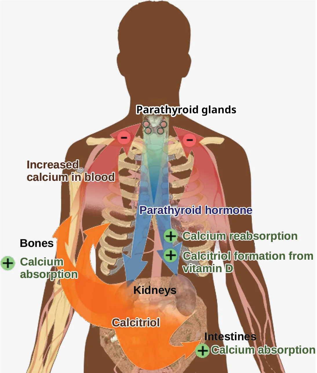
Extended description
At the top, ‘Parathyroid glands’ labels a small illustration of the thyroid and parathyroid glands in the neck, flanked by two red circles each marked with a white minus sign. Below, blue text reads ‘Parathyroid hormone’ beside a wide blue arrow that runs down from the neck through the chest to a label reading ‘Kidneys’ near the bottom center. Partway down, two green plus-sign icons branch off the blue arrow, labeled ‘Calcium reabsorption’ and ‘Calcitriol formation from vitamin D.’ At the upper left, the text ‘Increased calcium in blood’ sits above a wide orange arrow that curves up from a label reading ‘Bones’ at the lower left, beside which a green plus-sign icon reads ‘Calcium absorption.’ Below ‘Kidneys,’ the label ‘Calcitriol’ sits above a second wide orange arrow that curves right to a label reading ‘Intestines’ at the lower right, beside which a second green plus-sign icon also reads ‘Calcium absorption.’
Hyperparathyroidism results from an overproduction of parathyroid hormone. This results in excessive calcium being removed from bones and introduced into blood circulation, producing structural weakness of the bones, which can lead to deformation and fractures, plus nervous system impairment due to high blood calcium levels. Hypoparathyroidism, the underproduction of PTH, results in extremely low levels of blood calcium, which causes impaired muscle function and may result in tetany (severe sustained muscle contraction).
The hormone calcitonin, which is produced by the parafollicular or C cells of the thyroid, has the opposite effect on blood calcium levels as does PTH. Calcitonin decreases blood calcium levels by inhibiting osteoclasts, stimulating osteoblasts, and stimulating calcium excretion by the kidneys. This results in calcium being added to the bones to promote structural integrity. Calcitonin is most important in children (when it stimulates bone growth), during pregnancy (when it reduces bone loss), and during prolonged starvation (because it reduces bone mass loss). In healthy nonpregnant, unstarved adults, the role of calcitonin is unclear.
Hormonal Regulation of Growth
Hormonal regulation is required for the growth and replication of most cells in the body. Growth hormone (GH), produced by the anterior portion of the pituitary gland, accelerates the rate of protein synthesis, particularly in skeletal muscle and bones. Growth hormone has direct and indirect mechanisms of action. The first direct action of GH is stimulation of triglyceride breakdown (lipolysis) and release into the blood by adipocytes. This results in a switch by most tissues from utilizing glucose as an energy source to utilizing fatty acids. This process is called a glucose-sparing effect. In another direct mechanism, GH stimulates glycogen breakdown in the liver; the glycogen is then released into the blood as glucose. Blood glucose levels increase as most tissues are utilizing fatty acids instead of glucose for their energy needs. The GH mediated increase in blood glucose levels is called a diabetogenic effect because it is similar to the high blood glucose levels seen in diabetes mellitus.
The indirect mechanism of GH action is mediated by insulin-like growth factors (IGFs) or somatomedins, which are a family of growth-promoting proteins produced by the liver, which stimulates tissue growth. IGFs stimulate the uptake of amino acids from the blood, allowing the formation of new proteins, particularly in skeletal muscle cells, cartilage cells, and other target cells, as shown below. This is especially important after a meal, when glucose and amino acid concentration levels are high in the blood. GH levels are regulated by two hormones produced by the hypothalamus. GH release is stimulated by growth hormone-releasing hormone (GHRH) and is inhibited by growth hormone-inhibiting hormone (GHIH), also called somatostatin.
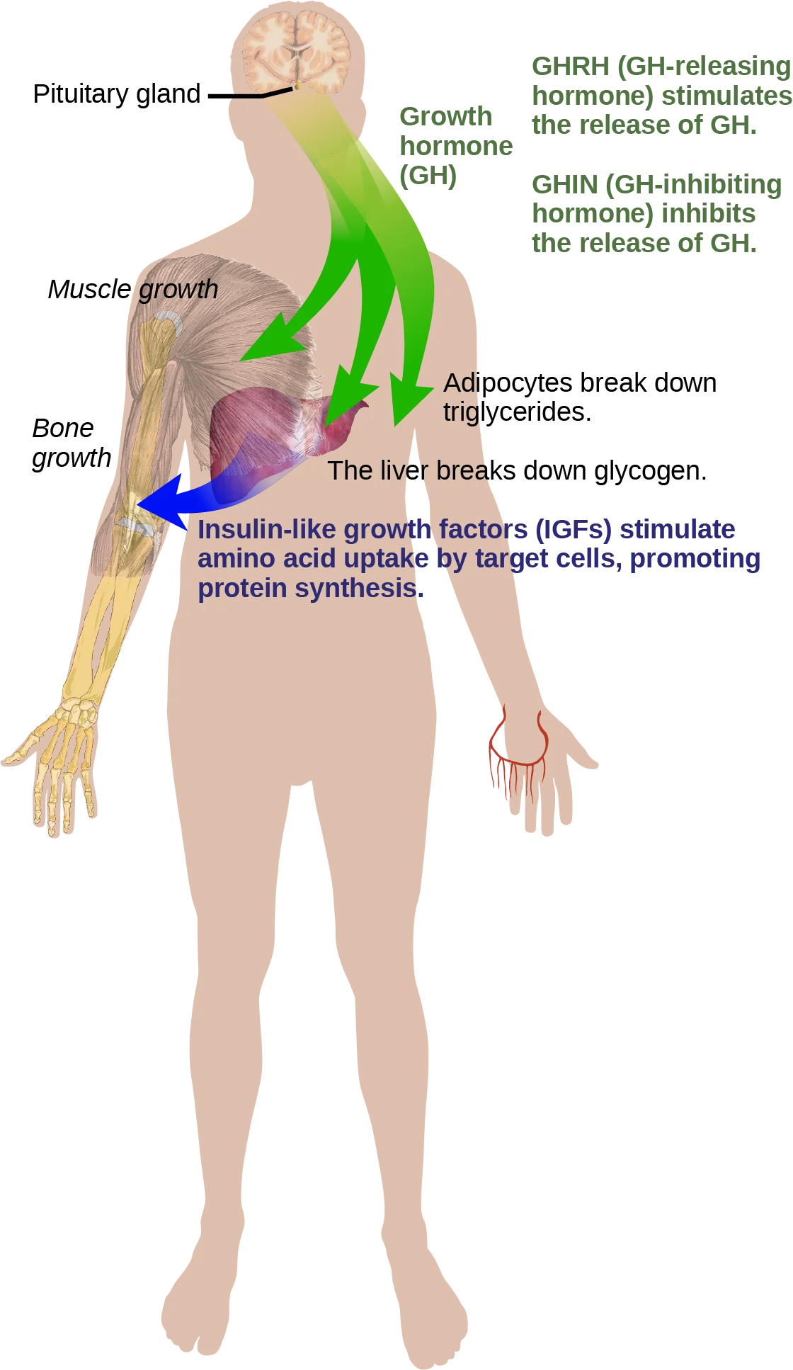
Extended description
At the upper left, ‘Pituitary gland’ labels a leader line to a brain cross-section at the top of the figure. Below it, bold green text reads ‘Growth hormone (GH),’ beside a broad green ribbon flowing down from the head that splits into three arrows: one curves left into the shoulder, ending at a muscle illustration beside the italic label ‘Muscle growth’; the other two point down and right, ending at the labels ‘Adipocytes break down triglycerides.’ and ‘The liver breaks down glycogen.’ At the upper right, green text reads ‘GHRH (GH-releasing hormone) stimulates the release of GH.’ and, below it, ‘GHIH (GH-inhibiting hormone) inhibits the release of GH.’ Lower on the figure, a separate blue arrow points left from the text ‘Insulin-like growth factors (IGFs) stimulate amino acid uptake by target cells, promoting protein synthesis,’ ending at a forearm-bone illustration beside the italic label ‘Bone growth.’
A balanced production of growth hormone is critical for proper development. Underproduction of GH in adults does not appear to cause any abnormalities, but in children it can result in pituitary dwarfism, resulting in abnormally short stature with normal body proportions. The height of such individuals is most frequently less than 4 feet (122 cm) in height. Oversecretion of growth hormone can lead to gigantism in children, causing excessive growth. In some documented cases, individuals can reach heights of over eight feet. In adults, excessive GH can lead to acromegaly, a condition in which there is enlargement of bones in the face, hands, and feet that are still capable of growth.
Hormonal Regulation of Stress
When a threat or danger is perceived, the body responds by releasing hormones that will ready it for the “fight-or-flight” response. The effects of this response are familiar to anyone who has been in a stressful situation: increased heart rate, dry mouth, and hair standing up.
Evolution Connection. Fight-or-Flight Response.
Interactions of the endocrine hormones have evolved to ensure the body’s internal environment remains stable. Stressors are stimuli that disrupt homeostasis. The sympathetic division of the vertebrate autonomic nervous system has evolved the fight-or-flight response to counter stress-induced disruptions of homeostasis. In the initial alarm phase, the sympathetic nervous system stimulates an increase in energy levels through increased blood glucose levels. This prepares the body for physical activity that may be required to respond to stress: to either fight for survival or to flee from danger.
However, some stresses, such as illness or injury, can last for a long time. Glycogen reserves, which provide energy in the short-term response to stress, are exhausted after several hours and cannot meet long-term energy needs. If glycogen reserves were the only energy source available, neural functioning could not be maintained once the reserves became depleted due to the nervous system’s high requirement for glucose. In this situation, the body has evolved a response to counter long-term stress through the actions of the glucocorticoids, which ensure that long-term energy requirements can be met. The glucocorticoids mobilize lipid and protein reserves, stimulate gluconeogenesis, conserve glucose for use by neural tissue, and stimulate the conservation of salts and water. The mechanisms to maintain homeostasis that are described here are those observed in the human body. However, the fight-or-flight response exists in some form in all vertebrates.
The sympathetic nervous system regulates the stress response via the hypothalamus. Stressful stimuli cause the hypothalamus to signal the adrenal medulla (which mediates short-term stress responses) via nerve impulses, and the adrenal cortex, which mediates long-term stress responses, via the hormone adrenocorticotropic hormone (ACTH), which is produced by the anterior pituitary.
Short-term Stress Response
When presented with a stressful situation, the body responds by calling for the release of hormones that provide a burst of energy. The hormones epinephrine (also known as adrenaline) and norepinephrine (also known as noradrenaline) are released by the adrenal medulla. How do these hormones provide a burst of energy? Epinephrine and norepinephrine increase blood glucose levels by stimulating the liver and skeletal muscles to break down glycogen and by stimulating glucose release by liver cells. Additionally, these hormones increase oxygen availability to cells by increasing the heart rate and dilating the bronchioles. The hormones also prioritize body function by increasing blood supply to essential organs such as the heart, brain, and skeletal muscles, while restricting blood flow to organs not in immediate need, such as the skin, digestive system, and kidneys. Epinephrine and norepinephrine are collectively called catecholamines.
Link to Learning
Watch a Discovery Channel animation describing the fight-or-flight response.
Long-term Stress Response
Long-term stress response differs from short-term stress response. The body cannot sustain the bursts of energy mediated by epinephrine and norepinephrine for long times. Instead, other hormones come into play. In a long-term stress response, the hypothalamus triggers the release of ACTH from the anterior pituitary gland. The adrenal cortex is stimulated by ACTH to release steroid hormones called corticosteroids. Corticosteroids turn on transcription of certain genes in the nuclei of target cells. They change enzyme concentrations in the cytoplasm and affect cellular metabolism. There are two main corticosteroids: glucocorticoids such as cortisol, and mineralocorticoids such as aldosterone. These hormones target the breakdown of fat into fatty acids in the adipose tissue. The fatty acids are released into the bloodstream for other tissues to use for ATP production. The glucocorticoids primarily affect glucose metabolism by stimulating glucose synthesis. Glucocorticoids also have anti-inflammatory properties through inhibition of the immune system. For example, cortisone is used as an anti-inflammatory medication; however, it cannot be used long term as it increases susceptibility to disease due to its immune-suppressing effects.
Mineralocorticoids function to regulate ion and water balance of the body. The hormone aldosterone stimulates the reabsorption of water and sodium ions in the kidney, which results in increased blood pressure and volume.
Hypersecretion of glucocorticoids can cause a condition known as Cushing’s disease, characterized by a shifting of fat storage areas of the body. This can cause the accumulation of adipose tissue in the face and neck, and excessive glucose in the blood. Hyposecretion of the corticosteroids can cause Addison’s disease, which may result in bronzing of the skin, hypoglycemia, and low electrolyte levels in the blood.
Summary
Water levels in the body are controlled by antidiuretic hormone (ADH), which is produced in the hypothalamus and triggers the reabsorption of water by the kidneys. Underproduction of ADH can cause diabetes insipidus. Aldosterone, a hormone produced by the adrenal cortex of the kidneys, enhances Na⁺ reabsorption from the extracellular fluids and subsequent water reabsorption by diffusion. The renin-angiotensin-aldosterone system is one way that aldosterone release is controlled.
The reproductive system is controlled by the gonadotropins follicle-stimulating hormone (FSH) and luteinizing hormone (LH), which are produced by the pituitary gland. Gonadotropin release is controlled by the hypothalamic hormone gonadotropin-releasing hormone (GnRH). FSH stimulates the maturation of sperm cells in testes and is inhibited by the hormone inhibin, while LH stimulates the production of the androgen testosterone. FSH stimulates egg maturation in ovaries, while LH stimulates the production of estrogens and progesterone. Estrogens are a group of steroid hormones produced by the ovaries that trigger increased development of breast tissue, redistribution of fat towards hips, legs, and breast, the maturation of the uterus and vagina, as well as control the maturation of the ova. Within ovarian reproductive systems, the pituitary also produces prolactin, which stimulates milk production after childbirth, and oxytocin, which stimulates uterine contraction during childbirth and milk let-down during suckling.
Insulin is produced by the pancreas in response to rising blood glucose levels and allows cells to utilize blood glucose and store excess glucose for later use. Diabetes mellitus is caused by reduced insulin activity and causes high blood glucose levels, or hyperglycemia. Glucagon is released by the pancreas in response to low blood glucose levels and stimulates the breakdown of glycogen into glucose, which can be used by the body. The body’s basal metabolic rate is controlled by the thyroid hormones thyroxine (T₄) and triiodothyronine (T₃). The anterior pituitary produces thyroid stimulating hormone (TSH), which controls the release of T₃ and T₄ from the thyroid gland. Iodine is necessary in the production of thyroid hormone, and the lack of iodine can lead to a condition called goiter.
Parathyroid hormone (PTH) is produced by the parathyroid glands in response to low blood Ca²⁺ levels. The parafollicular cells of the thyroid produce calcitonin, which reduces blood Ca²⁺ levels. Growth hormone (GH) is produced by the anterior pituitary and controls the growth rate of muscle and bone. GH action is indirectly mediated by insulin-like growth factors (IGFs). Short-term stress causes the hypothalamus to trigger the adrenal medulla to release epinephrine and norepinephrine, which trigger the fight or flight response. Long-term stress causes the hypothalamus to trigger the anterior pituitary to release adrenocorticotropic hormone (ACTH), which causes the release of corticosteroids, glucocorticoids, and mineralocorticoids, from the adrenal cortex.
Key terms
- acromegaly — condition caused by overproduction of GH in adults.
- Addison’s disease — disorder caused by the hyposecretion of corticosteroids.
- adrenocorticotropic hormone (ACTH) — hormone released by the anterior pituitary, which stimulates the adrenal cortex to release corticosteroids during the long-term stress response.
- aldosterone — steroid hormone produced by the adrenal cortex that stimulates the reabsorption of Na⁺ from extracellular fluids and secretion of K⁺.
- androgen — male sex hormone such as testosterone.
- antidiuretic hormone (ADH) — hormone produced by the hypothalamus and released by the posterior pituitary that increases water reabsorption by the kidneys.
- calcitonin — hormone produced by the parafollicular cells of the thyroid gland that functions to lower blood Ca²⁺ levels and promote bone growth.
- corticosteroid — hormone released by the adrenal cortex in response to long-term stress.
- cortisol — glucocorticoid produced in response to stress.
- Cushing’s disease — disorder caused by the hypersecretion of glucocorticoids.
- diabetes insipidus — disorder caused by underproduction of ADH.
- diabetes mellitus — disorder caused by low levels of insulin activity.
- diabetogenic effect — effect of GH that causes blood glucose levels to rise similar to diabetes mellitus.
- epinephrine — hormone released by the adrenal medulla in response to a short term stress.
- estrogens — a group of steroid hormones, including estradiol and several others, that are produced by the ovaries and elicit a suite of characteristics (including increased development of breast tissue, redistribution of fat towards hips, legs, and breast, and the maturation of the uterus and vagina) as well as control the maturation of the ova.
- follicle-stimulating hormone (FSH) — hormone produced by the anterior pituitary that stimulates gamete production.
- gigantism — condition caused by overproduction of GH in children.
- glucagon — hormone produced by the alpha cells of the pancreas in response to low blood sugar; functions to raise blood sugar levels.
- glucocorticoid — corticosteroid that affects glucose metabolism.
- gluconeogenesis — synthesis of glucose from amino acids.
- glucose-sparing effect — effect of GH that causes tissues to use fatty acids instead of glucose as an energy source.
- glycogenolysis — breakdown of glycogen into glucose.
- goiter — enlargement of the thyroid gland caused by insufficient dietary iodine levels.
- gonadotropin — hormone that regulates the gonads, including FSH and LH.
- growth hormone (GH) — hormone produced by the anterior pituitary that promotes protein synthesis and body growth.
- growth hormone-inhibiting hormone (GHIH) — hormone produced by the hypothalamus that inhibits growth hormone production, also called somatostatin.
- growth hormone-releasing hormone (GHRH) — hormone released by the hypothalamus that triggers the release of GH.
- hyperglycemia — high blood sugar level.
- hyperthyroidism — overactivity of the thyroid gland.
- hypoglycemia — low blood sugar level.
- hypothyroidism — underactivity of the thyroid gland.
- insulin — hormone produced by the beta cells of the pancreas in response to high blood glucose levels; functions to lower blood glucose levels.
- insulin-like growth factor (IGF) — growth-promoting protein produced by the liver.
- mineralocorticoid — corticosteroid that affects ion and water balance.
- norepinephrine — hormone released by the adrenal medulla in response to a short-term stress.
- osmoreceptor — receptor in the hypothalamus that monitors the concentration of electrolytes in the blood.
- oxytocin — hormone released by the posterior pituitary to stimulate uterine contractions during childbirth and milk let-down in the mammary glands.
- parathyroid hormone (PTH) — hormone produced by the parathyroid glands in response to low blood Ca²⁺ levels; functions to raise blood Ca²⁺ levels.
- pituitary dwarfism — condition caused by underproduction of GH in children.
- prolactin (PRL) — hormone produced by the anterior pituitary that stimulates milk production.
- prolactin-inhibiting hormone — hormone produced by the hypothalamus that inhibits the release of prolactin.
- prolactin-releasing hormone — hormone produced by the hypothalamus that stimulates the release of prolactin.
- renin — enzyme produced by the juxtaglomerular apparatus of the kidneys that reacts with angiotensinogen to cause the release of aldosterone.
- thyroglobulin — glycoprotein found in the thyroid that is converted into thyroid hormone.
- thyroid-stimulating hormone (TSH) — hormone produced by the anterior pituitary that controls the release of T₃ and T₄ from the thyroid gland.
- thyroxine (tetraiodothyronine, T₄) — thyroid hormone containing 4 iodines that controls the basal metabolic rate.
- triiodothyronine (T₃) — thyroid hormone containing 3 iodines that controls the basal metabolic rate.
Practice
Explain how hormones regulate the excretory system
Drinking alcoholic beverages causes an increase in urine output. This most likely occurs because alcohol:
ADH’s whole job is to make the kidneys hold on to water — think about what has to happen to that hormone for more water to leave the body as urine instead.The hormone produced by the hypothalamus and released by the posterior pituitary that increases water reabsorption by the kidneys is called ________.
This hormone is inserted into kidney tubules as temporary water channels called aquaporins, and its release is reduced by drinking alcohol.The steroid hormone produced by the adrenal cortex that stimulates the reabsorption of Na⁺ from extracellular fluids and secretion of K⁺ is called ________.
This hormone’s release is triggered by the renin-angiotensin system when blood pressure drops.A corticosteroid that affects ion and water balance is called a ________.
Aldosterone is the section’s example of this class of adrenal-cortex hormone.The enzyme produced by the juxtaglomerular apparatus of the kidneys that reacts with angiotensinogen to cause the release of aldosterone is called ________.
This enzyme starts the pathway that converts angiotensinogen into angiotensin I.The disorder caused by underproduction of ADH is called ________.
Without enough of this hormone, the kidneys cannot retain water, so it must be continually replaced by drinking.Discuss the role of hormones in the reproductive system
FSH and LH release from the anterior pituitary is stimulated by ________.
This releasing hormone comes from the hypothalamus, not from the anterior pituitary itself, and its production is inhibited by rising gonad-hormone levels.How would mutations that completely ablate the function of the androgen receptor impact the phenotypic development of humans with XY chromosomes?
If target tissues cannot respond to androgens at all, work out what secondary sex characteristics are left to develop by default.Name and describe a function of one hormone produced by the anterior pituitary and one hormone produced by the posterior pituitary.
Show model answer
Did your answer mention:
A hormone that regulates the gonads, including FSH and LH, is called a ________.
This category of pituitary hormone is named for the reproductive organs it acts on.A group of steroid hormones, including estradiol, that are produced by the ovaries and control the maturation of the ova is called ________.
Low levels of this hormone group have been connected to osteoporosis, while high levels correlate with increased cancer risk.The hormone produced by the anterior pituitary that stimulates milk production is called ________.
This anterior-pituitary hormone’s release is inhibited by dopamine, acting here as a hypothalamic inhibiting hormone.The hormone released by the posterior pituitary to stimulate uterine contractions during childbirth and milk let-down in the mammary glands is called ________.
This hormone’s release during labor is a rare example in the body of a positive, rather than negative, feedback loop.Describe how hormones regulate metabolism
What hormone is produced by beta cells of the pancreas?
This is the hormone whose release rises after a meal and lowers blood glucose by increasing cellular uptake and storage.Describe one direct action of growth hormone (GH).
Show model answer
Did your answer mention:
The hormone produced by the alpha cells of the pancreas in response to low blood sugar that functions to raise blood sugar levels is called ________.
This hormone stimulates the breakdown of glycogen into glucose, the opposite effect of insulin.A high blood sugar level is called ________.
This is the condition the kidneys struggle to fully recover glucose from, so some is lost in the urine.A low blood sugar level is called ________.
Oversecretion of insulin can cause this condition, which can lead to muscle weakness or, if untreated, unconsciousness.The breakdown of glycogen into glucose is called ________.
Glucagon triggers this process in skeletal muscle cells and liver cells when blood glucose declines.The synthesis of glucose from amino acids is called ________.
Glucagon stimulates the liver to absorb amino acids from the blood and convert them by this process.The disorder caused by low levels of insulin activity is called ________.
This disorder’s main symptoms include excessive thirst and hunger, blurred vision, and glucose appearing in the urine.Explain the role of hormones in different diseases
When blood calcium levels are low, PTH stimulates:
PTH raises blood calcium by causing bone to be reabsorbed — think about which bone cell type breaks bone down rather than building it.Researchers have recently demonstrated that stressed people are more susceptible to contracting the common cold than people who are not stressed. What kind of stress must the infected patients be experiencing, and why does it make them more susceptible to the virus?
Show model answer
Did your answer mention:
The hormone produced by the parathyroid glands in response to low blood Ca²⁺ levels that functions to raise blood Ca²⁺ levels is called ________.
This hormone triggers the formation of calcitriol, an active form of vitamin D, to increase intestinal calcium absorption.The condition caused by underproduction of GH in children, resulting in abnormally short stature with normal body proportions, is called ________.
This condition most often leaves affected individuals less than 4 feet (122 cm) tall.The condition caused by overproduction of GH in children, causing excessive growth, is called ________.
Some documented cases of this childhood condition have reached heights of over eight feet.The condition caused by overproduction of GH in adults, resulting in enlargement of bones in the face, hands, and feet, is called ________.
Unlike the childhood overproduction condition, the bones affected here are ones still capable of growth in an adult.The disorder caused by the hypersecretion of glucocorticoids, characterized by a shifting of fat storage to the face and neck, is called ________.
This disorder is the opposite of the one caused by corticosteroid hyposecretion.The disorder caused by the hyposecretion of corticosteroids, which may result in bronzing of the skin and low electrolyte levels, is called ________.
This disorder is the opposite of the one caused by glucocorticoid hypersecretion.Overactivity of the thyroid gland is called ________.
This condition’s effects include weight loss, excess heat production, sweating, and an increased heart rate; Graves’ disease is one example.This section is adapted from Biology 2e, Section 37.3: Regulation of Body Processes by Mary Ann Clark, Jung Choi, Matthew Douglas, and OpenStax, © OpenStax, licensed under CC BY-NC-SA 4.0. Access the original for free at openstax.org. Changes: the norepinephrine Key terms entry ends at “short-term stress” where the source glossary runs on into a stray fragment, “stress hormone production by the gonads”, that belongs to no sentence (reported as a source defect); figures re-encoded as WebP; three figures re-kinded from the manifest’s file-extension “photo” guess to “diagram” (Figure_37_03_01b, Figure_37_03_04, and Figure_37_03_05 are hand-drawn flow charts and body diagrams, not captured photographs; Figure_B37_03_02, Figure_B37_03_05, and Figure_B37_03_06 were already correctly guessed as diagrams and are confirmed here) — only Figure_37_03_02 (the Jason Giambi photograph) is a true photo; a longdesc added to every diagram whose meaning is not carried by its caption alone — the renin-angiotensin-aldosterone pathway, the ovarian hormone-feedback illustration, the diabetes-symptom body map, the insulin/glucagon cycle, the parathyroid-hormone pathway, and the growth-hormone pathway — transcribing each drawing’s own printed labels and counting its boxes, arrows, and leader lines; letter-spaced text-to-speech alts in the source CNXML (e.g., “A D H,” “P T H,” “F S H and L H,” “upper case G lower case n…”) rewritten as plain prose alts, with the walk-through detail moved to each figure’s longdesc; the growth-hormone pathway’s longdesc corrects an artwork typo — the image itself prints “GHIN (GH-inhibiting hormone),” while the module’s own glossary and prose consistently name this hormone “growth hormone-inhibiting hormone (GHIH)” — reported as a source defect, with the artwork label left as printed; a corrupted-order phrase in the pinned CNXML — “describing the flight-or-flight response” in the second Link to Learning note, where every other instance in this section (including the note titled “Fight-or-Flight Response” two paragraphs above it) reads “fight-or-flight” — corrected to “fight-or-flight” and reported as a source defect; the Visual Connection question’s stem (“Which of the following statement about these two conditions is true?”, identical in both the body note and the exercise copy) corrected to “statements,” a grammar fix disclosed here since it is retained exercise text; in-text pointers to figures (“the figure below,” “Figure 37.9,” etc.) replaced with “shown below,” “illustrated below,” or “below,” since Hugo does not number figures; the note wrapping the pancreatic-tumor/type-I-diabetes Visual Connection rendered as its figure followed by a multiple choice, kept in the body in the Metabolism section — the note copy and the <exercise> copy print identical question and option wording (both quote “Which of the following statement…” and the same four lettered options), so no adjudication was needed, and the source’s own key (B, “will both cause hyperglycemia”) is kept as printed; the end-of-section Review Questions and Critical Thinking Questions adapted into the closing interactive Practice block (multiple choice and self-check respectively); rubric checkpoints added to each self-check, decomposing its model answer (the source solution) into check-off clauses with no new claims; the growth-hormone self-check’s model answer trims the source solution’s opening two sentences (“Hormonal regulation is required for the growth and replication of most cells in the body. Growth hormone (GH), produced by the anterior pituitary, accelerates the rate of protein synthesis, particularly in skeletal muscles and bones.”), which duplicate the Growth section’s own opening paragraph word for word; twenty-two key-term recall (textin) items added from the glossary, a representative subset of the section’s forty-seven glossary terms rather than all of them (insulin is additionally tested by a Review Question multiple choice; the rest appear only in the Key terms list and the prose); the section’s five Review Questions, three Critical Thinking Questions, and one Visual Connection are all used at least once across the body and Practice block.