Endocrine Glands
By the end of this section, you will be able to:
- Describe the role of different glands in the endocrine system
- Explain how the different glands work together to maintain homeostasis
Both the endocrine and nervous systems use chemical signals to communicate and regulate the body’s physiology. The endocrine system releases hormones that act on target cells to regulate development, growth, energy metabolism, reproduction, and many behaviors. The nervous system releases neurotransmitters or neurohormones that regulate neurons, muscle cells, and endocrine cells. Because the neurons can regulate the release of hormones, the nervous and endocrine systems work in a coordinated manner to regulate the body’s physiology.
Hypothalamic-Pituitary Axis
The hypothalamus in vertebrates integrates the endocrine and nervous systems. The hypothalamus is an endocrine organ located in the diencephalon of the brain. It receives input from the body and other brain areas and initiates endocrine responses to environmental changes. The hypothalamus acts as an endocrine organ, synthesizing hormones and transporting them along axons to the posterior pituitary gland. It synthesizes and secretes regulatory hormones that control the endocrine cells in the anterior pituitary gland. The hypothalamus contains autonomic centers that control endocrine cells in the adrenal medulla via neuronal control.
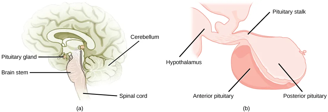
The pituitary gland, sometimes called the hypophysis or “master gland” is located at the base of the brain in the sella turcica, a groove of the sphenoid bone of the skull, shown below. It is attached to the hypothalamus via a stalk called the pituitary stalk (or infundibulum). The anterior portion of the pituitary gland is regulated by releasing or release-inhibiting hormones produced by the hypothalamus, and the posterior pituitary receives signals via neurosecretory cells to release hormones produced by the hypothalamus. The pituitary has two distinct regions—the anterior pituitary and the posterior pituitary—which between them secrete nine different peptide or protein hormones. The posterior lobe of the pituitary gland contains axons of the hypothalamic neurons.
Extended description
Two panels. (a) A shaded sagittal outline of the brain and upper spinal column. A leader line labeled ‘Pituitary gland’ points to a small oval structure hanging just below the brain; ‘Brain stem’ points to the stalk-like structure directly beneath it, which continues down into ‘Spinal cord’; ‘Cerebellum’ points to the folded, cauliflower-shaped structure at the back of the brain. (b) A close-up view: at upper left, a wedge-shaped ‘Hypothalamus’ narrows into the ‘Pituitary stalk,’ labeled at upper right, which leads down into the pituitary body. The larger, darker-shaded lobe on the left of that body is labeled ‘Anterior pituitary’; the lighter-shaded lobe on the right is labeled ‘Posterior pituitary.’
Anterior Pituitary
The anterior pituitary gland, or adenohypophysis, is surrounded by a capillary network that extends from the hypothalamus, down along the infundibulum, and to the anterior pituitary. This capillary network is a part of the hypophyseal portal system that carries substances from the hypothalamus to the anterior pituitary and hormones from the anterior pituitary into the circulatory system. A portal system carries blood from one capillary network to another; therefore, the hypophyseal portal system allows hormones produced by the hypothalamus to be carried directly to the anterior pituitary without first entering the circulatory system.
The anterior pituitary produces seven hormones: growth hormone (GH), prolactin (PRL), thyroid-stimulating hormone (TSH), melanin-stimulating hormone (MSH), adrenocorticotropic hormone (ACTH), follicle-stimulating hormone (FSH), and luteinizing hormone (LH). Anterior pituitary hormones are sometimes referred to as tropic hormones, because they control the functioning of other organs. While these hormones are produced by the anterior pituitary, their production is controlled by regulatory hormones produced by the hypothalamus. These regulatory hormones can be releasing hormones or inhibiting hormones, causing more or less of the anterior pituitary hormones to be secreted. These travel from the hypothalamus through the hypophyseal portal system to the anterior pituitary where they exert their effect. Negative feedback then regulates how much of these regulatory hormones are released and how much anterior pituitary hormone is secreted.
Posterior Pituitary
The posterior pituitary is significantly different in structure from the anterior pituitary. It is a part of the brain, extending down from the hypothalamus, and contains mostly nerve fibers and neuroglial cells, which support axons that extend from the hypothalamus to the posterior pituitary. The posterior pituitary and the infundibulum together are referred to as the neurohypophysis.
The hormones antidiuretic hormone (ADH), also known as vasopressin, and oxytocin are produced by neurons in the hypothalamus and transported within these axons along the infundibulum to the posterior pituitary. They are released into the circulatory system via neural signaling from the hypothalamus. These hormones are considered to be posterior pituitary hormones, even though they are produced by the hypothalamus, because that is where they are released into the circulatory system. The posterior pituitary itself does not produce hormones, but instead stores hormones produced by the hypothalamus and releases them into the bloodstream.
Thyroid Gland
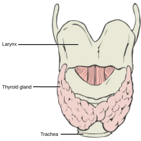
The thyroid gland is located in the neck, just below the larynx and in front of the trachea, as shown below. It is a butterfly-shaped gland with two lobes that are connected by the isthmus. It has a dark red color due to its extensive vascular system. When the thyroid swells due to dysfunction, it can be felt under the skin of the neck.
Extended description
A front view of the throat. ‘Larynx’ labels the pale, notched structure at the top. Below it, ‘Thyroid gland’ labels the pink, butterfly-shaped gland with two lobed sides joined across the midline. ‘Trachea’ labels the ringed tube continuing down from beneath the thyroid.
The thyroid gland is made up of many spherical thyroid follicles, which are lined with a simple cuboidal epithelium. These follicles contain a viscous fluid, called colloid, which stores the glycoprotein thyroglobulin, the precursor to the thyroid hormones. The follicles produce hormones that can be stored in the colloid or released into the surrounding capillary network for transport to the rest of the body via the circulatory system.
Thyroid follicle cells synthesize the hormone thyroxine, which is also known as T₄ because it contains four atoms of iodine, and triiodothyronine, also known as T₃ because it contains three atoms of iodine. Follicle cells are stimulated to release stored T₃ and T₄ by thyroid stimulating hormone (TSH), which is produced by the anterior pituitary. These thyroid hormones increase the rates of mitochondrial ATP production.
A third hormone, calcitonin, is produced by parafollicular cells of the thyroid. Calcitonin release is not controlled by TSH, but instead is released when calcium ion concentrations in the blood rise. Calcitonin functions to help regulate calcium concentrations in body fluids. It acts in the bones to inhibit osteoclast activity and in the kidneys to stimulate excretion of calcium. The combination of these two events lowers body fluid levels of calcium.
Parathyroid Glands
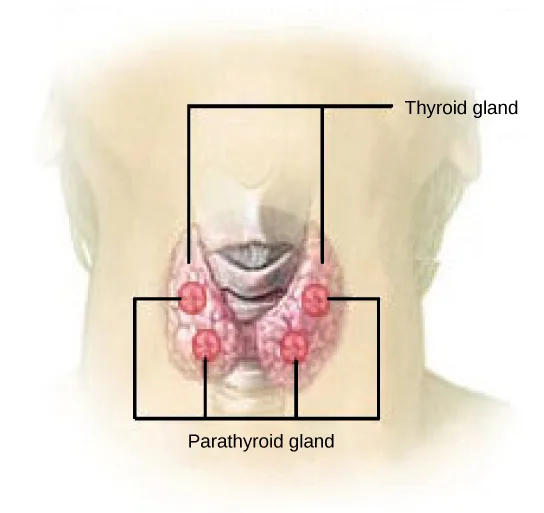
Most people have four parathyroid glands; however, the number can vary from two to six. These glands are located on the posterior surface of the thyroid gland, as shown below. Normally, there is a superior gland and an inferior gland associated with each of the thyroid’s two lobes. Each parathyroid gland is covered by connective tissue and contains many secretory cells that are associated with a capillary network.
Extended description
A view of the neck from behind. A bracket over the pink, butterfly-shaped gland at center is labeled ‘Thyroid gland.’ Four small red circles are embedded in the thyroid tissue, two on each lobe with one above the other; a second bracket spanning all four is labeled ‘Parathyroid gland.’
The parathyroid glands produce parathyroid hormone (PTH). PTH increases blood calcium concentrations when calcium ion levels fall below normal. PTH (1) enhances reabsorption of Ca²⁺ by the kidneys, (2) stimulates osteoclast activity and inhibits osteoblast activity, and (3) it stimulates synthesis and secretion of calcitriol by the kidneys, which enhances Ca²⁺ absorption by the digestive system. PTH is produced by chief cells of the parathyroid. PTH and calcitonin work in opposition to one another to maintain homeostatic Ca²⁺ levels in body fluids. Another type of cells, oxyphil cells, exist in the parathyroid but their function is not known.
Adrenal Glands
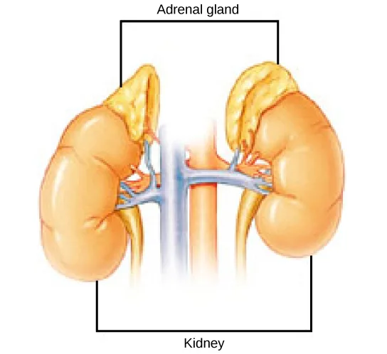
The adrenal glands are associated with the kidneys; one gland is located on top of each kidney as shown below. The adrenal glands consist of an outer adrenal cortex and an inner adrenal medulla. These regions secrete different hormones.
Extended description
Two bean-shaped kidneys sit side by side, joined by a central vertical blood-vessel trunk. A yellow, cap-shaped gland sits atop each kidney. A bracket over both yellow caps is labeled ‘Adrenal gland,’ and a bracket under both kidneys is labeled ‘Kidney.’
Adrenal Cortex
The adrenal cortex is made up of layers of epithelial cells and associated capillary networks. These layers form three distinct regions: an outer zona glomerulosa that produces mineralocorticoids, a middle zona fasciculata that produces glucocorticoids, and an inner zona reticularis that produces androgens.
The main mineralocorticoid is aldosterone, which regulates the concentration of Na⁺ ions in urine, sweat, and saliva. Aldosterone release from the adrenal cortex is stimulated by a decrease in blood concentrations of sodium ions, blood volume, or blood pressure, or by an increase in blood potassium levels.
The three main glucocorticoids are cortisol, corticosterone, and cortisone. The glucocorticoids stimulate the synthesis of glucose and gluconeogenesis (converting a non-carbohydrate to glucose) by liver cells and they promote the release of fatty acids from adipose tissue. These hormones increase blood glucose levels to maintain levels within a normal range between meals. These hormones are secreted in response to ACTH and levels are regulated by negative feedback.
The adrenal cortex also produces small amounts of testosterone precursor, although the role of this additional hormone production is not fully understood. Testosterone is a type of androgen that promotes a suite of characteristics such as the growth and development of the testes and penis, increased skeletal and muscular growth, enlargement of the larynx, increased growth and redistribution of body hair, and increased sexual drive. Testosterone secretion is regulated by both the hypothalamus and the anterior pituitary gland. The hypothalamus sends releasing hormones that stimulate the release of gonadotropins from the anterior pituitary gland. Testosterone produced in small amounts in the adrenal cortex may work with sex hormones released from the gonads.
Adrenal Medulla
The adrenal medulla contains large, irregularly shaped cells that are closely associated with blood vessels. These cells are innervated by preganglionic autonomic nerve fibers from the central nervous system.
The adrenal medulla contains two types of secretory cells: one that produces epinephrine (adrenaline) and another that produces norepinephrine (noradrenaline). Epinephrine is the primary adrenal medulla hormone accounting for 75 to 80 percent of its secretions. Epinephrine and norepinephrine increase heart rate, breathing rate, cardiac muscle contractions, blood pressure, and blood glucose levels. They also accelerate the breakdown of glucose in skeletal muscles and stored fats in adipose tissue.
The release of epinephrine and norepinephrine is stimulated by neural impulses from the sympathetic nervous system. Secretion of these hormones is stimulated by acetylcholine release from preganglionic sympathetic fibers innervating the adrenal medulla. These neural impulses originate from the hypothalamus in response to stress to prepare the body for the fight-or-flight response.
Pancreas
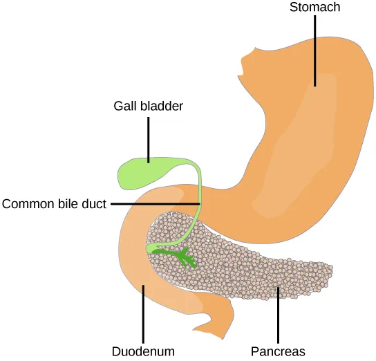
The pancreas, shown below, is an elongated organ that is located between the stomach and the proximal portion of the small intestine. It contains both exocrine cells that excrete digestive enzymes and endocrine cells that release hormones. It is sometimes referred to as a heterocrine gland because it has both endocrine and exocrine functions.
Extended description
At upper right, ‘Stomach’ labels a large orange, curved organ. At upper left, ‘Gall bladder’ labels a small green oval hanging beside it. ‘Common bile duct’ labels a thin green tube running from the gall bladder down past the stomach’s lower curve. Below, ‘Duodenum’ labels the pale tube looping at lower left, and ‘Pancreas’ labels the speckled, teardrop-shaped organ at lower right that the duodenum wraps around.
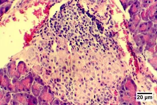
The endocrine cells of the pancreas form clusters called pancreatic islets or the islets of Langerhans, as visible in the micrograph shown below. The pancreatic islets contain two primary cell types: alpha cells, which produce the hormone glucagon, and beta cells, which produce the hormone insulin. These hormones regulate blood glucose levels. As blood glucose levels decline, alpha cells release glucagon to raise the blood glucose levels by increasing rates of glycogen breakdown and glucose release by the liver. When blood glucose levels rise, such as after a meal, beta cells release insulin to lower blood glucose levels by increasing the rate of glucose uptake in most body cells, and by increasing glycogen synthesis in skeletal muscles and the liver. Together, glucagon and insulin regulate blood glucose levels.
Pineal Gland
The pineal gland produces melatonin. The rate of melatonin production is affected by the photoperiod. Collaterals from the visual pathways innervate the pineal gland. During the day photoperiod, little melatonin is produced; however, melatonin production increases during the dark photoperiod (night). In some mammals, melatonin has an inhibitory effect on reproductive functions by decreasing production and maturation of sperm, oocytes, and reproductive organs. Melatonin is an effective antioxidant, protecting the CNS from free radicals such as nitric oxide and hydrogen peroxide. Lastly, melatonin is involved in biological rhythms, particularly circadian rhythms such as the sleep-wake cycle and eating habits.
Gonads
The gonads—the testes and ovaries—produce steroid hormones. The testes produce androgens, testosterone being the most prominent, which allow for the growth and development of the testes and penis, increased skeletal and muscular growth, enlargement of the larynx, increased growth and redistribution of body hair, and the production of sperm cells. The ovaries produce estradiol and progesterone, which cause secondary sex characteristics and prepare the body for childbirth.
Endocrine Glands and their Associated Hormones
| Endocrine Gland | Associated Hormones | Effect |
|---|---|---|
| Hypothalamus | releasing and inhibiting hormones | regulate hormone release from pituitary gland; produce oxytocin; produce uterine contractions and milk secretion in breast tissue |
| antidiuretic hormone (ADH) | water reabsorption from kidneys; vasoconstriction to increase blood pressure | |
| Pituitary (Anterior) | growth hormone (GH) | promotes growth of body tissues, protein synthesis; metabolic functions |
| prolactin (PRL) | promotes milk production | |
| thyroid stimulating hormone (TSH) | stimulates thyroid hormone release | |
| adrenocorticotropic hormone (ACTH) | stimulates hormone release by adrenal cortex, glucocorticoids | |
| follicle-stimulating hormone (FSH) | stimulates gamete production (both ova and sperm); secretion of estradiol | |
| luteinizing hormone (LH) | stimulates androgen production by gonads; ovulation, secretion of progesterone | |
| melanocyte-stimulating hormone (MSH) | stimulates melanocytes of the skin increasing melanin pigment production. | |
| Pituitary (Posterior) | antidiuretic hormone (ADH) | stimulates water reabsorption by kidneys |
| oxytocin | stimulates uterine contractions during childbirth; milk ejection; stimulates ductus deferens and prostate gland contraction during emission | |
| Thyroid | thyroxine, triiodothyronine | stimulate and maintain metabolism; growth and development |
| calcitonin | reduces blood Ca²⁺ levels | |
| Parathyroid | parathyroid hormone (PTH) | increases blood Ca²⁺ levels |
| Adrenal (Cortex) | aldosterone | increases blood Na⁺ levels; increases K⁺ secretion |
| cortisol, corticosterone, cortisone | increase blood glucose levels; anti-inflammatory effects | |
| Adrenal (Medulla) | epinephrine, norepinephrine | stimulate fight-or-flight response; increase blood glucose levels; increase metabolic activities |
| Pancreas | insulin | reduces blood glucose levels |
| glucagon | increases blood glucose levels | |
| Pineal gland | melatonin | regulates some biological rhythms and protects CNS from free radicals |
| Testes | androgens | regulate, promote, increase or maintain sperm production; a suite of characteristics including growth and development of the testes and penis, increased skeletal and muscular growth, enlargement of the larynx, and increased growth and redistribution of body hair |
| Ovaries | estrogen | promotes uterine lining growth; a suite of characteristics including increased development of breast tissue, redistribution of fat towards hips, legs, and breast, and the maturation of the uterus and vagina |
| progestins | promote and maintain uterine lining growth |
Organs with Secondary Endocrine Functions
There are several organs whose primary functions are non-endocrine but that also possess endocrine functions. These include the heart, kidneys, intestines, thymus, gonads, and adipose tissue.
The heart possesses endocrine cells in the walls of the atria that are specialized cardiac muscle cells. These cells release the hormone atrial natriuretic peptide (ANP) in response to increased blood volume. High blood volume causes the cells to be stretched, resulting in hormone release. ANP acts on the kidneys to reduce the reabsorption of Na⁺, causing Na⁺ and water to be excreted in the urine. ANP also reduces the amounts of renin released by the kidneys and aldosterone released by the adrenal cortex, further preventing the retention of water. In this way, ANP causes a reduction in blood volume and blood pressure, and reduces the concentration of Na⁺ in the blood.
The gastrointestinal tract produces several hormones that aid in digestion. The endocrine cells are located in the mucosa of the GI tract throughout the stomach and small intestine. Some of the hormones produced include gastrin, secretin, and cholecystokinin, which are secreted in the presence of food, and some of which act on other organs such as the pancreas, gallbladder, and liver. They trigger the release of gastric juices, which help to break down and digest food in the GI tract.
While the adrenal glands associated with the kidneys are major endocrine glands, the kidneys themselves also possess endocrine function. Renin is released in response to decreased blood volume or pressure and is part of the renin-angiotensin-aldosterone system that leads to the release of aldosterone. Aldosterone then causes the retention of Na⁺ and water, raising blood volume. The kidneys also release calcitriol, which aids in the absorption of Ca²⁺ and phosphate ions. Erythropoietin (EPO) is a protein hormone that triggers the formation of red blood cells in the bone marrow. EPO is released in response to low oxygen levels. Because red blood cells are oxygen carriers, increased production results in greater oxygen delivery throughout the body. EPO has been used by athletes to improve performance, as greater oxygen delivery to muscle cells allows for greater endurance. Because red blood cells increase the viscosity of blood, artificially high levels of EPO can cause severe health risks.
The thymus is found behind the sternum; it is most prominent in infants, becoming smaller in size through adulthood. The thymus produces hormones referred to as thymosins, which contribute to the development of the immune response.
Adipose tissue is a connective tissue found throughout the body. It produces the hormone leptin in response to food intake. Leptin increases the activity of anorexigenic neurons and decreases that of orexigenic neurons, producing a feeling of satiety after eating, thus affecting appetite and reducing the urge for further eating. Leptin is also associated with reproduction. It must be present for GnRH and gonadotropin synthesis to occur. Extremely thin females may enter puberty late; however, if adipose levels increase, more leptin will be produced, improving fertility.
Summary
The pituitary gland is located at the base of the brain and is attached to the hypothalamus by the infundibulum. The anterior pituitary receives products from the hypothalamus by the hypophyseal portal system and produces six hormones. The posterior pituitary is an extension of the brain and releases hormones (antidiuretic hormone and oxytocin) produced by the hypothalamus.
The thyroid gland is located in the neck and is composed of two lobes connected by the isthmus. The thyroid is made up of follicle cells that produce the hormones thyroxine and triiodothyronine. Parafollicular cells of the thyroid produce calcitonin. The parathyroid glands lie on the posterior surface of the thyroid gland and produce parathyroid hormone.
The adrenal glands are located on top of the kidneys and consist of the renal cortex and renal medulla. The adrenal cortex is the outer part of the adrenal gland and produces the corticosteroids, glucocorticoids, and mineralocorticoids. The adrenal medulla is the inner part of the adrenal gland and produces the catecholamines epinephrine and norepinephrine.
The pancreas lies in the abdomen between the stomach and the small intestine. Clusters of endocrine cells in the pancreas form the islets of Langerhans, which are composed of alpha cells that release glucagon and beta cells that release insulin.
Some organs possess endocrine activity as a secondary function but have another primary function. The heart produces the hormone atrial natriuretic peptide, which functions to reduce blood volume, pressure, and Na⁺ concentration. The gastrointestinal tract produces various hormones that aid in digestion. The kidneys produce renin, calcitriol, and erythropoietin. Adipose tissue produces leptin, which promotes satiety signals in the brain.
Key terms
- adrenal cortex — outer portion of adrenal glands that produces corticosteroids.
- adrenal gland — endocrine glands associated with the kidneys.
- adrenal medulla — inner portion of adrenal glands that produces epinephrine and norepinephrine.
- alpha cell — endocrine cell of the pancreatic islets that produces the hormone glucagon.
- anterior pituitary — portion of the pituitary gland that produces six hormones; also called adenohypophysis.
- atrial natriuretic peptide (ANP) — hormone produced by the heart to reduce blood volume, pressure, and Na⁺ concentration.
- beta cell — endocrine cell of the pancreatic islets that produces the hormone insulin.
- colloid — fluid inside the thyroid gland that contains the glycoprotein thyroglobulin.
- endocrine gland — gland that secretes hormones into the surrounding interstitial fluid, which then diffuse into blood and are carried to various organs and tissues within the body.
- erythropoietin (EPO) — hormone produced by the kidneys to stimulate red blood cell production in the bone marrow.
- hypophyseal portal system — system of blood vessels that carries hormones from the hypothalamus to the anterior pituitary.
- islets of Langerhans (pancreatic islets) — endocrine cells of the pancreas.
- isthmus — tissue mass that connects the two lobes of the thyroid gland.
- leptin — hormone produced by adipose tissue that promotes feelings of satiety and reduces hunger.
- pancreas — organ located between the stomach and the small intestine that contains exocrine and endocrine cells.
- parafollicular cell — thyroid cell that produces the hormone calcitonin.
- parathyroid gland — gland located on the surface of the thyroid that produces parathyroid hormone.
- pituitary gland — endocrine gland located at the base of the brain composed of an anterior and posterior region; also called hypophysis.
- pituitary stalk — (also, infundibulum) stalk that connects the pituitary gland to the hypothalamus.
- posterior pituitary — extension of the brain that releases hormones produced by the hypothalamus; along with the infundibulum, it is also referred to as the neurohypophysis.
- thymus — gland located behind the sternum that produces thymosin hormones that contribute to the development of the immune system.
- thyroid gland — endocrine gland located in the neck that produces thyroid hormones thyroxine and triiodothyronine.
Practice
Describe the role of different glands in the endocrine system
Which endocrine glands are associated with the kidneys?
This gland pair sits directly atop the organs named in the question, and the section’s own discussion of it opens with that same detail.Which of the following hormones is not produced by the anterior pituitary?
Three of these four are among the seven hormones the anterior pituitary itself produces; the fourth is produced by the hypothalamus and only stored and released by the pituitary’s other lobe.Recent studies suggest that blue light exposure can impact human circadian rhythms. This suggests that blue light disrupts the function of the _____ gland(s).
This gland’s hormone production is directly driven by light input from the visual pathways, and it governs the sleep-wake cycle.The adrenal medulla contains two types of secretory cells, what are they and what are their functions?
Show model answer
Did your answer mention:
The viscous fluid inside the thyroid gland that stores the glycoprotein thyroglobulin is called ________.
This fluid fills the spherical thyroid follicles and can either store thyroid hormone precursor or be released into the surrounding capillary network.The clusters of endocrine cells in the pancreas are called the ________.
These clusters contain alpha cells and beta cells, which release glucagon and insulin.Explain how the different glands work together to maintain homeostasis
What does aldosterone regulate, and how is it stimulated?
Show model answer
Did your answer mention:
How would damage to the posterior pituitary gland affect the production and release of ADH and inhibiting hormones?
Show model answer
Did your answer mention:
The system of blood vessels that carries hormones from the hypothalamus directly to the anterior pituitary, without first entering general circulation, is called the ________.
A ‘portal system’ carries blood from one capillary network to another rather than back to the heart first — this one links two endocrine structures directly.The hormone produced by the kidneys that stimulates red blood cell production in the bone marrow in response to low oxygen levels is called ________.
Some athletes have misused this hormone to boost their blood’s oxygen-carrying capacity, since greater red blood cell production allows for greater endurance.The hormone produced by the heart in response to increased blood volume, which acts on the kidneys to reduce blood volume, pressure, and sodium concentration, is called ________.
This hormone’s cells are stretched by high blood volume, and its effect on the kidneys works in the opposite direction of aldosterone’s.The hormone produced by adipose tissue in response to food intake, which promotes feelings of satiety and must be present for GnRH and gonadotropin synthesis to occur, is called ________.
Extremely thin individuals, who produce less of this hormone, may enter puberty later than typical — a link between energy stores and reproductive hormone release.This section is adapted from Biology 2e, Section 37.5: Endocrine Glands by Mary Ann Clark, Jung Choi, Matthew Douglas, and OpenStax, © OpenStax, licensed under CC BY-NC-SA 4.0. Access the original for free at openstax.org. Changes: the adrenal-cortex sentence on aldosterone reads “Na⁺ ions in urine, sweat, and saliva” where the source prints “urine, sweat, pancreas, and saliva” — the module’s own Critical Thinking solution lists “urine, sweat, and saliva”, and the pancreas is not a secreted fluid (reported as a source defect); figures re-encoded as WebP; five figures re-kinded from the manifest’s file-extension “photo” guess to “diagram” (Figure_37_05_01ab, Figure_37_05_02, Figure_37_05_03, Figure_37_05_04, and Figure_37_05_05 are all hand-drawn or digitally rendered anatomical illustrations, not captured photographs — Figure_37_05_06, the pancreatic-islet micrograph, is the one genuine photo and keeps that kind); a longdesc added to every diagram, walking each drawing’s own printed leader-line labels (the two-panel pituitary figure, the thyroid, the parathyroid, the adrenal, and the pancreas illustrations) — the micrograph needed none, since its caption and alt already carry its content; the pituitary figure’s alt rewritten from the source’s single run-on sentence to explicitly name what panel (a) and panel (b) each show, since the source alt did not distinguish them; in-text pointers to figures (“Figure 37.15” through “Figure 37.20”) replaced with “shown below” or “illustrated below,” since Hugo does not number figures; ion charges and iodine-count subscripts set as Unicode (Na⁺, K⁺, Ca²⁺, T₃, T₄) in place of the source’s <sup>/<sub> markup; the “Endocrine Glands and their Associated Hormones” table kept as a Markdown table in the body — its columns (gland, hormone, effect) name a gland/hormone pairing rather than categories, so it does not qualify for the sortbins “columns name categories” test and has no Practice-block counterpart; the end-of-section Review Questions and Critical Thinking Questions adapted into the closing interactive Practice block (multiple choice and self-check respectively); rubric checkpoints added to each self-check, decomposing its model answer (the source solution) into check-off clauses with no new claims; six key-term recall items added from the glossary (colloid, islets of Langerhans, hypophyseal portal system, erythropoietin, atrial natriuretic peptide, leptin), covering six of the section’s twenty-two glossary terms, the rest appearing only in the Key terms list and the prose.