Types of Skeletal Systems
By the end of this section, you will be able to:
- Discuss the different types of skeletal systems
- Explain the role of the human skeletal system
- Compare and contrast different skeletal systems
A skeletal system is necessary to support the body, protect internal organs, and allow for the movement of an organism. There are three different skeleton designs that fulfill these functions: hydrostatic skeleton, exoskeleton, and endoskeleton.
Hydrostatic Skeleton
A hydrostatic skeleton is a skeleton formed by a fluid-filled compartment within the body, called the coelom. The organs of the coelom are supported by the aqueous fluid, which also resists external compression. This compartment is under hydrostatic pressure because of the fluid and supports the other organs of the organism. This type of skeletal system is found in soft-bodied animals such as sea anemones, earthworms, Cnidaria, and other invertebrates (illustrated below).
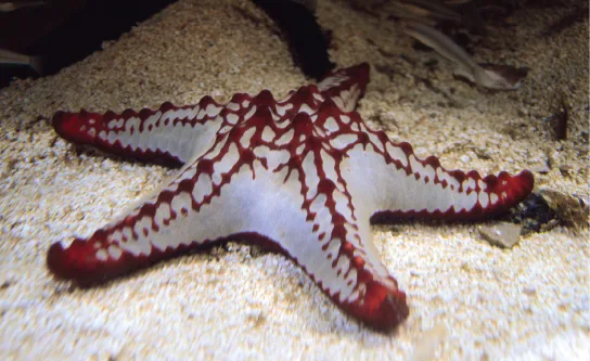
Movement in a hydrostatic skeleton is provided by muscles that surround the coelom. The muscles in a hydrostatic skeleton contract to change the shape of the coelom; the pressure of the fluid in the coelom produces movement. For example, earthworms move by waves of muscular contractions of the skeletal muscle of the body wall hydrostatic skeleton, called peristalsis, which alternately shorten and lengthen the body. Lengthening the body extends the anterior end of the organism. Most organisms have a mechanism to fix themselves in the substrate. Shortening the muscles then draws the posterior portion of the body forward. Although a hydrostatic skeleton is well-suited to invertebrate organisms such as earthworms and some aquatic organisms, it is not an efficient skeleton for terrestrial animals.
Exoskeleton
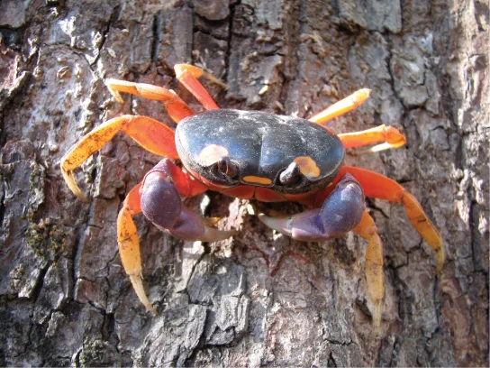
An exoskeleton is an external skeleton that consists of a hard encasement on the surface of an organism. For example, the shells of crabs and insects are exoskeletons (illustrated below). This skeleton type provides defence against predators, supports the body, and allows for movement through the contraction of attached muscles. As with vertebrates, muscles must cross a joint inside the exoskeleton. Shortening of the muscle changes the relationship of the two segments of the exoskeleton. Arthropods such as crabs and lobsters have exoskeletons that consist of 30–50 percent chitin, a polysaccharide derivative of glucose that is a strong but flexible material. Chitin is secreted by the epidermal cells. The exoskeleton is further strengthened by the addition of calcium carbonate in organisms such as the lobster. Because the exoskeleton is acellular, arthropods must periodically shed their exoskeletons because the exoskeleton does not grow as the organism grows.
Endoskeleton
An endoskeleton is a skeleton that consists of hard, mineralized structures located within the soft tissue of organisms. An example of a primitive endoskeletal structure is the spicules of sponges. The bones of vertebrates are composed of tissues, whereas sponges have no true tissues (illustrated below). Endoskeletons provide support for the body, protect internal organs, and allow for movement through contraction of muscles attached to the skeleton.
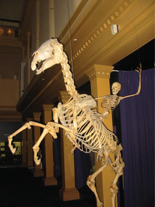
The human skeleton is an endoskeleton that consists of 206 bones in the adult. It has five main functions: providing support to the body, storing minerals and lipids, producing blood cells, protecting internal organs, and allowing for movement. The skeletal system in vertebrates is divided into the axial skeleton (which consists of the skull, vertebral column, and rib cage), and the appendicular skeleton (which consists of the shoulders, limb bones, the pectoral girdle, and the pelvic girdle).
Human Axial Skeleton
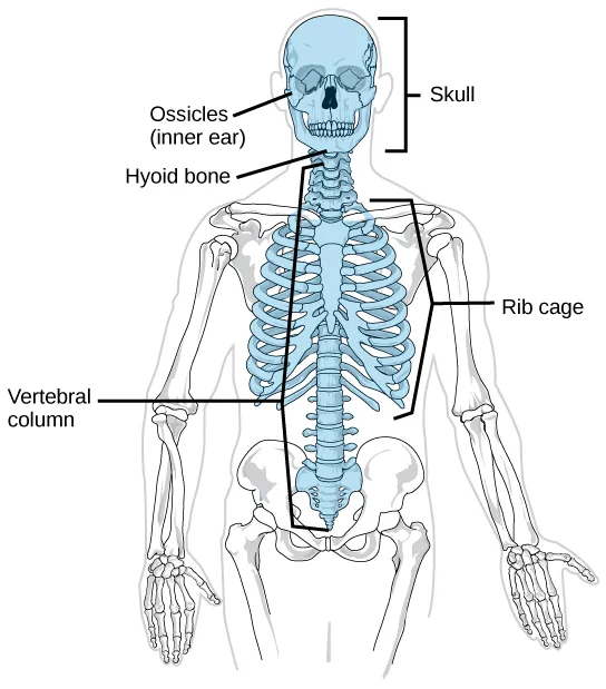
The axial skeleton forms the central axis of the body and includes the bones of the skull, ossicles of the middle ear, hyoid bone of the throat, vertebral column, and the thoracic cage (ribcage) (illustrated below). The function of the axial skeleton is to provide support and protection for the brain, the spinal cord, and the organs in the ventral body cavity. It provides a surface for the attachment of muscles that move the head, neck, and trunk, performs respiratory movements, and stabilizes parts of the appendicular skeleton.
Extended description
The illustration highlights the axial skeleton in blue against a pale outline of the rest of the skeleton. Reading top to bottom: ‘Ossicles (inner ear)’ and a ‘Skull’ bracket both label the head, one naming the inner-ear bones and the other the skull as a whole; ‘Hyoid bone’ labels a small bone at the front of the neck, below the jaw; ‘Vertebral column’ labels the spine running down the back; a ‘Rib cage’ bracket labels the ribs curving around the chest. Five labels in all.
The Skull
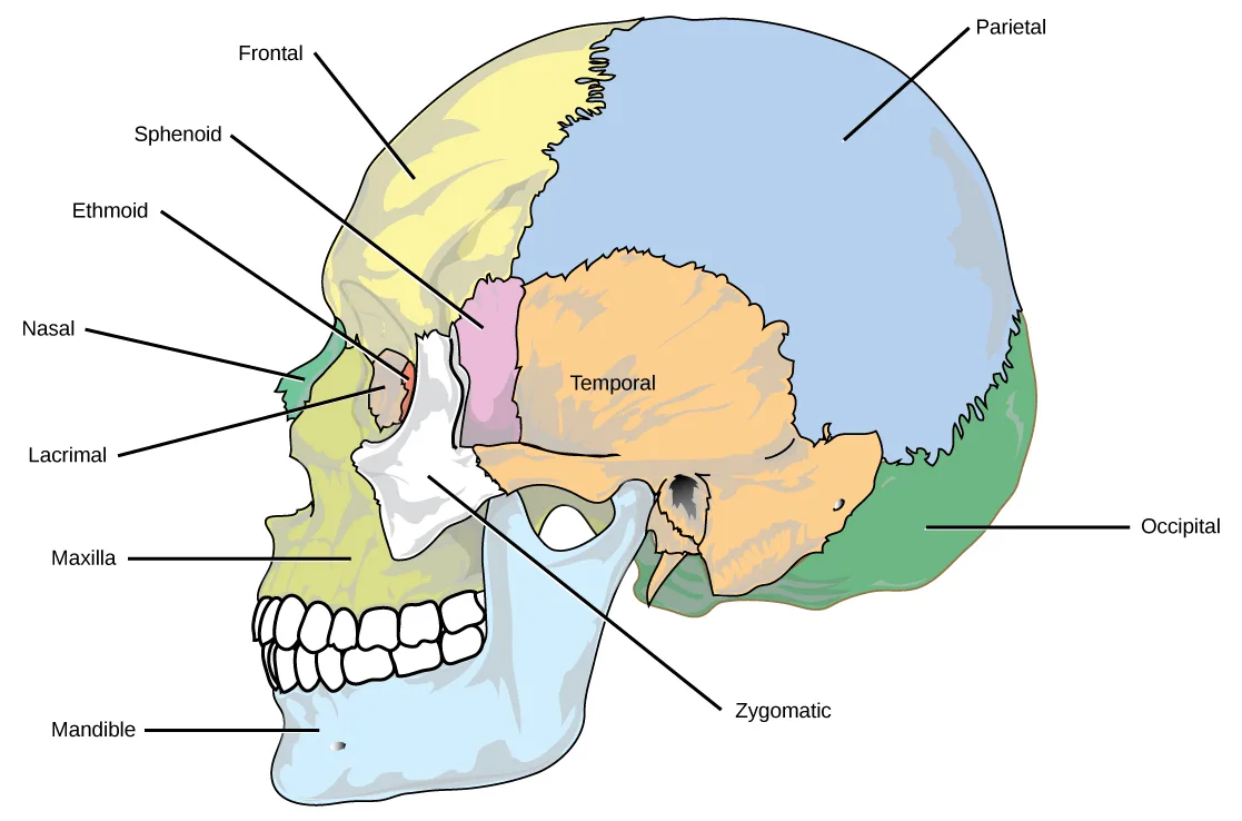
The bones of the skull support the structures of the face and protect the brain. The skull consists of 22 bones, which are divided into two categories: cranial bones and facial bones. The cranial bones are eight bones that form the cranial cavity, which encloses the brain and serves as an attachment site for the muscles of the head and neck. The eight cranial bones are the frontal bone, two parietal bones, two temporal bones, occipital bone, sphenoid bone, and the ethmoid bone. Although the bones developed separately in the embryo and fetus, in the adult, they are tightly fused with connective tissue and adjoining bones do not move (shown below).
Extended description
The skull is shown from the right side, colored by bone. Reading top to bottom on the left: ‘Frontal’ labels the pale yellow forehead region; ‘Sphenoid’ labels a small pink area behind the eye socket; ‘Ethmoid’ labels a thin green sliver in front of the sphenoid, near the nose; ‘Nasal’ labels the small bone at the bridge of the nose; ‘Lacrimal’ labels a tiny area inside the eye socket, near the nose; ‘Maxilla’ labels the white upper-jaw bone bearing the upper teeth; ‘Mandible’ labels the pale blue lower-jaw bone at the bottom, bearing the lower teeth — seven labels. On the right: ‘Parietal’ labels the large blue dome making up most of the skull roof; ‘Temporal’ is printed directly on the orange bone that makes up the side of the skull, below the parietal, with no leader line; ‘Zygomatic’ leads down to the small bone bridging the cheek to the ear region; ‘Occipital’ leads to the green area at the lower back of the skull — four more labels, eleven in all.
The auditory ossicles of the middle ear transmit sounds from the air as vibrations to the fluid-filled cochlea. The auditory ossicles consist of three bones each: the malleus, incus, and stapes. These are the smallest bones in the body and are unique to mammals.
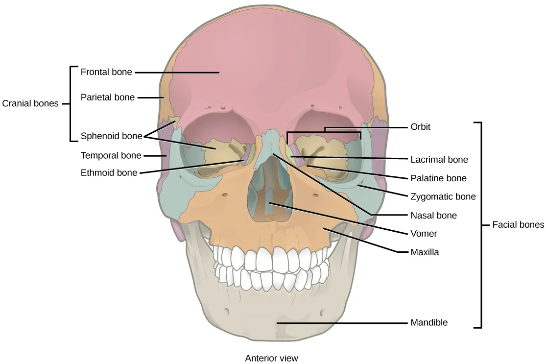
Fourteen facial bones form the face, provide cavities for the sense organs (eyes, mouth, and nose), protect the entrances to the digestive and respiratory tracts, and serve as attachment points for facial muscles. The 14 facial bones are the nasal bones, the maxillary bones, zygomatic bones, palatine, vomer, lacrimal bones, the inferior nasal conchae, and the mandible. All of these bones occur in pairs except for the mandible and the vomer (shown below).
Extended description
The skull is shown from the front. A bracket on the left labeled ‘Cranial bones’ gathers five leader-line labels, top to bottom: ‘Frontal bone’ (the pink dome across the top), ‘Parietal bone’ (tan, at the side above the temple, behind the frontal bone), ‘Sphenoid bone’ (the pale area just behind the eye socket), ‘Temporal bone’ (green, at the side), and ‘Ethmoid bone’ (a small pale sliver near the eye socket’s inner corner). A bracket on the right labeled ‘Facial bones’ gathers eight leader-line labels, top to bottom: ‘Orbit’ (the eye socket opening), ‘Lacrimal bone’ (a small bone at the inner corner of the eye socket), ‘Palatine bone’ (deep inside the nasal opening), ‘Zygomatic bone’ (the cheekbone beside the eye socket), ‘Nasal bone’ (the bridge of the nose), ‘Vomer’ (a thin bone in the center of the nasal opening), ‘Maxilla’ (the upper jaw bearing the upper teeth), and ‘Mandible’ (the lower jaw bearing the lower teeth, at the bottom) — thirteen leader-line labels in all, under the two group brackets. A caption beneath the drawing reads ‘Anterior view.’
Although it is not found in the skull, the hyoid bone is considered a component of the axial skeleton. The hyoid bone lies below the mandible in the front of the neck. It acts as a movable base for the tongue and is connected to muscles of the jaw, larynx, and tongue. The mandible articulates with the base of the skull. The mandible controls the opening to the airway and gut. In animals with teeth, the mandible brings the surfaces of the teeth in contact with the maxillary teeth.
The Vertebral Column
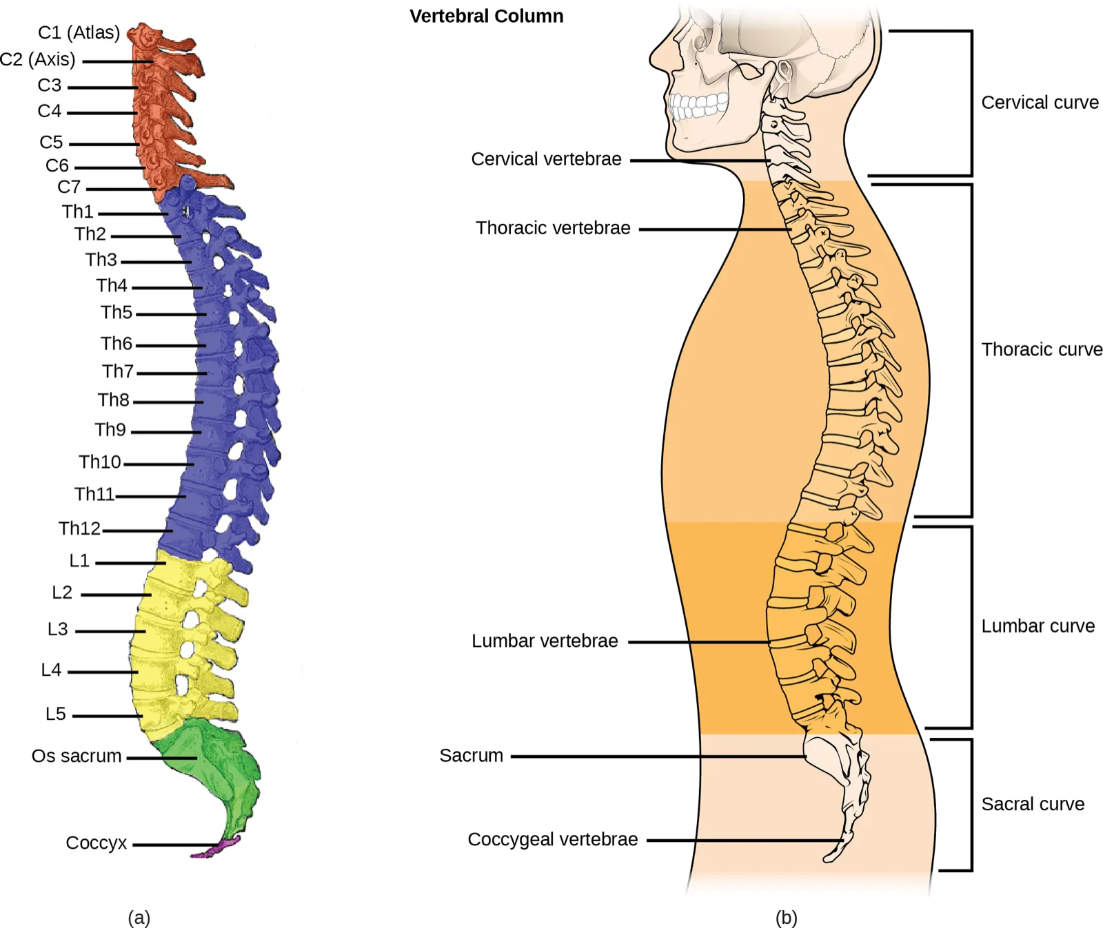
The vertebral column, or spinal column, surrounds and protects the spinal cord, supports the head, and acts as an attachment point for the ribs and muscles of the back and neck. The adult vertebral column comprises 26 bones: the 24 vertebrae, the sacrum, and the coccyx bones. In the adult, the sacrum is typically composed of five vertebrae that fuse into one. The coccyx is typically 3–4 vertebrae that fuse into one. Around the age of 70, the sacrum and the coccyx may fuse together. We begin life with approximately 33 vertebrae, but as we grow, several vertebrae fuse together. The adult vertebrae are further divided into the 7 cervical vertebrae, 12 thoracic vertebrae, and 5 lumbar vertebrae (illustrated below).
Extended description
(a) A vertical color-coded stack of vertebrae is labeled top to bottom, one leader line per bone: ‘C1 (Atlas),’ ‘C2 (Axis),’ ‘C3,’ ‘C4,’ ‘C5,’ ‘C6,’ ‘C7’ (seven cervical vertebrae, colored red-orange); ‘Th1’ through ‘Th12’ (twelve thoracic vertebrae, colored purple); ‘L1’ through ‘L5’ (five lumbar vertebrae, colored yellow); ‘Os sacrum’ (the fused triangular bone, colored green); and ‘Coccyx’ (the small tailbone below it, also green) — 26 labels in all. (b) A side view of a body outline shows the spine curving through the neck, chest, lower back, and pelvis, with five leader-line labels — ‘Cervical vertebrae,’ ‘Thoracic vertebrae,’ ‘Lumbar vertebrae,’ ‘Sacrum,’ and ‘Coccygeal vertebrae’ — pointing to the matching regions of the spine, and four bracket labels along the right margin — ‘Cervical curve,’ ‘Thoracic curve,’ ‘Lumbar curve,’ and ‘Sacral curve’ — spanning the same four regions from top to bottom.
Each vertebral body has a large hole in the center through which the nerves of the spinal cord pass. There is also a notch on each side through which the spinal nerves, which serve the body at that level, can exit from the spinal cord. The vertebral column is approximately 71 cm (28 inches) in adult male humans and is curved, which can be seen from a side view. The names of the spinal curves correspond to the region of the spine in which they occur. The thoracic and sacral curves are concave (curve inwards relative to the front of the body) and the cervical and lumbar curves are convex (curve outwards relative to the front of the body). The arched curvature of the vertebral column increases its strength and flexibility, allowing it to absorb shocks like a spring (illustrated above).
Intervertebral discs composed of fibrous cartilage lie between adjacent vertebral bodies from the second cervical vertebra to the sacrum. Each disc is part of a joint that allows for some movement of the spine and acts as a cushion to absorb shocks from movements such as walking and running. Intervertebral discs also act as ligaments to bind vertebrae together. The inner part of discs, the nucleus pulposus, hardens as people age and becomes less elastic. This loss of elasticity diminishes its ability to absorb shocks.
The Thoracic Cage
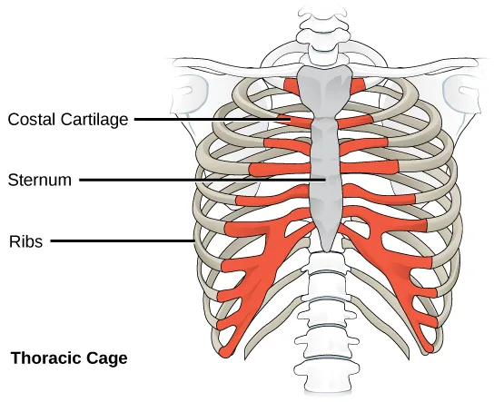
The thoracic cage, also known as the ribcage, is the skeleton of the chest, and consists of the ribs, sternum, thoracic vertebrae, and costal cartilages (illustrated below). The thoracic cage encloses and protects the organs of the thoracic cavity, including the heart and lungs. It also provides support for the shoulder girdles and upper limbs, and serves as the attachment point for the diaphragm, muscles of the back, chest, neck, and shoulders. Changes in the volume of the thorax enable breathing.
The sternum, or breastbone, is a long, flat bone located at the anterior of the chest. It is formed from three bones that fuse in the adult. The ribs are 12 pairs of long, curved bones that attach to the thoracic vertebrae and curve toward the front of the body, forming the ribcage. Costal cartilages connect the anterior ends of the ribs to the sternum, with the exception of rib pairs 11 and 12, which are free-floating ribs.
Extended description
Three leader lines point from left to right: ‘Costal Cartilage’ to the red segments joining the ribs to the sternum near the top; ‘Sternum’ to the flat, dagger-shaped pale gray bone down the center; ‘Ribs’ to the curved tan bones arcing around the chest, red only where their cartilage segments meet the sternum. Below the illustration, the bold label ‘Thoracic Cage’ names the whole structure — four labels in all.
Human Appendicular Skeleton
The appendicular skeleton is composed of the bones of the upper limbs (which function to grasp and manipulate objects) and the lower limbs (which permit locomotion). It also includes the pectoral girdle, or shoulder girdle, that attaches the upper limbs to the body, and the pelvic girdle that attaches the lower limbs to the body (illustrated below).
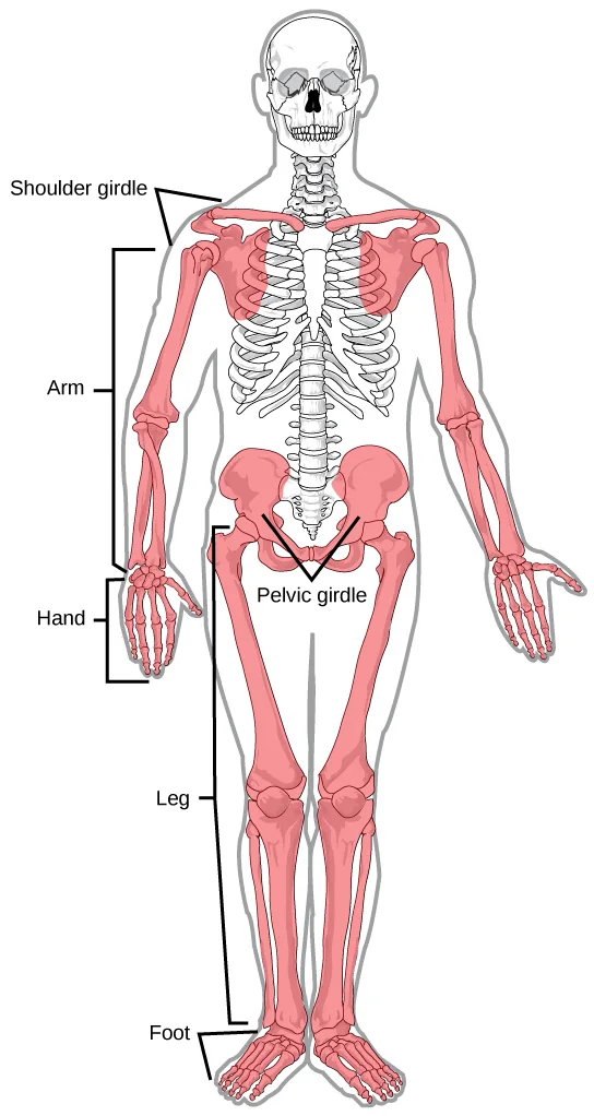
Extended description
The appendicular skeleton is highlighted in pink against a pale outline of the axial skeleton. Top to bottom: ‘Shoulder girdle’ labels the collarbone-and-shoulder-blade region; an ‘Arm’ bracket spans the upper arm and forearm bones on the reader’s left; a ‘Hand’ bracket spans the wrist and hand bones at the bottom of that same arm; ‘Pelvic girdle’ labels the hip bones at the body’s center; a ‘Leg’ bracket spans the thigh and lower-leg bones, also on the reader’s left; a ‘Foot’ bracket spans the ankle and foot bones at the bottom of that leg — six labels in all.
The Pectoral Girdle
The pectoral girdle bones provide the points of attachment of the upper limbs to the axial skeleton. The human pectoral girdle consists of the clavicle (or collarbone) in the anterior, and the scapula (or shoulder blades) in the posterior (illustrated below).
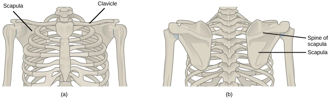
Extended description
(a) In the anterior (front) view, two leader lines label the pale tan bones on either side of the ribcage: ‘Scapula’ points to the shoulder blade at the side, and ‘Clavicle’ points to the long collarbone running above the ribcage to the shoulder. (b) In the posterior (back) view of the same bones, ‘Spine of scapula’ points to the raised ridge running across the shoulder blade, and ‘Scapula’ points to the shoulder blade itself — four labels in all.
The clavicles are S-shaped bones that position the arms on the body. The clavicles lie horizontally across the front of the thorax (chest) just above the first rib. These bones are fairly fragile and are susceptible to fractures. For example, a fall with the arms outstretched causes the force to be transmitted to the clavicles, which can break if the force is excessive. The clavicle articulates with the sternum and the scapula.
The scapulae are flat, triangular bones that are located at the back of the pectoral girdle. They support the muscles crossing the shoulder joint. A ridge, called the spine, runs across the back of the scapula and can easily be felt through the skin (illustrated above). The spine of the scapula is a good example of a bony protrusion that facilitates a broad area of attachment for muscles to bone.
The Upper Limb
The upper limb contains 30 bones in three regions: the arm (shoulder to elbow), the forearm (ulna and radius), and the wrist and hand (illustrated below).
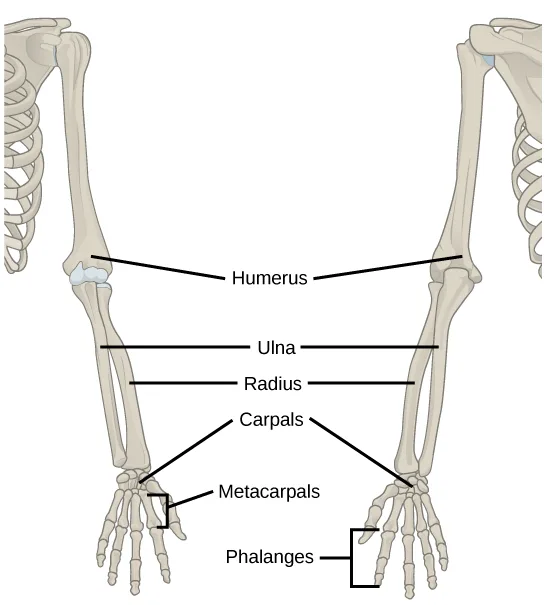
Extended description
Two upper limbs are shown side by side, one from the front and one from behind, sharing one set of leader-line labels that point to both: ‘Humerus’ labels the single long bone of the upper arm; ‘Ulna’ and ‘Radius’ label the two forearm bones, the ulna on the pinky-finger side and the radius on the thumb side; ‘Carpals’ labels the small wrist bones; ‘Metacarpals’ labels the bones of the palm; ‘Phalanges’ labels the finger bones at the bottom — six labels in all.
An articulation is any place at which two bones are joined. The humerus is the largest and longest bone of the upper limb and the only bone of the arm. It articulates with the scapula at the shoulder and with the forearm at the elbow. The forearm extends from the elbow to the wrist and consists of two bones: the ulna and the radius. The radius is located along the lateral (thumb) side of the forearm and articulates with the humerus at the elbow. The ulna is located on the medial aspect (pinky-finger side) of the forearm. It is longer than the radius. The ulna articulates with the humerus at the elbow. The radius and ulna also articulate with the carpal bones and with each other, which in vertebrates enables a variable degree of rotation of the carpus with respect to the long axis of the limb. The hand includes the eight bones of the carpus (wrist), the five bones of the metacarpus (palm), and the 14 bones of the phalanges (digits). Each digit consists of three phalanges, except for the thumb, when present, which has only two.
The Pelvic Girdle
The pelvic girdle bones provide the points of attachment of the lower limbs to the axial skeleton. Because it is responsible for bearing the weight of the body and for locomotion, the pelvic girdle is securely attached to the axial skeleton by strong ligaments. It also has deep sockets with robust ligaments to securely attach the femur to the body. The pelvic girdle is further strengthened by two large hip bones. In adults, the hip bones, or coxal bones, are formed by the fusion of three pairs of bones: the ilium, ischium, and pubis. The pelvis joins together in the anterior of the body at a joint called the pubic symphysis and with the bones of the sacrum at the posterior of the body.
The female pelvis is slightly different from the male pelvis. Over generations of evolution, females with a wider pubic angle and larger diameter pelvic canal reproduced more successfully. Therefore, their offspring also had pelvic anatomy that enabled successful childbirth (illustrated below).
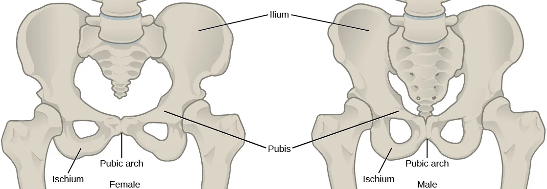
Extended description
Two pelvises are shown side by side: (a) the female pelvis on the left, (b) the male pelvis on the right, each captioned below by sex. Two labels span both pelvises with leader lines to each: ‘Ilium’ points to the wide, curved upper bone on both sides, and ‘Pubis’ points to the bones meeting at the front center of each pelvis. Each pelvis also carries its own pair of labels: ‘Ischium’ points to the lower rear part of the hip bone, and ‘Pubic arch’ points to the rounded gap below the pubic bones — the female pelvis’s arch is visibly wider than the male’s.
The Lower Limb
The lower limb consists of the thigh, the leg, and the foot. The bones of the lower limb are the femur (thigh bone), patella (kneecap), tibia and fibula (bones of the leg), tarsals (bones of the ankle), and metatarsals and phalanges (bones of the foot) (illustrated below). The bones of the lower limbs are thicker and stronger than the bones of the upper limbs because of the need to support the entire weight of the body and the resulting forces from locomotion. In addition to evolutionary fitness, the bones of an individual will respond to forces exerted upon them.
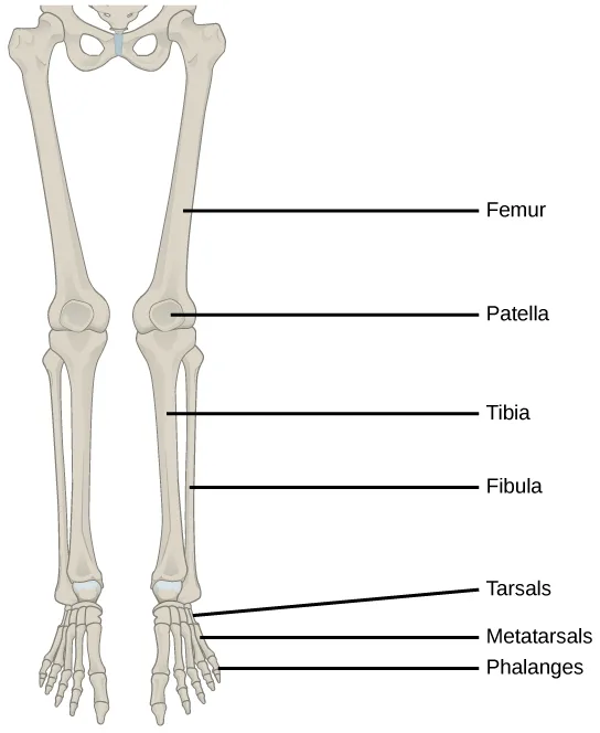
Extended description
Two legs are shown side by side, sharing one set of leader-line labels that point to both: ‘Femur’ labels the thigh bone; ‘Patella’ labels the kneecap; ‘Tibia’ labels the thicker, inner bone of the lower leg; ‘Fibula’ labels the thinner, outer bone of the lower leg; ‘Tarsals’ labels the ankle bones; ‘Metatarsals’ labels the bones of the foot; ‘Phalanges’ labels the toe bones at the bottom — seven labels in all.
The femur, or thighbone, is the longest, heaviest, and strongest bone in the body. The femur and pelvis form the hip joint at the proximal end. At the distal end, the femur, tibia, and patella form the knee joint. The patella, or kneecap, is a triangular bone that lies anterior to the knee joint. The patella is embedded in the tendon of the femoral extensors (quadriceps). It improves knee extension by reducing friction. The tibia, or shinbone, is a large bone of the leg that is located directly below the knee. The tibia articulates with the femur at its proximal end, with the fibula and the tarsal bones at its distal end. It is the second largest bone in the human body and is responsible for transmitting the weight of the body from the femur to the foot. The fibula, or calf bone, parallels and articulates with the tibia. It does not articulate with the femur and does not bear weight. The fibula acts as a site for muscle attachment and forms the lateral part of the ankle joint.
The tarsals are the seven bones of the ankle. The ankle transmits the weight of the body from the tibia and the fibula to the foot. The metatarsals are the five bones of the foot. The phalanges are the 14 bones of the toes. Each toe consists of three phalanges, except for the big toe that has only two (illustrated below). Variations exist in other species; for example, the horse’s metacarpals and metatarsals are oriented vertically and do not make contact with the substrate.
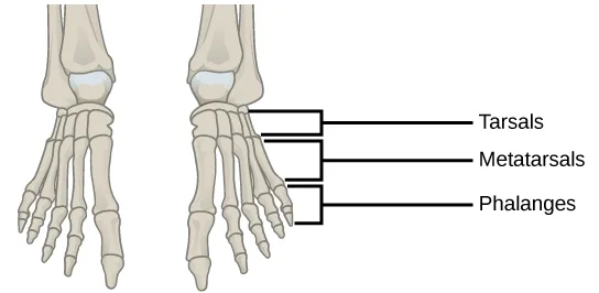
Extended description
Two feet are shown side by side. Three bracket labels on the right point to both: ‘Tarsals’ brackets the cluster of ankle bones at the back of the foot; ‘Metatarsals’ brackets the row of long bones across the middle of the foot; ‘Phalanges’ brackets the toe bones at the front — three labels in all.
Evolution Connection
Evolution of Body Design for Locomotion on Land
The transition of vertebrates onto land required a number of changes in body design, as movement on land presents a number of challenges for animals that are adapted to movement in water. The buoyancy of water provides a certain amount of lift, and a common form of movement by fish is lateral undulations of the entire body. This back and forth movement pushes the body against the water, creating forward movement. In most fish, the muscles of paired fins attach to girdles within the body, allowing for some control of locomotion. As certain fish began moving onto land, they retained their lateral undulation form of locomotion (anguilliform). However, instead of pushing against water, their fins or flippers became points of contact with the ground, around which they rotated their bodies.
The effect of gravity and the lack of buoyancy on land meant that body weight was suspended on the limbs, leading to increased strengthening and ossification of the limbs. The effect of gravity also required changes to the axial skeleton. Lateral undulations of land animal vertebral columns cause torsional strain. A firmer, more ossified vertebral column became common in terrestrial tetrapods because it reduces strain while providing the strength needed to support the body’s weight. In later tetrapods, the vertebrae began allowing for vertical motion rather than lateral flexion. Another change in the axial skeleton was the loss of a direct attachment between the pectoral girdle and the head. This reduced the jarring to the head caused by the impact of the limbs on the ground. The vertebrae of the neck also evolved to allow movement of the head independently of the body.
The appendicular skeleton of land animals is also different from aquatic animals. The shoulders attach to the pectoral girdle through muscles and connective tissue, thus reducing the jarring of the skull. Because of a lateral undulating vertebral column, in early tetrapods, the limbs were splayed out to the side and movement occurred by performing “push-ups.” The vertebrae of these animals had to move side-to-side in a similar manner to fish and reptiles. This type of motion requires large muscles to move the limbs toward the midline; it was almost like walking while doing push-ups, and it is not an efficient use of energy. Later tetrapods have their limbs placed under their bodies, so that each stride requires less force to move forward. This resulted in decreased adductor muscle size and an increased range of motion of the scapulae. This also restricts movement primarily to one plane, creating forward motion rather than moving the limbs upward as well as forward. The femur and humerus were also rotated, so that the ends of the limbs and digits were pointed forward, in the direction of motion, rather than out to the side. By placement underneath the body, limbs can swing forward like a pendulum to produce a stride that is more efficient for moving over land.
Summary
The three types of skeleton designs are hydrostatic skeletons, exoskeletons, and endoskeletons. A hydrostatic skeleton is formed by a fluid-filled compartment held under hydrostatic pressure; movement is created by the muscles producing pressure on the fluid. An exoskeleton is a hard external skeleton that protects the outer surface of an organism and enables movement through muscles attached on the inside. An endoskeleton is an internal skeleton composed of hard, mineralized tissue that also enables movement by attachment to muscles. The human skeleton is an endoskeleton that is composed of the axial and appendicular skeleton. The axial skeleton is composed of the bones of the skull, ossicles of the ear, hyoid bone, vertebral column, and ribcage. The skull consists of eight cranial bones and 14 facial bones. Six bones make up the ossicles of the middle ear, while the hyoid bone is located in the neck under the mandible. The vertebral column contains 26 bones, and it surrounds and protects the spinal cord. The thoracic cage consists of the sternum, ribs, thoracic vertebrae, and costal cartilages. The appendicular skeleton is made up of the limbs of the upper and lower limbs. The pectoral girdle is composed of the clavicles and the scapulae. The upper limb contains 30 bones in the arm, the forearm, and the hand. The pelvic girdle attaches the lower limbs to the axial skeleton. The lower limb includes the bones of the thigh, the leg, and the foot.
Key terms
- appendicular skeleton — composed of the bones of the upper limbs, which function to grasp and manipulate objects, and the lower limbs, which permit locomotion.
- articulation — any place where two bones are joined.
- auditory ossicle — (also, middle ear) transduces sounds from the air into vibrations in the fluid-filled cochlea.
- axial skeleton — forms the central axis of the body and includes the bones of the skull, the ossicles of the middle ear, the hyoid bone of the throat, the vertebral column, and the thoracic cage (ribcage).
- carpus — eight bones that comprise the wrist.
- clavicle — S-shaped bone that positions the arms laterally.
- coxal bone — hip bone.
- cranial bone — one of eight bones that form the cranial cavity that encloses the brain and serves as an attachment site for the muscles of the head and neck.
- endoskeleton — skeleton of living cells that produces a hard, mineralized tissue located within the soft tissue of organisms.
- exoskeleton — a secreted cellular product external skeleton that consists of a hard encasement on the surface of an organism.
- facial bone — one of the 14 bones that form the face; provides cavities for the sense organs (eyes, mouth, and nose) and attachment points for facial muscles.
- femur — (also, thighbone) longest, heaviest, and strongest bone in the body.
- fibula — (also, calf bone) parallels and articulates with the tibia.
- forearm — extends from the elbow to the wrist and consists of two bones: the ulna and the radius.
- humerus — only bone of the arm.
- hydrostatic skeleton — skeleton that consists of aqueous fluid held under pressure in a closed body compartment.
- hyoid bone — lies below the mandible in the front of the neck.
- intervertebral disc — composed of fibrous cartilage; lies between adjacent vertebrae from the second cervical vertebra to the sacrum.
- lower limb — consists of the thigh, the leg, and the foot.
- metacarpus — five bones that comprise the palm.
- metatarsal — one of the five bones of the foot.
- patella — (also, kneecap) triangular bone that lies anterior to the knee joint.
- pectoral girdle — bones that transmit the force generated by the upper limbs to the axial skeleton.
- phalange — one of the bones of the fingers or toes.
- pelvic girdle — bones that transmit the force generated by the lower limbs to the axial skeleton.
- radius — bone located along the lateral (thumb) side of the forearm; articulates with the humerus at the elbow.
- rib — one of 12 pairs of long, curved bones that attach to the thoracic vertebrae and curve toward the front of the body to form the ribcage.
- scapula — flat, triangular bone located at the posterior pectoral girdle.
- skull — bone that supports the structures of the face and protects the brain.
- sternum — (also, breastbone) long, flat bone located at the front of the chest.
- tarsal — one of the seven bones of the ankle.
- thoracic cage — (also, ribcage) skeleton of the chest, which consists of the ribs, thoracic vertebrae, sternum, and costal cartilages.
- tibia — (also, shinbone) large bone of the leg that is located directly below the knee.
- ulna — bone located on the medial aspect (pinky-finger side) of the forearm.
- vertebral column — (also, spine) surrounds and protects the spinal cord, supports the head, and acts as an attachment point for ribs and muscles of the back and neck.
Practice
Discuss the different types of skeletal systems
A skeleton that consists of aqueous fluid held under pressure in a closed body compartment is called a ________.
This is the skeleton design that lets soft-bodied invertebrates such as sea anemones and earthworms move using pressure from an internal fluid compartment, rather than any hard structure.A secreted, external skeleton that consists of a hard encasement on the surface of an organism is called a(n) ________.
Crabs, lobsters, and insects carry this hard casing on the outside of the body, and must periodically shed it because it does not grow as the animal does.A skeleton of living cells that produces a hard, mineralized tissue located within the soft tissue of organisms is called a(n) ________.
Humans and horses share this skeleton type, built from living, mineralized tissue that lies inside the body rather than surrounding it or replaced by an external casing.Explain the role of the human skeletal system
The forearm consists of the:
The humerus is named elsewhere in this section as the single bone of the upper arm, not the forearm — which two other bones does the section name as making up the forearm itself?The pectoral girdle consists of the:
The sternum and the coccyx are axial-skeleton bones the pectoral girdle attaches to or near, not girdle bones themselves — which two bones does the section name as making up the girdle?All of the following are groups of vertebrae except ________, which is a curvature.
Three of these terms name actual groups of vertebrae in the vertebral column; one instead names a different region of the skeleton.Which of these is a facial bone?
Frontal, occipital, and temporal are named among the section’s eight cranial bones — the facial bones are listed separately, just after them.What are the major differences between the male pelvis and female pelvis that permit childbirth in females?
Show model answer
Did your answer mention:
What are the major differences between the pelvic girdle and the pectoral girdle that allow the pelvic girdle to bear the weight of the body?
Show model answer
Did your answer mention:
Scoliosis is a medical condition where the spine develops a sideways curvature. How would this change interfere with the normal function of the spine?
Show model answer
Did your answer mention:
The bone that supports the structures of the face and protects the brain is called the ________.
This structure is made of 22 bones in total, split into cranial bones and facial bones.The structure that surrounds and protects the spinal cord, supports the head, and acts as an attachment point for the ribs and muscles of the back and neck is called the ________.
This structure comprises 26 bones in the adult — 24 individual vertebrae plus the fused sacrum and coccyx — running from the neck down the back.Compare and contrast different skeletal systems
Which of the following is not a true statement comparing exoskeletons and endoskeletons?
Re-read each statement against the section’s own sentences on growth, organism size, and defense — one topic here is never mentioned by the section at all.Both hydrostatic and exoskeletons can protect internal organs from harm. Contrast the ways the skeletons perform these functions.
Show model answer
Did your answer mention:
An endoskeleton is an internal skeleton composed of ________ that also enables movement by attachment to muscles.
Unlike a hydrostatic skeleton’s fluid or an exoskeleton’s outer casing, this skeleton type’s material is calcified and located inside the body.This section is adapted from Biology 2e, Section 38.1: Types of Skeletal Systems by Mary Ann Clark, Jung Choi, Matthew Douglas, and OpenStax, © OpenStax, licensed under CC BY-NC-SA 4.0. Access the original for free at openstax.org. Changes: figures re-encoded as WebP; ten figures re-kinded from the manifest’s file-extension “photo” guess to “diagram” (Figure_38_01_04 through Figure_38_01_06, Figure_38_01_08 through Figure_38_01_14 are hand-drawn, labeled line illustrations saved as JPEG, none a captured photograph); a longdesc added to every one of those eleven diagrams (the ten re-kinded plus Figure_B38_01_07ab, already guessed “diagram”), each counting and placing its own leader-line and bracket labels — the axial-skeleton overview, the two skull views, the two-panel vertebral column, the thoracic cage, the appendicular-skeleton overview, the two-panel pectoral girdle, the upper limb, the pelvis comparison, the lower limb, and the foot; in-text pointers to figures (“Figure 38.2” through “Figure 38.15”) replaced with “illustrated below,” “shown below,” or “illustrated above,” since Hugo does not number figures; the end-of-section Review Questions and Critical Thinking Questions adapted into the closing interactive Practice block (multiple choice and self-check respectively); rubric checkpoints added to each self-check, decomposing its model answer (the source solution) into check-off clauses with no new claims; the Review Question comparing exoskeletons and endoskeletons (eip-798) keeps the source-keyed option “Exoskeletons provide less mechanical leverage.” even though the section never discusses mechanical leverage at all — the other three options are each supported by the section’s own sentences on growth (only exoskeletons fail to grow with the organism), defense (exoskeletons “provide defence against predators”), and endoskeleton support, so the key is kept on source authority rather than reworded from outside knowledge; five key-term recall items added from the glossary (hydrostatic skeleton, exoskeleton, endoskeleton, skull, vertebral column, with “spine” and “spinal column” accepted as the module’s own synonyms for vertebral column), covering a representative subset of the section’s thirty-five glossary terms rather than all of them; one summary-derived textin cloze added to the comparison objective’s group (blanking “hard mineralized tissue” from the closing summary’s sentence on the endoskeleton’s composition — the comma dropped to keep the graded answer comma-free).