Joints and Skeletal Movement
By the end of this section, you will be able to:
- Classify the different types of joints on the basis of structure
- Explain the role of joints in skeletal movement
The point at which two or more bones meet is called a joint, or articulation. Joints are responsible for movement, such as the movement of limbs, and stability, such as the stability found in the bones of the skull.
Classification of Joints on the Basis of Structure
There are two ways to classify joints: on the basis of their structure or on the basis of their function. The structural classification divides joints into bony, fibrous, cartilaginous, and synovial joints depending on the material composing the joint and the presence or absence of a cavity in the joint.
Fibrous Joints
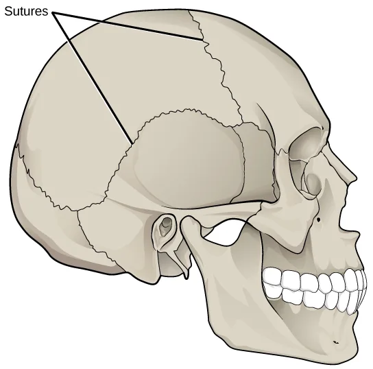
The bones of fibrous joints are held together by fibrous connective tissue. There is no cavity, or space, present between the bones and so most fibrous joints do not move at all, or are only capable of minor movements. There are three types of fibrous joints: sutures, syndesmoses, and gomphoses. Sutures are found only in the skull and possess short fibers of connective tissue that hold the skull bones tightly in place, shown below.
Extended description
A single label, ‘Sutures,’ has two leader lines pointing to two different jagged suture lines drawn on a side view of the skull: one crossing the crown of the skull from front to back, and a second, shorter one lower on the skull, near the ear opening.
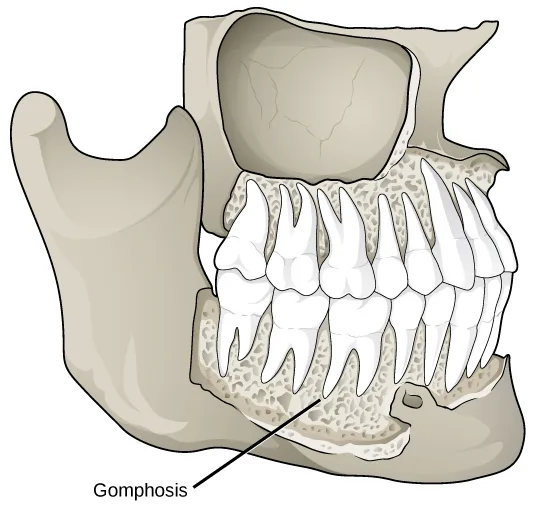
Syndesmoses are joints in which the bones are connected by a band of connective tissue, allowing for more movement than in a suture. An example of a syndesmosis is the joint of the tibia and fibula in the ankle. The amount of movement in these types of joints is determined by the length of the connective tissue fibers. Gomphoses occur between teeth and their sockets; the term refers to the way the tooth fits into the socket like a peg, shown below. The tooth is connected to the socket by a connective tissue referred to as the periodontal ligament.
Cartilaginous Joints
Cartilaginous joints are joints in which the bones are connected by cartilage. There are two types of cartilaginous joints: synchondroses and symphyses. In a synchondrosis, the bones are joined by hyaline cartilage. Synchondroses are found in the epiphyseal plates of growing bones in children. In symphyses, hyaline cartilage covers the end of the bone but the connection between bones occurs through fibrocartilage. Symphyses are found at the joints between vertebrae. Either type of cartilaginous joint allows for very little movement.
Synovial Joints
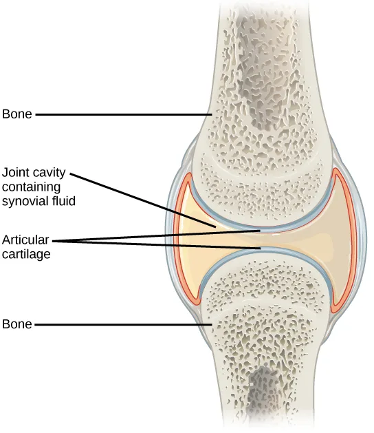
Synovial joints are the only joints that have a space between the adjoining bones, shown below. This space is referred to as the synovial (or joint) cavity and is filled with synovial fluid. Synovial fluid lubricates the joint, reducing friction between the bones and allowing for greater movement. The ends of the bones are covered with articular cartilage, a hyaline cartilage, and the entire joint is surrounded by an articular capsule composed of connective tissue that allows movement of the joint while resisting dislocation. Articular capsules may also possess ligaments that hold the bones together. Synovial joints are capable of the greatest movement of the three structural joint types; however, the more mobile a joint, the weaker the joint. Knees, elbows, and shoulders are examples of synovial joints.
Extended description
Two long bones meet end to end, drawn in cross-section. Top to bottom, four labels point into the joint: ‘Bone’ points to the upper bone’s spongy interior; ‘Joint cavity containing synovial fluid’ points to the pale central space between the bones; ‘Articular cartilage’ has two leader lines, one to the smooth cap on the upper bone’s end and one to the matching cap on the lower bone’s end; ‘Bone’ points to the lower bone’s spongy interior. A reddish-orange band encloses the joint on both sides, representing the surrounding capsule, though it carries no label of its own.
Classification of Joints on the Basis of Function
The functional classification divides joints into three categories: synarthroses, amphiarthroses, and diarthroses. A synarthrosis is a joint that is immovable. This includes sutures, gomphoses, and synchondroses. Amphiarthroses are joints that allow slight movement, including syndesmoses and symphyses. Diarthroses are joints that allow for free movement of the joint, as in synovial joints.
Movement at Synovial Joints
The wide range of movement allowed by synovial joints produces different types of movements. The movement of synovial joints can be classified as one of four different types: gliding, angular, rotational, or special movement.
Gliding Movement
Gliding movements occur as relatively flat bone surfaces move past each other. Gliding movements produce very little rotation or angular movement of the bones. The joints of the carpal and tarsal bones are examples of joints that produce gliding movements.
Angular Movement
Angular movements are produced when the angle between the bones of a joint changes. There are several different types of angular movements, including flexion, extension, hyperextension, abduction, adduction, and circumduction. Flexion, or bending, occurs when the angle between the bones decreases. Moving the forearm upward at the elbow or moving the wrist to move the hand toward the forearm are examples of flexion. Extension is the opposite of flexion in that the angle between the bones of a joint increases. Straightening a limb after flexion is an example of extension. Extension past the regular anatomical position is referred to as hyperextension. This includes moving the neck back to look upward, or bending the wrist so that the hand moves away from the forearm.
Abduction occurs when a bone moves away from the midline of the body. Examples of abduction are moving the arms or legs laterally to lift them straight out to the side. Adduction is the movement of a bone toward the midline of the body. Movement of the limbs inward after abduction is an example of adduction. Circumduction is the movement of a limb in a circular motion, as in moving the arm in a circular motion.
Rotational Movement
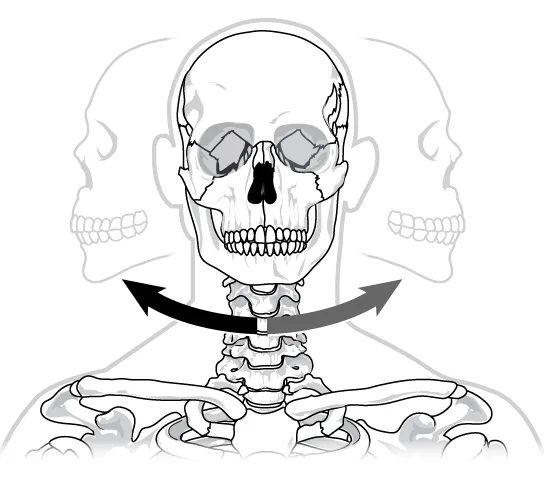
Rotational movement is the movement of a bone as it rotates around its longitudinal axis. Rotation can be toward the midline of the body, which is referred to as medial rotation, or away from the midline of the body, which is referred to as lateral rotation. Movement of the head from side to side is an example of rotation.
Special Movements
Some movements that cannot be classified as gliding, angular, or rotational are called special movements. Inversion involves the soles of the feet moving inward, toward the midline of the body. Eversion is the opposite of inversion, movement of the sole of the foot outward, away from the midline of the body. Protraction is the anterior movement of a bone in the horizontal plane. Retraction occurs as a joint moves back into position after protraction. Protraction and retraction can be seen in the movement of the mandible as the jaw is thrust outwards and then back inwards. Elevation is the movement of a bone upward, such as when the shoulders are shrugged, lifting the scapulae. Depression is the opposite of elevation—movement downward of a bone, such as after the shoulders are shrugged and the scapulae return to their normal position from an elevated position. Dorsiflexion is a bending at the ankle such that the toes are lifted toward the knee. Plantar flexion is a bending at the ankle when the heel is lifted, such as when standing on the toes. Supination is the movement of the radius and ulna bones of the forearm so that the palm faces forward. Pronation is the opposite movement, in which the palm faces backward. Opposition is the movement of the thumb toward the fingers of the same hand, making it possible to grasp and hold objects.
Types of Synovial Joints
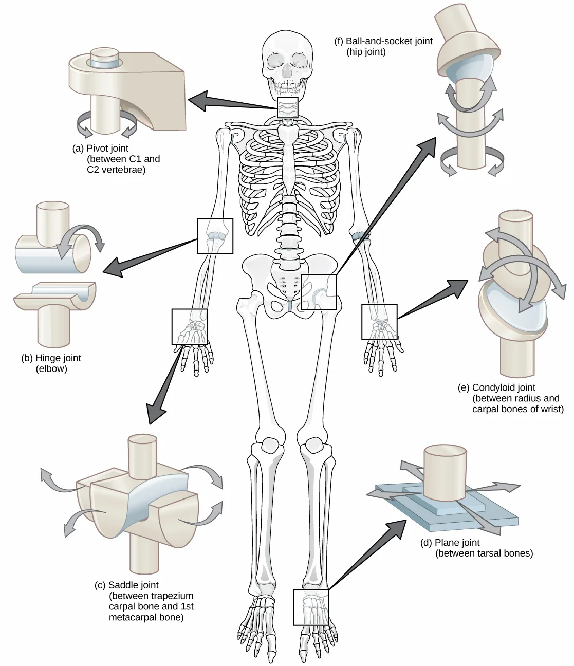
Synovial joints are further classified into six different categories on the basis of the shape and structure of the joint. The shape of the joint affects the type of movement permitted by the joint, shown below. These joints can be described as planar, hinge, pivot, condyloid, saddle, or ball-and-socket joints.
Extended description
Six lettered inset diagrams surround a central front-facing skeleton, each linked to a boxed region on the skeleton by a dark arrow. (a) Pivot joint, upper left, linked from a box over the neck’s C1–C2 vertebrae: a cylindrical peg fits into a collar, with a curved arrow wrapping the shaft to show rotation. (b) Hinge joint, left, linked from a box over the elbow: a rod rests in a curved trough, with a single curved arrow sweeping over the rod in one plane. (c) Saddle joint, lower left, linked from a box over the thumb base: two saddle-shaped pieces cross at right angles, with four curved arrows, one at each of the four ends, pointing outward to show movement along two perpendicular axes. (d) Plane joint, lower right, linked from a box over the ankle region: a peg sits on a flat layered plate, with two straight arrows pointing toward opposite corners of the plate to show back-and-forth gliding. (e) Condyloid joint, right, linked from a box over the other wrist: an oval ball sits in an oval socket, with two curved arrows crossing around it — one running top to bottom, one side to side — to show movement along two axes without rotation. (f) Ball-and-socket joint, upper right, linked from a box over the hip: a sphere sits in a deep cup on a shaft, with curved arrows around the sphere in multiple directions plus a curved arrow wrapping the shaft’s base, to show movement in every direction, including rotation.
Planar Joints
Planar joints have bones with articulating surfaces that are flat or slightly curved faces. These joints allow for gliding movements, and so the joints are sometimes referred to as gliding joints. The range of motion is limited in these joints and does not involve rotation. Planar joints are found in the carpal bones in the hand and the tarsal bones of the foot, as well as between vertebrae, shown below.
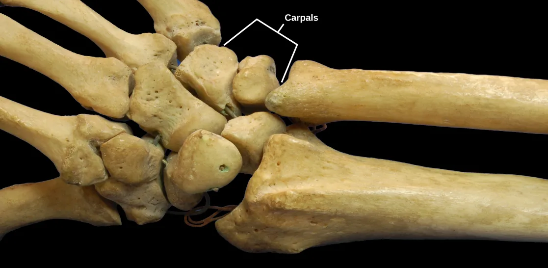
Extended description
A bracket labeled ‘Carpals’ encloses a cluster of small, knobby wrist bones at the center of the photograph, between the two long forearm bones entering from the right and the metacarpal and finger bones extending to the left.
Hinge Joints
In hinge joints, the slightly rounded end of one bone fits into the slightly hollow end of the other bone. In this way, one bone moves while the other remains stationary, like the hinge of a door. The elbow is an example of a hinge joint. The knee is sometimes classified as a modified hinge joint, shown below.
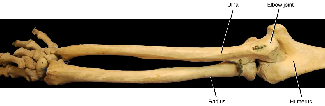
Extended description
Four labels point to the photographed arm bones. At the far left, unlabeled wrist and hand bones lead into two parallel forearm bones: ‘Ulna’ labels the upper of the two, and ‘Radius’ labels the lower. Both extend right to ‘Elbow joint,’ which labels the junction where the forearm bones meet the wider upper-arm bone; ‘Humerus’ labels that upper-arm bone, continuing to the right edge of the photograph.
Pivot Joints
Pivot joints consist of the rounded end of one bone fitting into a ring formed by the other bone. This structure allows rotational movement, as the rounded bone moves around its own axis. An example of a pivot joint is the joint of the first and second vertebrae of the neck that allows the head to move back and forth, shown below. The joint of the wrist that allows the palm of the hand to be turned up and down is also a pivot joint.
Condyloid Joints
Condyloid joints consist of an oval-shaped end of one bone fitting into a similarly oval-shaped hollow of another bone, shown below. This is also sometimes called an ellipsoidal joint. This type of joint allows angular movement along two axes, as seen in the joints of the wrist and fingers, which can move both side to side and up and down.
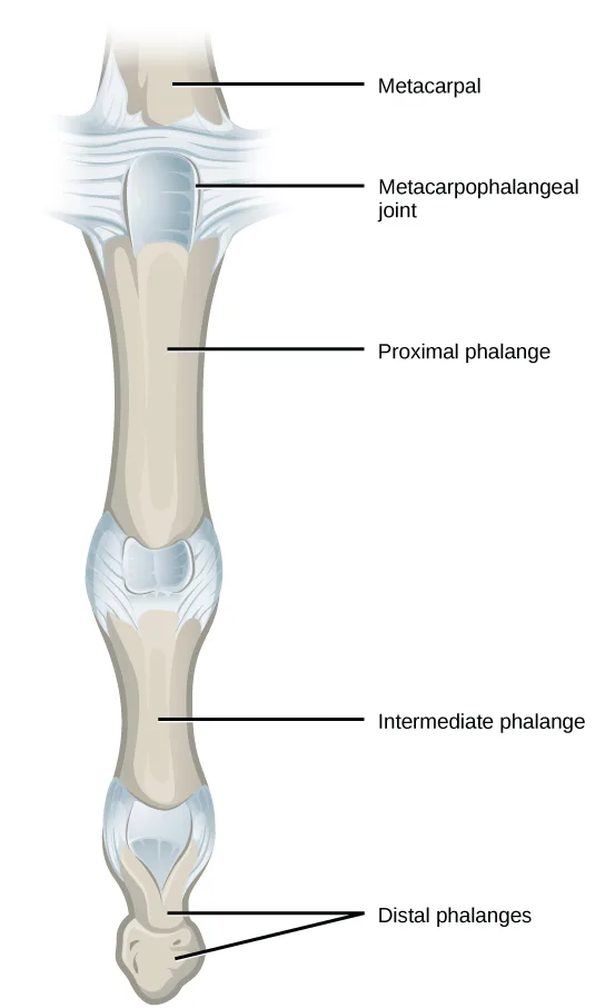
Extended description
Five labels run top to bottom down a single finger, illustrated in cross-section from the hand: ‘Metacarpal’ labels the wide bone at the top; ‘Metacarpophalangeal joint’ labels the rounded joint just below it; ‘Proximal phalange’ labels the long bone below that joint; ‘Intermediate phalange’ labels the next long bone down, separated from the proximal phalange by an unlabeled joint; ‘Distal phalanges’ has two leader lines pointing to the small bone at the fingertip.
Saddle Joints
Saddle joints are so named because the ends of each bone resemble a saddle, with concave and convex portions that fit together. Saddle joints allow angular movements similar to condyloid joints but with a greater range of motion. An example of a saddle joint is the thumb joint, which can move back and forth and up and down, but more freely than the wrist or fingers, shown below.
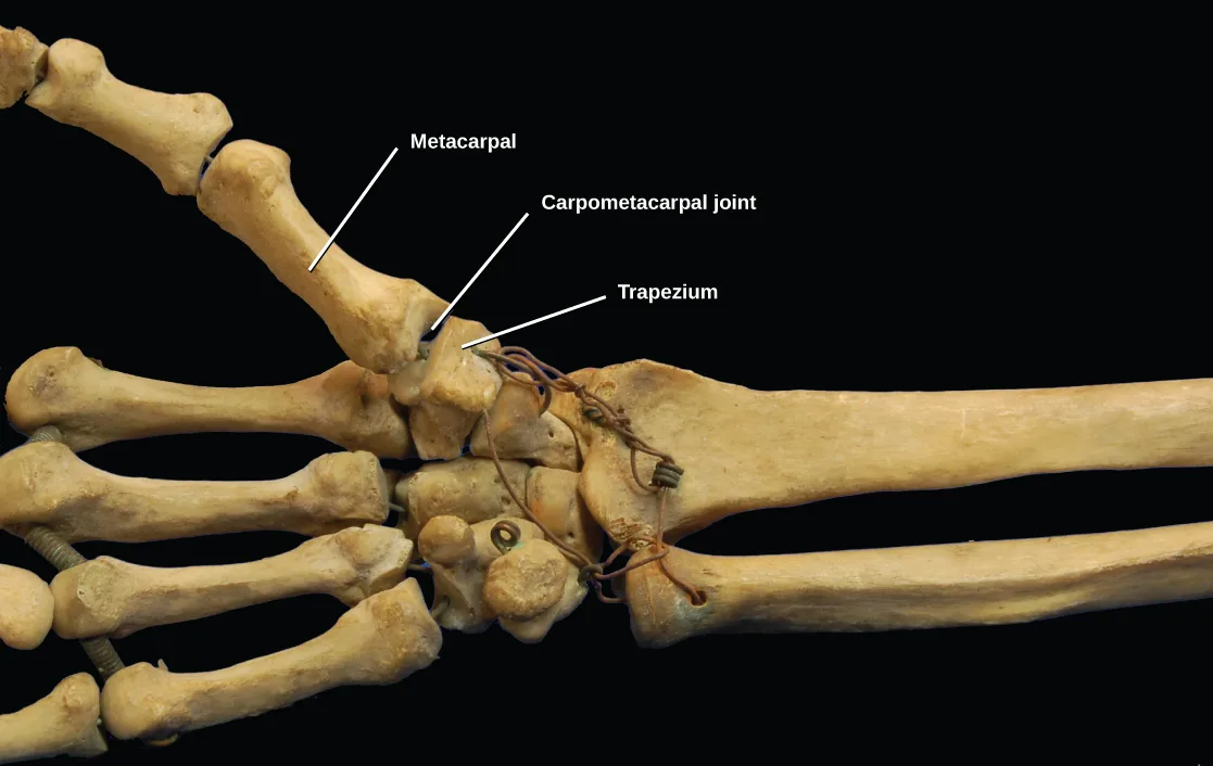
Extended description
Three labels point to the photographed thumb and wrist bones: ‘Metacarpal’ labels the long bone of the thumb at upper left; ‘Carpometacarpal joint’ labels the junction where it meets the wrist; ‘Trapezium’ labels the wrist bone just below the joint that the metacarpal articulates with, among the other carpal and metacarpal bones of the hand visible around them.
Ball-and-Socket Joints
Ball-and-socket joints possess a rounded, ball-like end of one bone fitting into a cuplike socket of another bone. This organization allows the greatest range of motion, as all movement types are possible in all directions. Examples of ball-and-socket joints are the shoulder and hip joints, shown below.
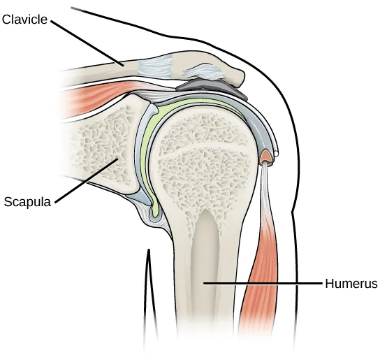
Extended description
Three labels point to the drawn shoulder cross-section: ‘Clavicle’ labels the collarbone at the upper left, running toward the joint; ‘Scapula’ labels the shoulder blade forming the socket below and to the left; ‘Humerus’ labels the upper-arm bone whose rounded head sits in the socket, with the surrounding joint capsule, cartilage, and a muscle tendon drawn but not individually labeled.
Link to Learning
Watch an animation showing the six types of synovial joints.
Career Connection Rheumatologist
Rheumatologists are medical doctors who specialize in the diagnosis and treatment of disorders of the joints, muscles, and bones. They diagnose and treat diseases such as arthritis, musculoskeletal disorders, osteoporosis, and autoimmune diseases such as ankylosing spondylitis and rheumatoid arthritis.
Rheumatoid arthritis (RA) is an inflammatory disorder that primarily affects the synovial joints of the hands, feet, and cervical spine. Affected joints become swollen, stiff, and painful. Although it is known that RA is an autoimmune disease in which the body’s immune system mistakenly attacks healthy tissue, the cause of RA remains unknown. Immune cells from the blood enter joints and the synovium causing cartilage breakdown, swelling, and inflammation of the joint lining. Breakdown of cartilage causes bones to rub against each other causing pain. RA is more common in females than males and the age of onset is usually 40–50 years of age.
Rheumatologists can diagnose RA on the basis of symptoms such as joint inflammation and pain, X-ray and MRI imaging, and blood tests. Arthrography is a type of medical imaging of joints that uses a contrast agent, such as a dye, that is opaque to X-rays. This allows the soft tissue structures of joints—such as cartilage, tendons, and ligaments—to be visualized. An arthrogram differs from a regular X-ray by showing the surface of soft tissues lining the joint in addition to joint bones. An arthrogram allows early degenerative changes in joint cartilage to be detected before bones become affected.
There is currently no cure for RA; however, rheumatologists have a number of treatment options available. Early stages can be treated with rest of the affected joints by using a cane or by using joint splints that minimize inflammation. When inflammation has decreased, exercise can be used to strengthen the muscles that surround the joint and to maintain joint flexibility. If joint damage is more extensive, medications can be used to relieve pain and decrease inflammation. Anti-inflammatory drugs such as aspirin, topical pain relievers, and corticosteroid injections may be used. Surgery may be required in cases in which joint damage is severe.
Summary
The structural classification of joints divides them into bony, fibrous, cartilaginous, and synovial joints. The bones of fibrous joints are held together by fibrous connective tissue; the three types of fibrous joints are sutures, syndesmoses, and gomphoses. Cartilaginous joints are joints in which the bones are connected by cartilage; the two types of cartilaginous joints are synchondroses and symphyses. Synovial joints are joints that have a space between the adjoining bones. The functional classification divides joints into three categories: synarthroses, amphiarthroses, and diarthroses. The movement of synovial joints can be classified as one of four different types: gliding, angular, rotational, or special movement. Gliding movements occur as relatively flat bone surfaces move past each other. Angular movements are produced when the angle between the bones of a joint changes. Rotational movement is the movement of a bone as it rotates around its own longitudinal axis. Special movements include inversion, eversion, protraction, retraction, elevation, depression, dorsiflexion, plantar flexion, supination, pronation, and opposition. Synovial joints are also classified into six different categories on the basis of the shape and structure of the joint: planar, hinge, pivot, condyloid, saddle, and ball-and-socket.
Key terms
- abduction — when a bone moves away from the midline of the body.
- adduction — movement of the limbs inward after abduction.
- amphiarthrosis — joint that allows slight movement; includes syndesmoses and symphyses.
- angular movement — produced when the angle between the bones of a joint changes.
- ball-and-socket joint — joint with a rounded, ball-like end of one bone fitting into a cuplike socket of another bone.
- cartilaginous joint — joint in which the bones are connected by cartilage.
- circumduction — movement of a limb in a circular motion.
- condyloid joint — oval-shaped end of one bone fitting into a similarly oval-shaped hollow of another bone.
- depression — movement downward of a bone, such as after the shoulders are shrugged and the scapulae return to their normal position from an elevated position; opposite of elevation.
- diarthrosis — joint that allows for free movement of the joint; found in synovial joints.
- dorsiflexion — bending at the ankle such that the toes are lifted toward the knee.
- elevation — movement of a bone upward, such as when the shoulders are shrugged, lifting the scapulae.
- eversion — movement of the sole of the foot outward, away from the midline of the body; opposite of inversion.
- extension — movement in which the angle between the bones of a joint increases; opposite of flexion.
- fibrous joint — joint held together by fibrous connective tissue.
- flexion — movement in which the angle between the bones decreases; opposite of extension.
- gliding movement — when relatively flat bone surfaces move past each other.
- gomphosis — the joint in which the tooth fits into the socket like a peg.
- hinge joint — slightly rounded end of one bone fits into the slightly hollow end of the other bone.
- hyperextension — extension past the regular anatomical position.
- inversion — soles of the feet moving inward, toward the midline of the body.
- joint — point at which two or more bones meet.
- lateral rotation — rotation away from the midline of the body.
- medial rotation — rotation toward the midline of the body.
- opposition — movement of the thumb toward the fingers of the same hand, making it possible to grasp and hold objects.
- plantar flexion — bending at the ankle such that the heel is lifted, such as when standing on the toes.
- planar joint — joint with bones whose articulating surfaces are flat.
- pivot joint — joint with the rounded end of one bone fitting into a ring formed by the other bone.
- pronation — movement in which the palm faces backward.
- protraction — anterior movement of a bone in the horizontal plane.
- retraction — movement in which a joint moves back into position after protraction.
- rotational movement — movement of a bone as it rotates around its own longitudinal axis.
- saddle joint — joint with concave and convex portions that fit together; named because the ends of each bone resemble a saddle.
- supination — movement of the radius and ulna bones of the forearm so that the palm faces forward.
- suture — short fiber of connective tissue that holds the skull bones tightly in place; found only in the skull.
- synarthrosis — joint that is immovable.
- symphysis — hyaline cartilage covers the end of the bone, but the connection between bones occurs through fibrocartilage; symphyses are found at the joints between vertebrae.
- synchondrosis — bones joined by hyaline cartilage; synchondroses are found in the epiphyseal plates of growing bones in children.
- syndesmosis — joint in which the bones are connected by a band of connective tissue, allowing for more movement than in a suture.
- synovial joint — only joint that has a space between the adjoining bones.
Practice
Classify the different types of joints on the basis of structure
Synchondroses and symphyses are:
Both terms name joints connected by cartilage — check which structural category that material puts them in.Which of the following is not a characteristic of the synovial fluid?
Three of these are the fluid’s actual jobs described in this section — think about what it does mechanically at the joint, then find the one function never mentioned.The elbow is an example of which type of joint?
Picture how the elbow moves like a door — motion in essentially one plane, with one bone staying fixed.A high ankle sprain is an injury caused by over-stretching the ligaments connecting the tibia and fibula. What type of joint is involved in this sprain?
This section names the type of fibrous joint that connects two bones with a band of connective tissue, allowing more movement than a suture but far less than a synovial joint.A joint held together by fibrous connective tissue is called a ________.
This structural category has no cavity between the bones, so most examples barely move at all.A short fiber of connective tissue that holds the skull bones tightly in place, found only in the skull, is called a ________.
This is the only fibrous joint type found nowhere else in the body.The joint in which a tooth fits into its socket like a peg is called a ________.
A periodontal ligament connects the tooth to its bony socket at this joint.A joint that is immovable is called a ________.
Sutures, gomphoses, and synchondroses all belong to this functional category.A joint in which the oval-shaped end of one bone fits into a similarly oval-shaped hollow of another bone is called a ________.
The wrist and finger joints that move both side to side and up and down are this type.A joint with a rounded, ball-like end of one bone fitting into a cuplike socket of another bone is called a ________.
The shoulder and hip are the two examples this section gives of this joint type.Explain the role of joints in skeletal movement
The movement of bone away from the midline of the body is called ________.
Think about which term describes moving a limb laterally away from the body, as when lifting an arm out to the side.What movements occur at the hip joint and knees as you bend down to touch your toes?
Show model answer
Did your answer mention:
What movement(s) occur(s) at the scapulae when you shrug your shoulders?
Show model answer
Did your answer mention:
Describe the joints and motions involved in taking a step forward if a person is initially standing still. Assume the person holds his foot at the same angle throughout the motion.
Show model answer
Did your answer mention:
Movement of the limbs inward after abduction is called ________.
This is the opposite of the movement that lifts a limb laterally out to the side.Movement of a limb in a circular motion is called ________.
Moving the whole arm in a circle, rather than back and forth in a single plane, is this movement.Extension past the regular anatomical position is called ________.
Tilting the neck back to look upward, or bending the wrist so the hand moves away from the forearm, are examples of this movement.Movement that occurs when relatively flat bone surfaces move past each other is called ________.
The carpal and tarsal bones are the section’s examples of joints that produce this kind of movement, with very little rotation or angular change.The movement of the soles of the feet inward, toward the midline of the body, is called ________.
This is the opposite of the movement that turns the sole of the foot outward, away from the midline.The movement of the forearm in which the palm faces backward is called ________.
This is the opposite of the forearm movement that turns the palm to face forward.This section is adapted from Biology 2e, Section 38.3: Joints and Skeletal Movement by Mary Ann Clark, Jung Choi, Matthew Douglas, and OpenStax, © OpenStax, licensed under CC BY-NC-SA 4.0. Access the original for free at openstax.org. Changes: figures re-encoded as WebP; seven of the ten figures re-kinded from the manifest’s file-extension “photo” guess to “diagram” (Figure_38_03_01, _02, _03, _04, _07, _08, and _10 are hand-drawn line illustrations, not captured photographs — the manifest guesses from the source JPEG extension alone); a longdesc added to every figure whose printed labels or panel layout are not fully carried by its one-line caption — the skull-suture drawing, the labeled synovial-joint cross-section, the six-panel joint-and-movement composite (counting all six lettered panels, their body location, and the schematic motion arrows each shows), the labeled wrist, elbow, finger, thumb, and shoulder anatomy — transcribing each drawing’s own printed labels in reading order; in-text pointers to figures (“Figure 38.20” and similar) replaced with “shown below,” since Hugo does not number figures; the two feature notes (the Link to Learning animation of the six synovial joint types, and the Career Connection profiling a rheumatologist) rendered as callouts, the Career Connection keeping its bold name, italic title, and all four paragraphs verbatim; the end-of-section Review Questions and Critical Thinking Questions adapted into the closing interactive Practice block (multiple choice and self-check respectively); rubric checkpoints added to each self-check, decomposing its model answer (the source solution) into check-off clauses with no new claims; thirteen key-term recall items added from the glossary (fibrous joint, suture, gomphosis, synarthrosis, condyloid joint, ball-and-socket joint, adduction, circumduction, hyperextension, gliding movement, inversion, pronation, plus abduction as a Review Question key), covering a representative subset of the section’s forty glossary terms rather than all of them; a typo in the pinned CNXML’s own Section Summary — “syndesomes” — corrected to “syndesmoses” to match every other occurrence of the term in the same module (body prose, the functional-classification paragraph, and the glossary), reported as a source defect; no Visual Connection question or comparison table appears in this module.