Systems of Gas Exchange
By the end of this section, you will be able to:
- Describe the passage of air from the outside environment to the lungs
- Explain how the lungs are protected from particulate matter
The primary function of the respiratory system is to deliver oxygen to the cells of the body’s tissues and remove carbon dioxide, a cell waste product. The main structures of the human respiratory system are the nasal cavity, the trachea, and lungs.
All aerobic organisms require oxygen to carry out their metabolic functions. Along the evolutionary tree, different organisms have devised different means of obtaining oxygen from the surrounding atmosphere. The environment in which the animal lives greatly determines how an animal respires. The complexity of the respiratory system is correlated with the size of the organism. As animal size increases, diffusion distances increase and the ratio of surface area to volume drops. In unicellular organisms, diffusion across the cell membrane is sufficient for supplying oxygen to the cell (shown below). Diffusion is a slow, passive transport process. In order for diffusion to be a feasible means of providing oxygen to an animal’s body, the rate of diffusion across the body surface must match the rate that oxygen is utilized. In other words, if the body were very large or thick, diffusion would not be able to provide oxygen quickly enough to the cells of the body. Therefore, dependence on diffusion as a means of obtaining oxygen and removing carbon dioxide remains feasible only for small organisms or those with highly-flattened bodies, such as many flatworms (Platyhelminthes). Larger organisms had to evolve specialized respiratory tissues, such as gills, lungs, and respiratory passages accompanied by complex circulatory systems, to transport oxygen throughout their entire body.
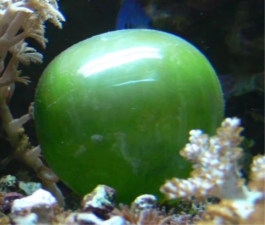
Direct Diffusion
For small multicellular organisms, diffusion across the outer membrane is sufficient to meet their oxygen needs. Gas exchange by direct diffusion across surface membranes is efficient for organisms less than 1 mm in diameter. In simple organisms, such as cnidarians and flatworms, every cell in the body is close to the external environment. Their cells are kept moist and gases diffuse quickly via direct diffusion. Flatworms are small, literally flat worms, which “breathe” through diffusion across the outer membrane (shown below). The flat shape of these organisms increases the surface area for diffusion, ensuring that each cell within the body is close to the outer membrane surface and has access to oxygen. If the flatworm had a cylindrical body, then the cells in the center would not be able to get oxygen.
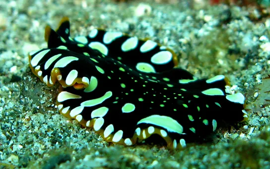
Skin and Gills
Earthworms and amphibians use their skin (integument) as a respiratory organ. A dense network of capillaries lies just below the skin and facilitates gas exchange between the external environment and the circulatory system. The respiratory surface must be kept moist in order for the gases to dissolve and diffuse across cell membranes.
Organisms that live in water need to obtain oxygen from the water. Oxygen dissolves in water but at a lower concentration than in the atmosphere. The atmosphere has roughly 21 percent oxygen. In water, the oxygen concentration is much lower than that. Fish and many other aquatic organisms have evolved gills to take up the dissolved oxygen from water (shown below). Gills are thin tissue filaments that are highly branched and folded. When water passes over the gills, the dissolved oxygen in water rapidly diffuses across the gills into the bloodstream. The circulatory system can then carry the oxygenated blood to the other parts of the body. In animals that contain coelomic fluid instead of blood, oxygen diffuses across the gill surfaces into the coelomic fluid. Gills are found in mollusks, annelids, and crustaceans.
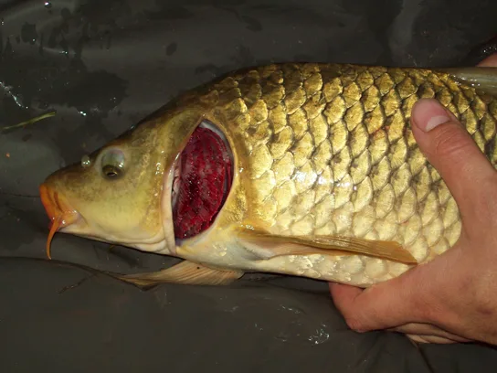
The folded surfaces of the gills provide a large surface area to ensure that the fish gets sufficient oxygen. Diffusion is a process in which material travels from regions of high concentration to low concentration until equilibrium is reached. In this case, blood with a low concentration of oxygen molecules circulates through the gills. The concentration of oxygen molecules in water is higher than the concentration of oxygen molecules in gills. As a result, oxygen molecules diffuse from water (high concentration) to blood (low concentration), as shown below. Similarly, carbon dioxide molecules in the blood diffuse from the blood (high concentration) to water (low concentration).
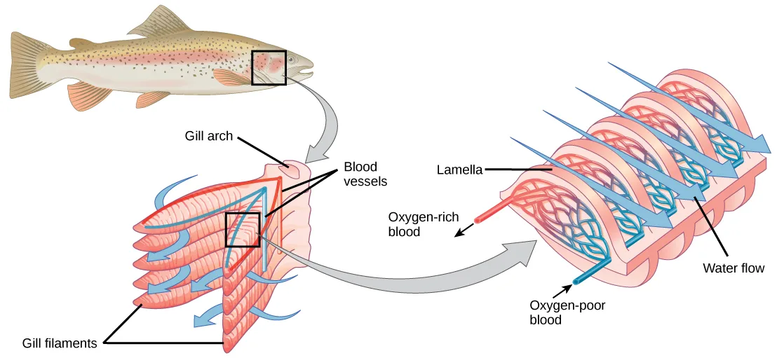
Extended description
At upper left, a small fish icon has a black box outlining the gill region just behind its head, with a gray arrow leading down to a close-up of one gill. That close-up is labeled Gill arch (the central structure the gill attaches to), Blood vessels (the red and blue lines running along the arch), and Gill filaments (the row of comb-like projections extending from the arch), with small blue arrows showing water flowing down and out between the filaments. A second gray arrow leads right to a further close-up of the lamellae: stacked, feather-like structures labeled Lamella, with an arrow labeled Oxygen-rich blood leaving toward the fish’s body and an arrow labeled Oxygen-poor blood entering from the body, while several arrows labeled Water flow pass over the lamellae from the opposite direction.
Tracheal Systems
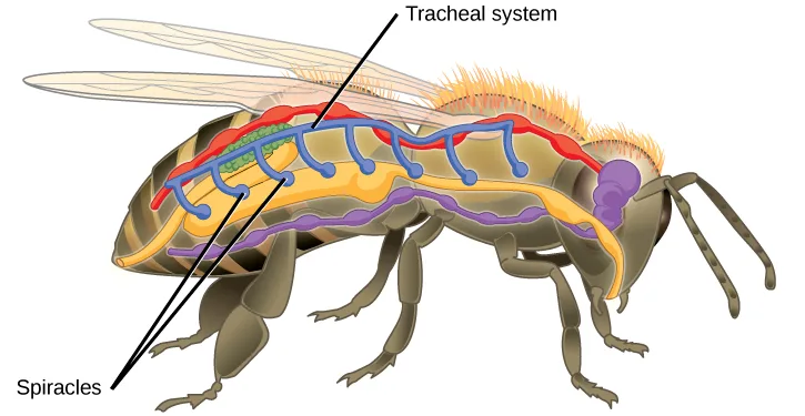
Insect respiration is independent of its circulatory system; therefore, the blood does not play a direct role in oxygen transport. Insects have a highly specialized type of respiratory system called the tracheal system, which consists of a network of small tubes that carries oxygen to the entire body. The tracheal system is the most direct and efficient respiratory system in active animals. The tubes in the tracheal system are made of a polymeric material called chitin.
Insect bodies have openings, called spiracles, along the thorax and abdomen. These openings connect to the tubular network, allowing oxygen to pass into the body (shown below) and regulating the diffusion of CO₂ and water vapor. Air enters and leaves the tracheal system through the spiracles. Some insects can ventilate the tracheal system with body movements.
Extended description
One leader line from the label Tracheal system points to the network of vertical tubes branching from a main tube that runs along the top of the body from front to back. Two leader lines from the label Spiracles point to two of the small openings along the lower body wall, in the thorax and abdomen, where the tracheal tubes originate.
Mammalian Systems
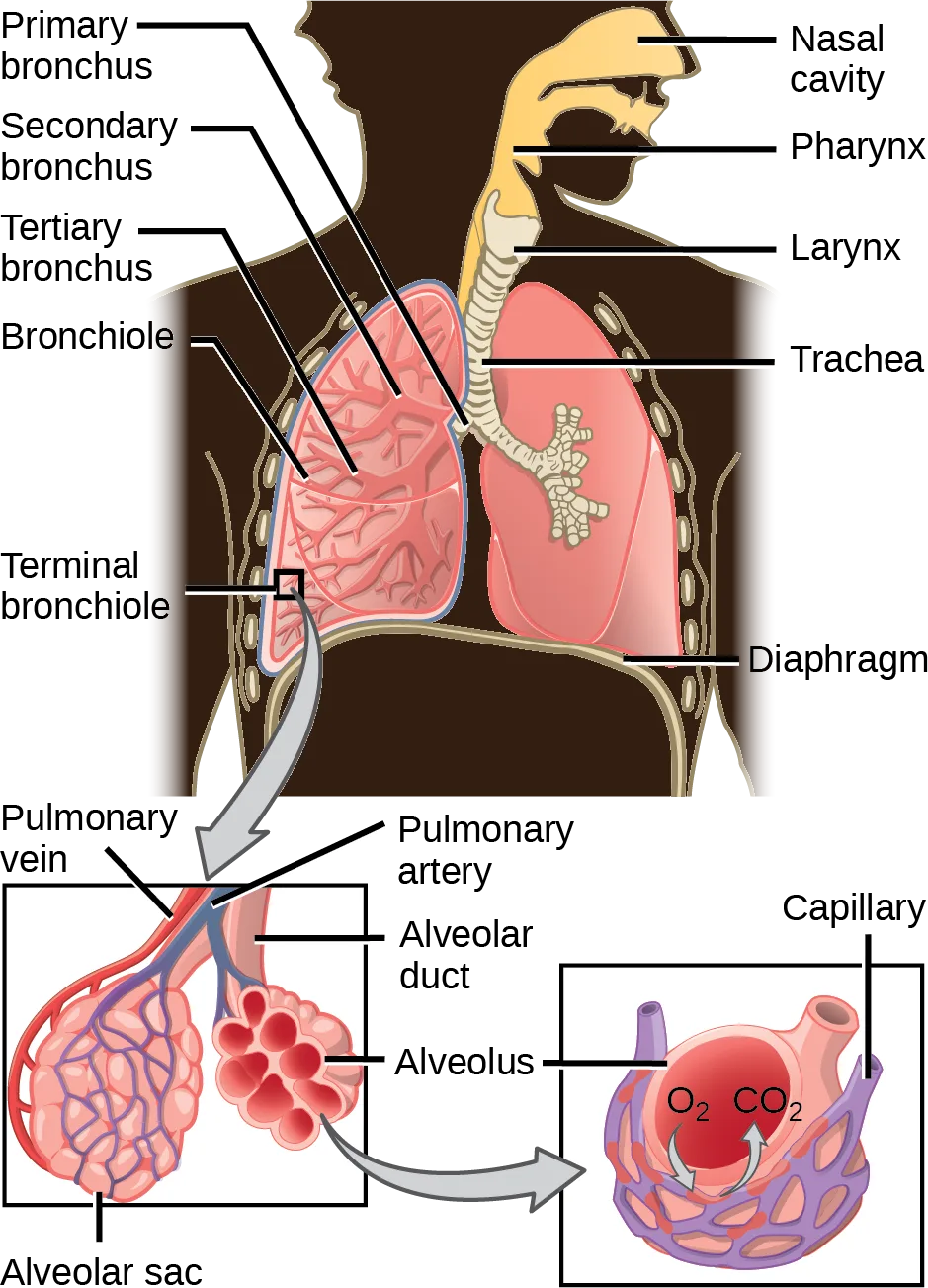
In mammals, pulmonary ventilation occurs via inhalation (breathing). During inhalation, air enters the body through the nasal cavity located just inside the nose (shown below). As air passes through the nasal cavity, the air is warmed to body temperature and humidified. The respiratory tract is coated with mucus to seal the tissues from direct contact with air. Mucus is high in water. As air crosses these surfaces of the mucous membranes, it picks up water. These processes help equilibrate the air to the body conditions, reducing any damage that cold, dry air can cause. Particulate matter that is floating in the air is removed in the nasal passages via mucus and cilia. The processes of warming, humidifying, and removing particles are important protective mechanisms that prevent damage to the trachea and lungs. Thus, inhalation serves several purposes in addition to bringing oxygen into the respiratory system.
Extended description
The upper drawing is a chest cutaway. On its right side, one leader line each labels Nasal cavity, Pharynx, Larynx, Trachea, and Diaphragm, top to bottom. On its left side, one leader line each labels Primary bronchus, Secondary bronchus, Tertiary bronchus, Bronchiole, and Terminal bronchiole, also top to bottom, tracing the airway from where the trachea first forks down to its finest branches. A gray arrow leads from the terminal bronchiole down to two insets below. The left inset labels Pulmonary vein and Pulmonary artery on the vessels running alongside an Alveolar duct, which opens into a grape-like cluster labeled Alveolar sac made up of individual units labeled Alveolus. A second gray arrow leads to the right inset, a single alveolus wrapped in a network labeled Capillary, where the alveolus shows O₂ and CO₂ each on its own curved arrow, marking the two gases diffusing between the alveolus and the capillary.
Which of the following statements about the mammalian respiratory system is false?
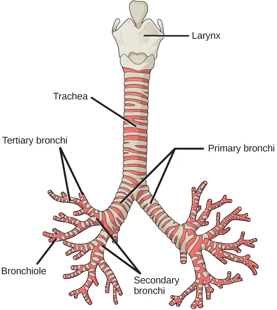
Trace the airway in the order it’s drawn — from the nasal cavity through the pharynx, larynx, and trachea, into the bronchi, and on to the bronchioles and alveolar ducts — and check whether each statement matches that branching direction.From the nasal cavity, air passes through the pharynx (throat) and the larynx (voice box), as it makes its way to the trachea (shown above). The main function of the trachea is to funnel the inhaled air to the lungs and the exhaled air back out of the body. The human trachea is a cylinder about 10 to 12 cm long and 2 cm in diameter that sits in front of the esophagus and extends from the larynx into the chest cavity where it divides into the two primary bronchi at the midthorax. It is made of incomplete rings of hyaline cartilage and smooth muscle (shown below). The trachea is lined with mucus-producing goblet cells and ciliated epithelia. The cilia propel foreign particles trapped in the mucus toward the pharynx. The cartilage provides strength and support to the trachea to keep the passage open. The smooth muscle can contract, decreasing the trachea’s diameter, which causes expired air to rush upwards from the lungs at a great force. The forced exhalation helps expel mucus when we cough. Smooth muscle can contract or relax, depending on stimuli from the external environment or the body’s nervous system.
Extended description
Reading top to bottom: one leader line labels Larynx, the wide structure capping the tube; one leader line labels Trachea, the central ringed tube below it. Where the trachea forks, two leader lines from the label Primary bronchi point to the first branch, one into each lung. Two leader lines from Tertiary bronchi point to finer branch points, both within the left lung, which is drawn with more visible branching than the right. Two leader lines from Secondary bronchi point to branch points between the primary and tertiary branches, one in each lung. One leader line from Bronchiole points to the finest, thread-like branch at the lower left of the left lung.
Lungs: Bronchi and Alveoli
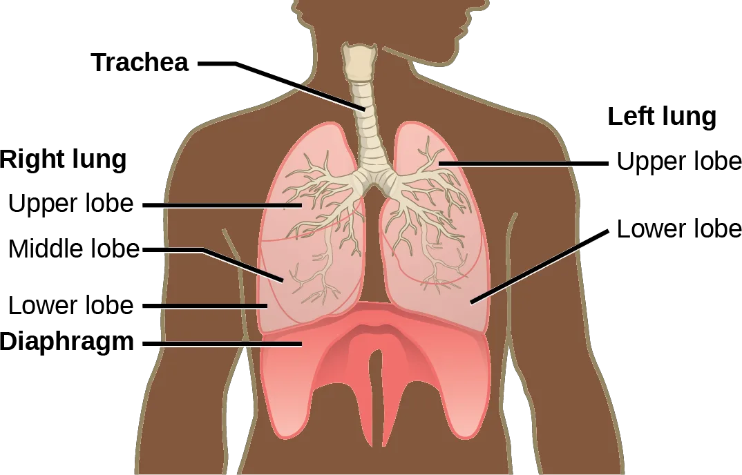
The end of the trachea bifurcates (divides) to the right and left lungs. The lungs are not identical. The right lung is larger and contains three lobes, whereas the smaller left lung contains two lobes (shown below). The muscular diaphragm, which facilitates breathing, is inferior to (below) the lungs and marks the end of the thoracic cavity.
Extended description
One leader line labels Trachea, the ringed tube descending from the neck. On the body’s right side, the bold label Right lung introduces three leader lines labeling Upper lobe, Middle lobe, and Lower lobe, top to bottom. On the body’s left side, the bold label Left lung introduces two leader lines labeling Upper lobe and Lower lobe. A leader line from the bold label Diaphragm points to the dome-shaped structure beneath both lungs.
In the lungs, air is diverted into smaller and smaller passages, or bronchi. Air enters the lungs through the two primary (main) bronchi (singular: bronchus). Each bronchus divides into secondary bronchi, then into tertiary bronchi, which in turn divide, creating smaller and smaller diameter bronchioles as they split and spread through the lung. Like the trachea, the bronchi are made of cartilage and smooth muscle. At the bronchioles, the cartilage is replaced with elastic fibers. Bronchi are innervated by nerves of both the parasympathetic and sympathetic nervous systems that control muscle contraction (parasympathetic) or relaxation (sympathetic) in the bronchi and bronchioles, depending on the nervous system’s cues. In humans, bronchioles with a diameter smaller than 0.5 mm are the respiratory bronchioles. They lack cartilage and therefore rely on inhaled air to support their shape. As the passageways decrease in diameter, the relative amount of smooth muscle increases.
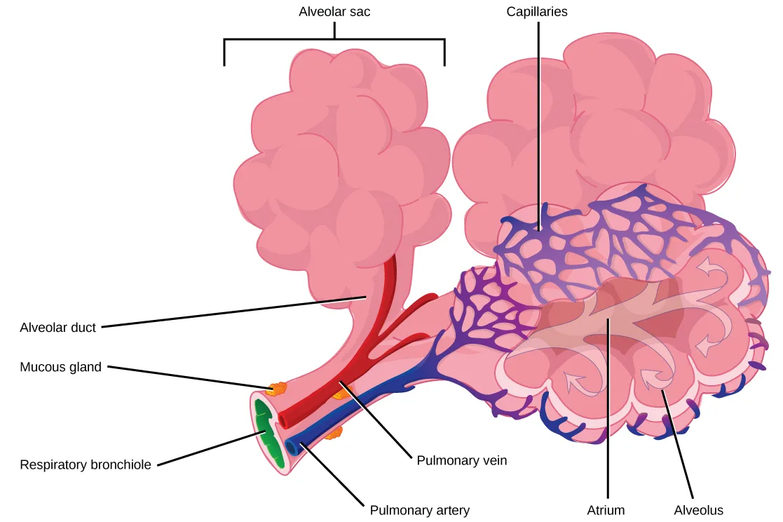
The terminal bronchioles subdivide into microscopic branches called respiratory bronchioles. The respiratory bronchioles subdivide into several alveolar ducts. Numerous alveoli and alveolar sacs surround the alveolar ducts. The alveolar sacs resemble bunches of grapes tethered to the end of the bronchioles (shown below). In the acinar region, the alveolar ducts are attached to the end of each bronchiole. At the end of each duct are approximately 100 alveolar sacs, each containing 20 to 30 alveoli that are 200 to 300 microns in diameter. Gas exchange occurs only in alveoli. Alveoli are made of thin-walled parenchymal cells, typically one-cell thick, that look like tiny bubbles within the sacs. Alveoli are in direct contact with capillaries (one-cell thick) of the circulatory system. Such intimate contact ensures that oxygen will diffuse from alveoli into the blood and be distributed to the cells of the body. In addition, the carbon dioxide that was produced by cells as a waste product will diffuse from the blood into alveoli to be exhaled. The anatomical arrangement of capillaries and alveoli emphasizes the structural and functional relationship of the respiratory and circulatory systems. Because there are so many alveoli (~300 million per lung) within each alveolar sac and so many sacs at the end of each alveolar duct, the lungs have a sponge-like consistency. This organization produces a very large surface area that is available for gas exchange. The surface area of alveoli in the lungs is approximately 75 m². This large surface area, combined with the thin-walled nature of the alveolar parenchymal cells, allows gases to easily diffuse across the cells.
Extended description
On the left, a bracket labeled Alveolar sac spans the pink, grape-like cluster of sacs. Below it, one leader line each labels Alveolar duct (the central red tube leading into the cluster), Mucous gland (small orange shapes at its base), Respiratory bronchiole (the green tube at the very base), Pulmonary vein, and Pulmonary artery (the red and blue vessels running alongside the duct). On the right, a matching cluster is shown in cutaway: one leader line labels Capillaries, the purple network covering its surface; one leader line labels Atrium, the open central airspace; and one leader line labels Alveolus, one of the individual grape-like sacs opening into that airspace.
Link to Learning
Protective Mechanisms
The air that organisms breathe contains particulate matter such as dust, dirt, viral particles, and bacteria that can damage the lungs or trigger allergic immune responses. The respiratory system contains several protective mechanisms to avoid problems or tissue damage. In the nasal cavity, hairs and mucus trap small particles, viruses, bacteria, dust, and dirt to prevent their entry.
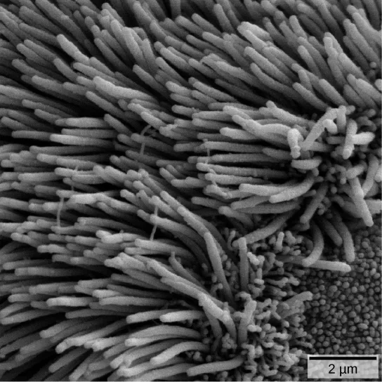
If particulates do make it beyond the nose, or enter through the mouth, the bronchi and bronchioles of the lungs also contain several protective devices. The lungs produce mucus—a sticky substance made of mucin, a complex glycoprotein, as well as salts and water—that traps particulates. The bronchi and bronchioles contain cilia, small hair-like projections that line the walls of the bronchi and bronchioles (shown below). These cilia beat in unison and move mucus and particles out of the bronchi and bronchioles back up to the throat where it is swallowed and eliminated via the esophagus.
In humans, for example, tar and other substances in cigarette smoke destroy or paralyze the cilia, making the removal of particles more difficult. In addition, smoking causes the lungs to produce more mucus, which the damaged cilia are not able to move. This causes a persistent cough, as the lungs try to rid themselves of particulate matter, and makes smokers more susceptible to respiratory ailments.
Summary
Animal respiratory systems are designed to facilitate gas exchange. In mammals, air is warmed and humidified in the nasal cavity. Air then travels down the pharynx, through the trachea, and into the lungs. In the lungs, air passes through the branching bronchi, reaching the respiratory bronchioles, which house the first site of gas exchange. The respiratory bronchioles open into the alveolar ducts, alveolar sacs, and alveoli. Because there are so many alveoli and alveolar sacs in the lung, the surface area for gas exchange is very large. Several protective mechanisms are in place to prevent damage or infection. These include the hair and mucus in the nasal cavity that trap dust, dirt, and other particulate matter before they can enter the system. In the lungs, particles are trapped in a mucus layer and transported via cilia up to the esophageal opening at the top of the trachea to be swallowed.
Key terms
- alveolar duct — duct that extends from the terminal bronchiole to the alveolar sac.
- alveolar sac — structure consisting of two or more alveoli that share a common opening.
- alveolus — (plural: alveoli) (also, air sac) terminal region of the lung where gas exchange occurs.
- bronchus — (plural: bronchi) smaller branch of cartilaginous tissue that stems off of the trachea; air is funneled through the bronchi to the region where gas exchange occurs in alveoli.
- bronchiole — airway that extends from the main tertiary bronchi to the alveolar sac.
- diaphragm — domed-shaped skeletal muscle located under lungs that separates the thoracic cavity from the abdominal cavity.
- larynx — voice box, a short passageway connecting the pharynx and the trachea.
- mucin — complex glycoprotein found in mucus.
- mucus — sticky protein-containing fluid secretion in the lung that traps particulate matter to be expelled from the body.
- nasal cavity — opening of the respiratory system to the outside environment.
- particulate matter — small particle such as dust, dirt, viral particles, and bacteria that are in the air.
- pharynx — throat; a tube that starts in the internal nares and runs partway down the neck, where it opens into the esophagus and the larynx.
- primary bronchus — (also, main bronchus) region of the airway within the lung that attaches to the trachea and bifurcates to each lung where it branches into secondary bronchi.
- respiratory bronchiole — terminal portion of the bronchiole tree that is attached to the terminal bronchioles and alveolar ducts, alveolar sacs, and alveoli.
- terminal bronchiole — region of bronchiole that attaches to the respiratory bronchioles.
- trachea — cartilaginous tube that transports air from the larynx to the primary bronchi.
Practice
Describe the passage of air from the outside environment to the lungs
The respiratory system ________.
The first paragraph says the system delivers one gas to the tissues and removes a different one as waste — which of the four options names only the delivered gas?Which is the order of airflow during inhalation?
The nasal cavity leads to the throat and voice box before air ever reaches the windpipe — check where the larynx falls relative to the trachea in each option.Describe the function of these terms and describe where they are located: main bronchus, trachea, alveoli, and acinus.
Show model answer
Did your answer mention:
How does the structure of alveoli maximize gas exchange?
Show model answer
Did your answer mention:
The cartilaginous tube that transports air from the larynx to the primary bronchi is called the ________.
This structure sits between the voice box and the point where the airway first splits toward each lung — its walls are held open by incomplete rings of stiff tissue.The terminal region of the lung where gas exchange occurs is called a(n) ________.
This is the single tiny sac at the very end of the branching airway — millions of them together give the lung its sponge-like texture.Explain how the lungs are protected from particulate matter
Air is warmed and humidified in the nasal passages. This helps to ________.
The text calls warming, humidifying, and particle removal together by one protective name — think about what that name implies these processes guard against, rather than what they cause.Small particles such as dust, dirt, viral particles, and bacteria that are in the air are known as ________.
The nasal hairs and mucus are the first line of defense trapping this before it can reach deeper into the airway.The sticky, protein-containing fluid secretion in the lung that traps particulate matter to be expelled from the body is called ________.
Cilia sweep this substance, along with whatever it has trapped, back up toward the throat to be swallowed.The complex glycoprotein found in mucus is called ________.
Along with salts and water, this glycoprotein is one of the components that makes the lung’s protective secretion sticky.This section is adapted from Biology 2e, Section 39.1: Systems of Gas Exchange by Mary Ann Clark, Jung Choi, Matthew Douglas, and OpenStax, © OpenStax, licensed under CC BY-NC-SA 4.0. Access the original for free at openstax.org. Changes: the respiratory bronchiole Key terms entry reads “alveolar ducts” where the source glossary prints “alveoli ducts” (reported as a source defect); figures re-encoded as WebP; four figures re-kinded from the manifest’s file-extension “photo” guess to “diagram” (Figure_B39_01_04, Figure_39_01_05, Figure_39_01_07, and Figure_39_01_09 are each a hand-drawn line illustration, not a captured photograph); a longdesc added to every diagram whose meaning is not carried by its one-line caption — the gill-anatomy illustration, the bee’s tracheal system, the mammalian-respiratory-system Visual Connection figure, the trachea/bronchi illustration, the lung-lobes illustration, and the alveolar duct/sac illustration — transcribing each drawing’s own printed labels and leader-line counts, in reading order; in-text pointers to figures (“Figure 39.1” through “Figure 39.11”) replaced with “shown below,” “shown above,” or “illustrated below,” since Hugo does not number figures; the note wrapping the mammalian-respiratory-system Visual Connection rendered as its figure followed by a multiple choice, kept in the body in the Mammalian Systems section — the note copy and the <exercise> copy print identical question and option wording, so no adjudication was needed; the interactive interactive-long note rendered as a Link to Learning callout, keeping the module’s own openstax.org/l/lungs_pulmonary redirect URL; the end-of-section Review Questions and Critical Thinking Questions adapted into the closing interactive Practice block (multiple choice and self-check respectively), one Review Question left in the body as the Visual Connection’s multiple choice; rubric checkpoints added to each self-check, decomposing its model answer (the source solution) into check-off clauses with no new claims; five key-term recall items added from the glossary (trachea, alveolus, particulate matter, mucus, mucin), covering five of the section’s sixteen glossary terms — the other eleven (alveolar duct, alveolar sac, bronchus, bronchiole, diaphragm, larynx, nasal cavity, pharynx, primary bronchus, respiratory bronchiole, terminal bronchiole) appear only in the Key terms list and the prose, several of them as the bolded defining term of their own paragraph.