Transport of Gases in Human Bodily Fluids
By the end of this section, you will be able to:
- Describe how oxygen is bound to hemoglobin and transported to body tissues
- Explain how carbon dioxide is transported from body tissues to the lungs
Once the oxygen diffuses across the alveoli, it enters the bloodstream and is transported to the tissues where it is unloaded, and carbon dioxide diffuses out of the blood and into the alveoli to be expelled from the body. Although gas exchange is a continuous process, the oxygen and carbon dioxide are transported by different mechanisms.
Transport of Oxygen in the Blood
Although oxygen dissolves in blood, only a small amount of oxygen is transported this way. Only 1.5 percent of oxygen in the blood is dissolved directly into the blood itself. Most oxygen—98.5 percent—is bound to a protein called hemoglobin and carried to the tissues.
Hemoglobin
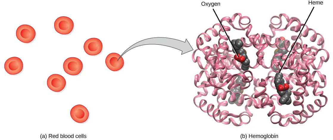
Hemoglobin, or Hb, is a protein molecule found in red blood cells (erythrocytes) made of four subunits: two alpha subunits and two beta subunits (shown below). Each subunit surrounds a central heme group that contains iron and binds one oxygen molecule, allowing each hemoglobin molecule to bind four oxygen molecules. Molecules with more oxygen bound to the heme groups are brighter red. As a result, oxygenated arterial blood where the Hb is carrying four oxygen molecules is bright red, while venous blood that is deoxygenated is darker red.
Extended description
Panel (a), left, captioned ‘Red blood cells’: seven identical red discs are scattered above the caption; a curved gray arrow leads from the rightmost disc to panel (b). Panel (b), right, captioned ‘Hemoglobin’: the molecule is drawn as pink ribbon coils forming two mirror-image halves, left and right, each a bundle of helices. A leader line labeled ‘Oxygen’ points down to a small red-and-gray space-filling cluster nested in the left half; a second leader line labeled ‘Heme’ points to a similar gray cluster in the right half. Matching gray/red clusters are visible in both halves, showing the molecule’s symmetry, though only the left cluster is labeled Oxygen and only the right one is labeled Heme.
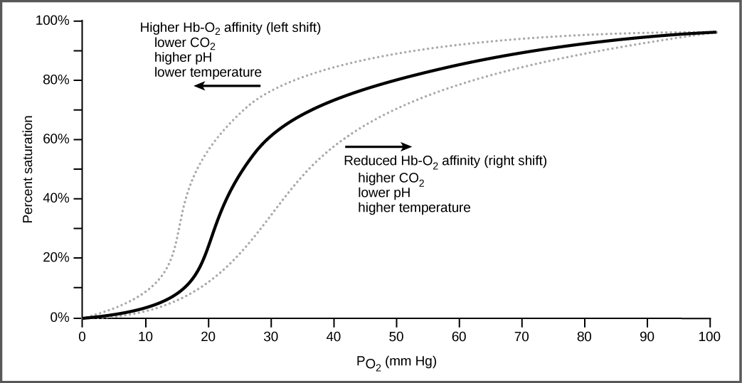
It is easier to bind a second and third oxygen molecule to Hb than the first molecule. This is because the hemoglobin molecule changes its shape, or conformation, as oxygen binds. The fourth oxygen is then more difficult to bind. The binding of oxygen to hemoglobin can be plotted as a function of the partial pressure of oxygen in the blood (x-axis) versus the relative Hb-oxygen saturation (y-axis). The resulting graph—an oxygen dissociation curve—is sigmoidal, or S-shaped (shown below). As the partial pressure of oxygen increases, the hemoglobin becomes increasingly saturated with oxygen.
Extended description
The x-axis is labeled oxygen partial pressure (PO₂) in mm Hg, from 0 to 100; the y-axis is labeled Percent saturation, from 0% to 100%. Three curves are drawn. The main, solid curve is S-shaped: it climbs slowly below about 10 mm Hg, rises quickly from roughly 20% saturation at 20 mm Hg to about 60% saturation at 30 mm Hg, then levels out, reaching nearly 100% saturation by 100 mm Hg. A dotted curve running above and to the left of the solid curve is labeled ‘Higher Hb-O₂ affinity (left shift),’ with a leftward arrow, and lists three causes beneath it: lower CO₂, higher pH, lower temperature. A second dotted curve running below and to the right of the solid curve is labeled ‘Reduced Hb-O₂ affinity (right shift),’ with a rightward arrow, and lists three causes beneath it: higher CO₂, lower pH, higher temperature.
The kidneys are responsible for removing excess H⁺ ions from the blood. If the kidneys fail, what would happen to blood pH and to hemoglobin affinity for oxygen?
Show model answer
Did your answer mention:
Factors That Affect Oxygen Binding
The oxygen-carrying capacity of hemoglobin determines how much oxygen is carried in the blood. In addition to PO₂, other environmental factors and diseases can affect oxygen carrying capacity and delivery.
Carbon dioxide levels, blood pH, and body temperature affect oxygen-carrying capacity (shown above). When carbon dioxide is in the blood, it reacts with water to form bicarbonate (HCO₃⁻) and hydrogen ions (H⁺). As the level of carbon dioxide in the blood increases, more H⁺ is produced and the pH decreases. This increase in carbon dioxide and subsequent decrease in pH reduce the affinity of hemoglobin for oxygen. The oxygen dissociates from the Hb molecule, shifting the oxygen dissociation curve to the right. Therefore, more oxygen is needed to reach the same hemoglobin saturation level as when the pH was higher. A similar shift in the curve also results from an increase in body temperature. Increased temperature, such as from increased activity of skeletal muscle, causes the affinity of hemoglobin for oxygen to be reduced.
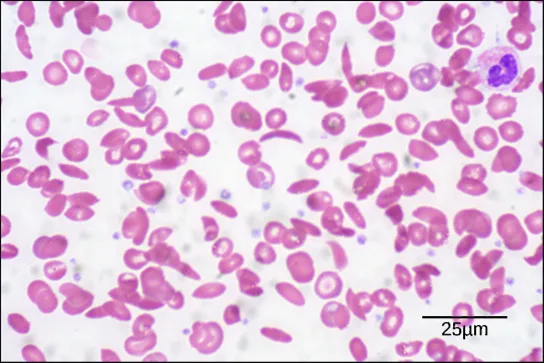
Diseases like sickle cell anemia and thalassemia decrease the blood’s ability to deliver oxygen to tissues and its oxygen-carrying capacity. In sickle cell anemia, the shape of the red blood cell is crescent-shaped, elongated, and stiffened, reducing its ability to deliver oxygen (shown below). In this form, red blood cells cannot pass through the capillaries. This is painful when it occurs. Thalassemia is a rare genetic disease caused by a defect in either the alpha or the beta subunit of Hb. Patients with thalassemia produce a high number of red blood cells, but these cells have lower-than-normal levels of hemoglobin. Therefore, the oxygen-carrying capacity is diminished.
Transport of Carbon Dioxide in the Blood
Carbon dioxide molecules are transported in the blood from body tissues to the lungs by one of three methods: dissolution directly into the blood, binding to hemoglobin, or carried as a bicarbonate ion. Several properties of carbon dioxide in the blood affect its transport. First, carbon dioxide is more soluble in blood than oxygen. About 5 to 7 percent of all carbon dioxide is dissolved in the plasma. Second, carbon dioxide can bind to plasma proteins or can enter red blood cells and bind to hemoglobin. This form transports about 10 percent of the carbon dioxide. When carbon dioxide binds to hemoglobin, a molecule called carbaminohemoglobin is formed. Binding of carbon dioxide to hemoglobin is reversible. Therefore, when it reaches the lungs, the carbon dioxide can freely dissociate from the hemoglobin and be expelled from the body.
Third, the majority of carbon dioxide molecules (85 percent) are carried as part of the bicarbonate buffer system. In this system, carbon dioxide diffuses into the red blood cells. Carbonic anhydrase (CA) within the red blood cells quickly converts the carbon dioxide into carbonic acid (H₂CO₃). Carbonic acid is an unstable intermediate molecule that immediately dissociates into bicarbonate ions (HCO₃⁻) and hydrogen (H⁺) ions. Since carbon dioxide is quickly converted into bicarbonate ions, this reaction allows for the continued uptake of carbon dioxide into the blood down its concentration gradient. It also results in the production of H⁺ ions. If too much H⁺ is produced, it can alter blood pH. However, hemoglobin binds to the free H⁺ ions and thus limits shifts in pH. The newly synthesized bicarbonate ion is transported out of the red blood cell into the liquid component of the blood in exchange for a chloride ion (Cl⁻); this is called the chloride shift. When the blood reaches the lungs, the bicarbonate ion is transported back into the red blood cell in exchange for the chloride ion. The H⁺ ion dissociates from the hemoglobin and binds to the bicarbonate ion. This produces the carbonic acid intermediate, which is converted back into carbon dioxide through the enzymatic action of CA. The carbon dioxide produced is expelled through the lungs during exhalation.
CO₂ + H₂O ↔ H₂CO₃ (carbonic acid) ↔ HCO₃⁻ + H⁺ (bicarbonate)
The benefit of the bicarbonate buffer system is that carbon dioxide is “soaked up” into the blood with little change to the pH of the system. This is important because it takes only a small change in the overall pH of the body for severe injury or death to result. The presence of this bicarbonate buffer system also allows for people to travel and live at high altitudes: When the partial pressure of oxygen and carbon dioxide change at high altitudes, the bicarbonate buffer system adjusts to regulate carbon dioxide while maintaining the correct pH in the body.
Carbon Monoxide Poisoning
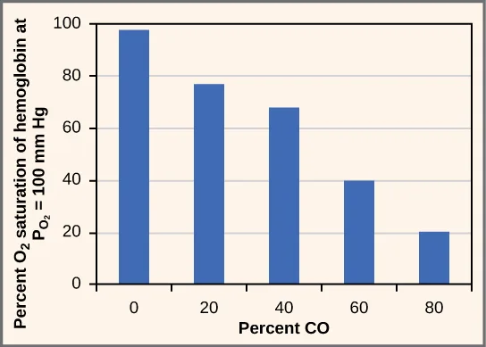
While carbon dioxide can readily associate and dissociate from hemoglobin, other molecules such as carbon monoxide (CO) cannot. Carbon monoxide has a greater affinity for hemoglobin than oxygen. Therefore, when carbon monoxide is present, it binds to hemoglobin preferentially over oxygen. As a result, oxygen cannot bind to hemoglobin, so very little oxygen is transported through the body (shown below). Carbon monoxide is a colorless, odorless gas and is therefore difficult to detect. It is produced by gas-powered vehicles and tools. Carbon monoxide can cause headaches, confusion, and nausea; long-term exposure can cause brain damage or death. Administering 100 percent (pure) oxygen is the usual treatment for carbon monoxide poisoning. Administration of pure oxygen speeds up the separation of carbon monoxide from hemoglobin.
Extended description
Five bars, one at each labeled value of percent CO — 0, 20, 40, 60, and 80 — decrease steadily in height: about 98% oxygen saturation at 0% CO, 77% at 20%, 68% at 40%, 40% at 60%, and 20% at 80%.
Summary
Hemoglobin is a protein found in red blood cells that is comprised of two alpha and two beta subunits that surround an iron-containing heme group. Oxygen readily binds this heme group. The ability of oxygen to bind increases as more oxygen molecules are bound to heme. Disease states and altered conditions in the body can affect the binding ability of oxygen, and increase or decrease its ability to dissociate from hemoglobin.
Carbon dioxide can be transported through the blood via three methods. It is dissolved directly in the blood, bound to plasma proteins or hemoglobin, or converted into bicarbonate. The majority of carbon dioxide is transported as part of the bicarbonate system. Carbon dioxide diffuses into red blood cells. Inside, carbonic anhydrase converts carbon dioxide into carbonic acid (H₂CO₃), which is subsequently hydrolyzed into bicarbonate (HCO₃⁻) and H⁺. The H⁺ ion binds to hemoglobin in red blood cells, and bicarbonate is transported out of the red blood cells in exchange for a chloride ion. This is called the chloride shift. Bicarbonate leaves the red blood cells and enters the blood plasma. In the lungs, bicarbonate is transported back into the red blood cells in exchange for chloride. The H⁺ dissociates from hemoglobin and combines with bicarbonate to form carbonic acid with the help of carbonic anhydrase, which further catalyzes the reaction to convert carbonic acid back into carbon dioxide and water. The carbon dioxide is then expelled from the lungs.
Key terms
- bicarbonate buffer system — system in the blood that absorbs carbon dioxide and regulates pH levels.
- bicarbonate (HCO₃⁻) ion — ion created when carbonic acid dissociates into H⁺ and HCO₃⁻.
- carbaminohemoglobin — molecule that forms when carbon dioxide binds to hemoglobin.
- carbonic anhydrase (CA) — enzyme that catalyzes carbon dioxide and water into carbonic acid.
- chloride shift — exchange of chloride for bicarbonate into or out of the red blood cell.
- heme group — centralized iron-containing group that is surrounded by the alpha and beta subunits of hemoglobin.
- hemoglobin — molecule in red blood cells that can bind oxygen, carbon dioxide, and carbon monoxide.
- oxygen-carrying capacity — amount of oxygen that can be transported in the blood.
- oxygen dissociation curve — curve depicting the affinity of oxygen for hemoglobin.
- sickle cell anemia — genetic disorder that affects the shape of red blood cells, and their ability to transport oxygen and move through capillaries.
- thalassemia — rare genetic disorder that results in mutation of the alpha or beta subunits of hemoglobin, creating smaller red blood cells with less hemoglobin.
Practice
Describe how oxygen is bound to hemoglobin and transported to body tissues
Which of the following will NOT facilitate the transfer of oxygen to tissues?
Three of these conditions shift the oxygen dissociation curve to the right, releasing more oxygen to the tissues — find the one that shifts it the other way.The majority of oxygen in the blood is transported by ________.
Recall what percentage of oxygen dissolves directly in the blood versus how much attaches to the iron-containing protein in red blood cells.How does the administration of 100 percent oxygen save a patient from carbon monoxide poisoning? Why wouldn’t giving carbon dioxide work?
Show model answer
Did your answer mention:
The centralized iron-containing structure that is surrounded by the alpha and beta subunits of hemoglobin and binds a single oxygen molecule is called the ________.
Each of hemoglobin’s four subunits surrounds one of these; together they hold the iron that gives blood its red color.The genetic disorder that makes red blood cells crescent-shaped, elongated, and stiffened, so they can no longer pass through capillaries, is called ________.
This disorder gets its name from the curved farm tool its affected red blood cells resemble.Explain how carbon dioxide is transported from body tissues to the lungs
The majority of carbon dioxide in the blood is transported by ________.
This route carries about 85 percent of the CO₂ that leaves the tissues — compare that share with the other two transport methods.What would happen if no carbonic anhydrase were present in red blood cells?
Show model answer
Did your answer mention:
The enzyme inside red blood cells that quickly converts carbon dioxide and water into carbonic acid is called ________.
Without this enzyme, carbon dioxide could not be hydrolyzed into the unstable intermediate that dissociates into bicarbonate.The molecule that forms when carbon dioxide, rather than oxygen, binds directly to hemoglobin is called ________.
This binding is reversible, so the molecule releases its carbon dioxide again once it reaches the lungs.The system that carries the majority of carbon dioxide out of the tissues while limiting the change to blood pH is called the ________.
It converts CO₂ into an unstable acid inside red blood cells, then into an ion exchanged for chloride at the tissues and back again at the lungs.This section is adapted from Biology 2e, Section 39.4: Transport of Gases in Human Bodily Fluids by Mary Ann Clark, Jung Choi, Matthew Douglas, and OpenStax, © OpenStax, licensed under CC BY-NC-SA 4.0. Access the original for free at openstax.org. Changes: the carbon-monoxide Critical Thinking question keeps both of its printed sentences while its model answer, like the source solution, addresses only the first (why 100 percent oxygen works) — the source never answers why carbon dioxide would not, and no answer was invented (reported as a source defect); figures re-encoded as WebP; two figures re-kinded from the manifest’s file-extension “photo” guess to “diagram” — Figure_39_04_01-8367 (a composite illustration of red blood cells and a hemoglobin ribbon diagram, not a photograph) and Figure_39_04_04 (a bar graph, not a photograph); a longdesc added to the hemoglobin-structure illustration, the oxygen dissociation curve, and the carbon-monoxide bar graph, each counted from the image (seven red blood cells and two labeled leader lines on the first; a solid curve plus two labeled, directional dotted shift-curves with three causes each on the second; five bars with their five values on the third); the inline PO2 math markup rendered as PO₂ and the ion notation (HCO₃⁻, H⁺, Cl⁻) set as Unicode throughout, per house notation; the display carbonic-acid reaction set as a Unicode text paragraph, keeping the source’s ↔ arrows, rather than as KaTeX; in-text figure pointers (“Figure 39.19” through “Figure 39.22”) replaced with “shown above”/“shown below,” since Hugo does not number figures; the note.visual-connection rendered as its figure immediately followed by a body self-check, since the module keys it with a prose solution rather than a lettered choice; the end-of-section Review Questions and Critical Thinking Questions adapted into the closing interactive Practice block (multiple choice and self-check respectively), using every keyed exercise in the module; five key-term recall items added from the glossary (heme group, sickle cell anemia, carbonic anhydrase, carbaminohemoglobin, bicarbonate buffer system); the other six glossary terms (bicarbonate ion, chloride shift, hemoglobin, oxygen-carrying capacity, oxygen dissociation curve, thalassemia) appear only in the Key terms list and the prose; rubric checkpoints added to each self-check, decomposing its model answer (the source solution) into check-off clauses with no new claims.