Overview of the Circulatory System
By the end of this section, you will be able to:
- Describe an open and closed circulatory system
- Describe interstitial fluid and hemolymph
- Compare and contrast the organization and evolution of the vertebrate circulatory system
In all animals, except a few simple types, the circulatory system is used to transport nutrients and gases through the body. Simple diffusion allows some water, nutrient, waste, and gas exchange into primitive animals that are only a few cell layers thick; however, bulk flow is the only method by which the entire body of larger more complex organisms is accessed.
Circulatory System Architecture
The circulatory system is effectively a network of cylindrical vessels: the arteries, veins, and capillaries that emanate from a pump, the heart. In all vertebrate organisms, as well as some invertebrates, this is a closed-loop system, in which the blood is not free in a cavity. In a closed circulatory system, blood is contained inside blood vessels and circulates unidirectionally from the heart around the systemic circulatory route, then returns to the heart again, as illustrated below in (a). As opposed to a closed system, arthropods—including insects, crustaceans, and most mollusks—have an open circulatory system, as illustrated below in (b). In an open circulatory system, the blood is not enclosed in the blood vessels but is pumped into a cavity called a hemocoel and is called hemolymph because the blood mixes with the interstitial fluid. As the heart beats and the animal moves, the hemolymph circulates around the organs within the body cavity and then reenters the hearts through openings called ostia. This movement allows for gas and nutrient exchange. An open circulatory system does not use as much energy as a closed system to operate or to maintain; however, there is a trade-off with the amount of blood that can be moved to metabolically active organs and tissues that require high levels of oxygen. In fact, one reason that insects with wing spans of up to two feet wide (70 cm) are not around today is probably because they were outcompeted by the arrival of birds 150 million years ago. Birds, having a closed circulatory system, are thought to have moved more agilely, allowing them to get food faster and possibly to prey on the insects.
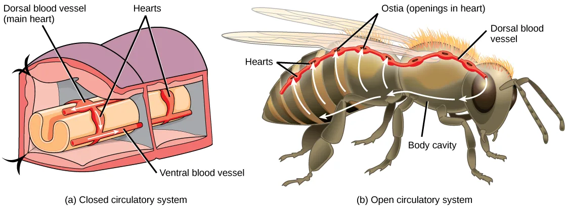
Extended description
(a) One leader line labels Dorsal blood vessel (main heart), the tube along the top of the cutaway, where a white arrow shows blood flowing toward the front of the segment. Two leader lines from the label Hearts point to two ring-like vessels looping down from the dorsal vessel to the ventral vessel — one in the near segment, one in the segment behind it — with a white arrow showing blood pumped downward through the near one. One leader line labels Ventral blood vessel, the tube along the bottom, where a white arrow shows blood flowing toward the rear of the body. (b) One leader line labels Dorsal blood vessel, the vessel narrowing toward the head. Two leader lines from the label Ostia (openings in heart) point to two openings on the vessel near the thorax-abdomen boundary, and two leader lines from the label Hearts point to two openings further back along the abdomen. One leader line labels Body cavity, the open space beneath the thorax. White arrows trace hemolymph leaving the vessel’s open tip near the head, sweeping backward beneath the wings and thorax into the body cavity, then rising through five points along the abdomen back into the dorsal vessel, which carries it forward again.
Circulatory System Variation in Animals
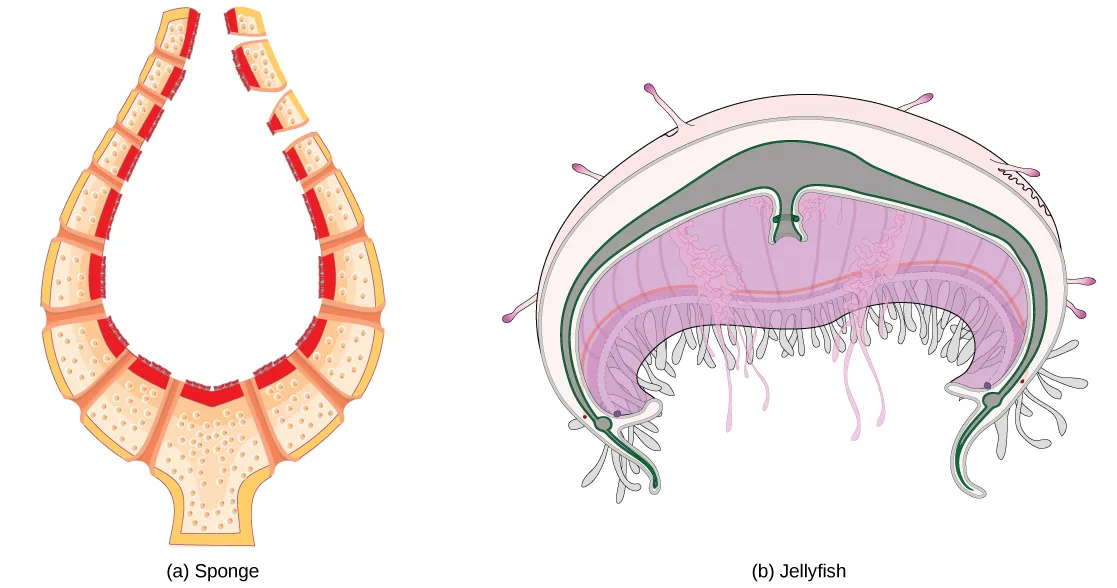
The circulatory system varies from simple systems in invertebrates to more complex systems in vertebrates. The simplest animals, such as the sponges (Porifera) and rotifers (Rotifera), do not need a circulatory system because diffusion allows adequate exchange of water, nutrients, and waste, as well as dissolved gases, as shown below in (a). Organisms that are more complex but still only have two layers of cells in their body plan, such as jellies (Cnidaria) and comb jellies (Ctenophora) also use diffusion through their epidermis and internally through the gastrovascular compartment. Both their internal and external tissues are bathed in an aqueous environment and exchange fluids by diffusion on both sides, as illustrated below in (b). Exchange of fluids is assisted by the pulsing of the jellyfish body.
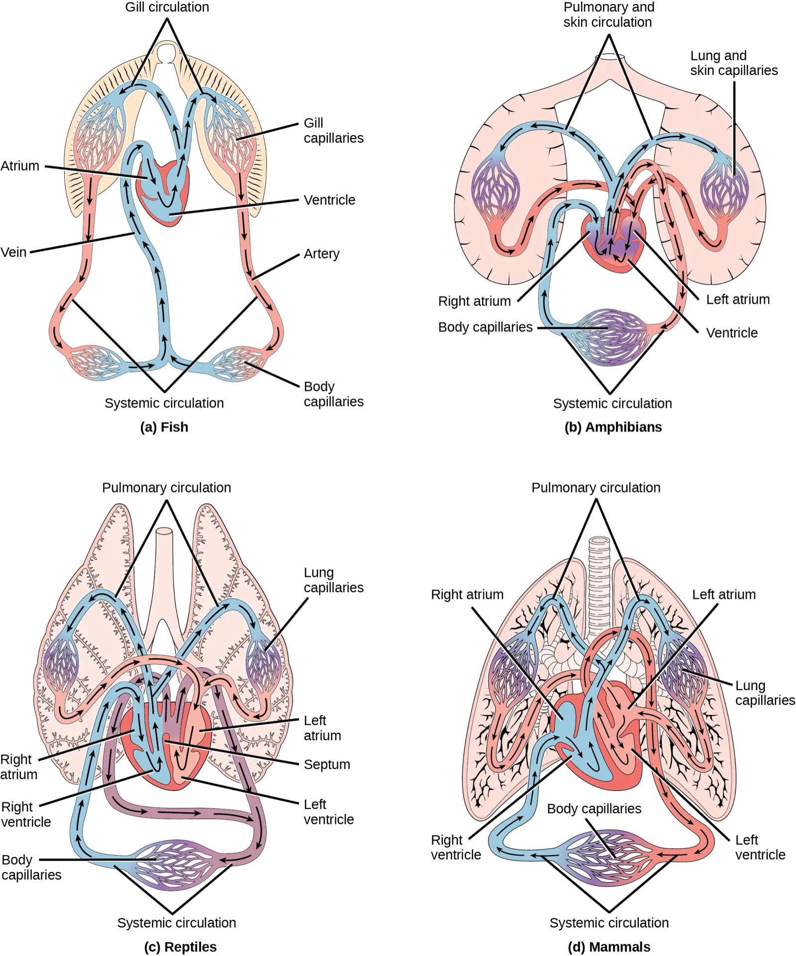
For more complex organisms, diffusion is not efficient for cycling gases, nutrients, and waste effectively through the body; therefore, more complex circulatory systems evolved. Most arthropods and many mollusks have open circulatory systems. In an open system, an elongated beating heart pushes the hemolymph through the body and muscle contractions help to move fluids. The larger more complex crustaceans, including lobsters, have developed arterial-like vessels to push blood through their bodies, and the most active mollusks, such as squids, have evolved a closed circulatory system and are able to move rapidly to catch prey. Closed circulatory systems are a characteristic of vertebrates; however, there are significant differences in the structure of the heart and the circulation of blood between the different vertebrate groups due to adaptation during evolution and associated differences in anatomy. The figure below illustrates the basic circulatory systems of some vertebrates: fish, amphibians, reptiles, and mammals.
Extended description
(a) Fish: one leader line labels Atrium, the chamber blood enters from the Vein (one leader line, left), and one leader line labels Ventricle, the chamber below it. Arrows trace blood from the atrium into the ventricle, up through both branches of a bracket labeled Gill circulation to the two Gill capillaries (one leader line, right), then down through both branches of a bracket labeled Artery (one leader line, right) to a bracket labeled Systemic circulation and into the two Body capillaries (one leader line, right) at the bottom, and back up through the vein to the atrium — one continuous loop. (b) Amphibians: one leader line each labels Right atrium and Left atrium, the two upper chambers, and one leader line labels Ventricle, the single lower chamber where they join. One leader line labels Body capillaries, the single capillary bed at the bottom, reached through a bracket labeled Systemic circulation. One leader line labels Lung and skin capillaries, one of the two bilateral capillary beds at the top, reached through a bracket labeled Pulmonary and skin circulation. Arrows show blood rising from the body capillaries through the right atrium into the ventricle, out to both lung-and-skin capillary beds, back through the left atrium into the ventricle again, and out to the body capillaries, the two loops’ arrows crossing where they meet in the ventricle. (c) Reptiles: labeled the same as amphibians (Right atrium, Left atrium, Body capillaries, Lung capillaries, and brackets for Pulmonary circulation and Systemic circulation) plus one leader line labeling Septum, the partial wall drawn between Right ventricle and Left ventricle (one leader line each) at the base of the heart. Arrows trace the same two-loop pattern as the amphibian heart, but with a labeled partial septum between the two ventricles and a purple blend where the blue and red ventricle colours meet above it, showing partial mixing. (d) Mammals: one leader line each labels Right atrium, Left atrium, Right ventricle, and Left ventricle — four separate chambers, with no gap drawn between the ventricles and so no septum label. Lung capillaries and Body capillaries each carry one leader line, and brackets label Pulmonary circulation and Systemic circulation as in the other panels. Arrows trace blood entering the right atrium and right ventricle, out to both lung capillary beds, back through the left atrium and left ventricle, and out to the body capillaries, with no arrow crossing between the two sides.
As illustrated above in (a), fish have a single circuit for blood flow and a two-chambered heart that has only a single atrium and a single ventricle. The atrium collects blood that has returned from the body and the ventricle pumps the blood to the gills where gas exchange occurs and the blood is re-oxygenated; this is called gill circulation. The blood then continues through the rest of the body before arriving back at the atrium; this is called systemic circulation. This unidirectional flow of blood produces a gradient of oxygenated to deoxygenated blood around the fish’s systemic circuit. The result is a limit in the amount of oxygen that can reach some of the organs and tissues of the body, reducing the overall metabolic capacity of fish.
In amphibians, reptiles, birds, and mammals, blood flow is directed in two circuits: one through the lungs and back to the heart, which is called pulmonary circulation, and the other throughout the rest of the body and its organs including the brain (systemic circulation). In amphibians, gas exchange also occurs through the skin during pulmonary circulation and is referred to as pulmocutaneous circulation.
As shown above in (b), amphibians have a three-chambered heart that has two atria and one ventricle rather than the two-chambered heart of fish. The two atria (superior heart chambers) receive blood from the two different circuits (the lungs and the systems), and then there is some mixing of the blood in the heart’s ventricle (inferior heart chamber), which reduces the efficiency of oxygenation. The advantage to this arrangement is that high pressure in the vessels pushes blood to the lungs and body. The mixing is mitigated by a ridge within the ventricle that diverts oxygen-rich blood through the systemic circulatory system and deoxygenated blood to the pulmocutaneous circuit. For this reason, amphibians are often described as having double circulation.
Most reptiles also have a three-chambered heart similar to the amphibian heart that directs blood to the pulmonary and systemic circuits, as shown above in (c). The ventricle is divided more effectively by a partial septum, which results in less mixing of oxygenated and deoxygenated blood. Some reptiles (alligators and crocodiles) are the most primitive animals to exhibit a four-chambered heart. Crocodilians have a unique circulatory mechanism where the heart shunts blood from the lungs toward the stomach and other organs during long periods of submergence, for instance, while the animal waits for prey or stays underwater waiting for prey to rot. One adaptation includes two main arteries that leave the same part of the heart: one takes blood to the lungs and the other provides an alternate route to the stomach and other parts of the body. Two other adaptations include a hole in the heart between the two ventricles, called the foramen of Panizza, which allows blood to move from one side of the heart to the other, and specialized connective tissue that slows the blood flow to the lungs. Together these adaptations have made crocodiles and alligators one of the most evolutionarily successful animal groups on earth.
In mammals and birds, the heart is also divided into four chambers: two atria and two ventricles, as illustrated above in (d). The oxygenated blood is separated from the deoxygenated blood, which improves the efficiency of double circulation and is probably required for the warm-blooded lifestyle of mammals and birds. The four-chambered heart of birds and mammals evolved independently from a three-chambered heart. The independent evolution of the same or a similar biological trait is referred to as convergent evolution.
Summary
In most animals, the circulatory system is used to transport blood through the body. Some primitive animals use diffusion for the exchange of water, nutrients, and gases. However, complex organisms use the circulatory system to carry gases, nutrients, and waste through the body. Circulatory systems may be open (mixed with the interstitial fluid) or closed (separated from the interstitial fluid). Closed circulatory systems are a characteristic of vertebrates; however, there are significant differences in the structure of the heart and the circulation of blood between the different vertebrate groups due to adaptations during evolution and associated differences in anatomy. Fish have a two-chambered heart with unidirectional circulation. Amphibians have a three-chambered heart, which has some mixing of the blood, and they have double circulation. Most non-avian reptiles have a three-chambered heart, but have little mixing of the blood; they have double circulation. Mammals and birds have a four-chambered heart with no mixing of the blood and double circulation.
Key terms
- atrium — (plural: atria) chamber of the heart that receives blood from the veins and sends blood to the ventricles.
- closed circulatory system — system in which the blood is separated from the bodily interstitial fluid and contained in blood vessels.
- double circulation — flow of blood in two circuits: the pulmonary circuit through the lungs and the systemic circuit through the organs and body.
- gill circulation — circulatory system that is specific to animals with gills for gas exchange; the blood flows through the gills for oxygenation.
- hemocoel — cavity into which blood is pumped in an open circulatory system.
- hemolymph — mixture of blood and interstitial fluid that is found in insects and other arthropods as well as most mollusks.
- interstitial fluid — fluid between cells.
- open circulatory system — system in which the blood is mixed with interstitial fluid and directly covers the organs.
- ostium — (plural: ostia) holes between blood vessels that allow the movement of hemolymph through the body of insects, arthropods, and mollusks with open circulatory systems.
- pulmocutaneous circulation — circulatory system in amphibians; the flow of blood to the lungs and the moist skin for gas exchange.
- pulmonary circulation — flow of blood away from the heart through the lungs where oxygenation occurs and then returns to the heart again.
- systemic circulation — flow of blood away from the heart to the brain, liver, kidneys, stomach, and other organs, the limbs, and the muscles of the body, and then the return of this blood to the heart.
- unidirectional circulation — flow of blood in a single circuit; occurs in fish where the blood flows through the gills, then past the organs and the rest of the body, before returning to the heart.
- ventricle — (heart) large inferior chamber of the heart that pumps blood into arteries.
Practice
Describe an open and closed circulatory system
Why are open circulatory systems advantageous to some animals?
The passage on wing-spanned insects and birds explains this as a trade-off: a closed system can move more blood to active tissues, but there’s a cost to keeping blood confined to vessels — think about what the open system saves by skipping that.Some animals use diffusion instead of a circulatory system. Examples include:
Check each pair of animals against what this section actually says about them — does each one truly lack a circulatory system, or does it have an open or closed one instead?Describe a closed circulatory system.
Show model answer
Did your answer mention:
A system in which blood is separated from the bodily interstitial fluid and contained in blood vessels is called a ________.
This is the opposite of the system found in insects, where blood mixes with body fluid in an open cavity.A system in which blood mixes with interstitial fluid and directly bathes the organs is called a(n) ________.
Insects, crustaceans, and most mollusks use this kind of system, where the pumped fluid enters a body cavity rather than staying inside vessels.Describe interstitial fluid and hemolymph
The fluid found between cells is called ________.
In an open circulatory system, this fluid mixes directly with the circulating blood.The mixture of blood and interstitial fluid found in insects, other arthropods, and most mollusks is called ________.
This fluid forms because these animals’ circulatory systems are open rather than closed.The cavity into which blood is pumped in an open circulatory system is called the ________.
This is the body cavity the pumped fluid fills as it circulates around an insect’s organs before reentering the heart.A single hole between blood vessels that allows hemolymph to move through an insect’s body is called an ________.
There are several of these small openings along an insect’s dorsal blood vessel, each one letting hemolymph flow back into the heart.Compare and contrast the organization and evolution of the vertebrate circulatory system
Blood flow that is directed through the lungs and back to the heart is called ________.
This is one of two circuits described for animals with lungs — the other circuit serves the brain, organs, and muscles rather than a gas-exchange surface.Describe systemic circulation.
Show model answer
Did your answer mention:
The heart chamber that receives blood from the veins and sends it to the ventricle is called the ________.
Fish have a single one of these chambers; mammals have two, one on each side of the heart.The large inferior heart chamber that pumps blood into the arteries is called the ________.
This chamber sits below the atrium and does the heavier pumping work of the two.Blood flow organized into two circuits — one through the lungs and one through the rest of the body — is called ________.
Amphibians, reptiles, and mammals are all described this way, in contrast to the single circuit of fish.The circulatory route through the gills, where blood is oxygenated in animals that have gills, is called ________.
Fish rely on this single route rather than a separate lung-specific one.This section is adapted from Biology 2e, Section 40.1: Overview of the Circulatory System by Mary Ann Clark, Jung Choi, Matthew Douglas, and OpenStax, © OpenStax, licensed under CC BY-NC-SA 4.0. Access the original for free at openstax.org. Changes: figures re-encoded as WebP; two figures re-kinded from the manifest’s file-extension “photo” guess to “diagram” (Figure_40_01_01ab and Figure_40_01_02ab are each a hand-drawn line illustration, not a captured photograph); a longdesc added to the earthworm/bee circulatory-system illustration and to the four-panel vertebrate circulatory-system diagram, transcribing each drawing’s own printed labels, leader lines, and arrow directions in reading order; in-text pointers to figures (“Figure 40.2” through “Figure 40.4”) replaced with “shown below,” “shown above,” or “illustrated below,” and inline references to sub-panels kept as “(a)"–"(d),” since Hugo does not number figures; the end-of-section Review Questions and Critical Thinking Questions adapted into the closing interactive Practice block (multiple choice and self-check respectively); rubric checkpoints added to each self-check, decomposing its model answer (the source solution) into check-off clauses with no new claims; ten key-term recall items added from the glossary (closed circulatory system, open circulatory system, interstitial fluid, hemolymph, hemocoel, ostium, atrium, ventricle, double circulation, gill circulation) — of the remaining four glossary terms, systemic circulation and pulmonary circulation are each tested by a retained Review Question or Critical Thinking Question, and pulmocutaneous circulation and unidirectional circulation appear only in the Key terms list and the prose.